Tag: vision

Study Finds that Perception of Colour Fades with Age

Photo by Mari Lezhava on Unsplash

There is a difference between how the brains of healthy older adults perceive colour compared to younger adults, finds a new study led by UCL researchers.

The research, published in Scientific Reports, compared how the pupils of younger and older people reacted to different aspects of colour in the environment.

The team recruited 17 healthy young adults with an average age of 27.7, and 20 healthy older adults with an average age of 64.4.

Participants were placed in a blackout room and shown 26 different colours for five seconds each, while the researchers measured the diameter of their pupils.

Pupils constrict in response to increases in colour lightness and chroma (colourfulness).

The colours shown included dark, muted, saturated and light shades of magenta, blue, green, yellow and red, alongside two shades of orange and four greyscale colours.

Using a highly sensitive eye tracking camera*, which recorded the pupil diameter at 1000 times per second, the team found that the pupils of healthy older people constricted less in response to colour chroma compared with young adults. This was particularly marked for green and magenta hues.

However, both younger and older adults had similar responses to the ‘lightness’ of a colour shade.

The study is the first to use pupillometry to show that as we grow older, our brains become less sensitive to the intensity of colours in the world around us.

The findings of the study also complement previous behavioural research that showed that older adults perceive surface colours to be less colourful than young adults.

Lead author, Dr Janneke van Leeuwen (UCL Queen Square Institute of Neurology), said: “This work brings into question the long-held belief among scientists that colour perception remains relatively constant across the lifespan, and suggests instead that colours slowly fade as we age. Our findings might also help explain why our colour preferences may alter as we age – and why at least some older people may prefer to dress in bold colours.”

The researchers believe that as we get older there is a decline in the body’s sensitivity to the saturation levels of colours within the primary visual cortex – the part of the brain that receives, integrates, and processes visual information relayed from the retinas.

Previous research also showed this to be a feature of a rare form of dementia called posterior cortical atrophy (PCA), where noticeable difficulties and abnormalities in colour perception could be due to a significant decline in the brain’s sensitivity to certain colour tones (specifically green and magenta) in the primary visual cortex and it’s connected networks.

Co-corresponding author, Professor Jason Warren (UCL Queen Square Institute of Neurology), said: “Our findings could have wide implications for how we adapt fashion, décor and other colour ‘spaces’ for older people, and potentially even for our understanding of diseases of the ageing brain, such as dementia. People with dementia can show changes in colour preferences and other symptoms relating to the visual brain – to interpret these correctly, we first need to gauge the effects of healthy ageing on colour perception. Further research is therefore needed to delineate the functional neuroanatomy of our findings, as higher cortical areas might also be involved.”

Source: University College London

Gene Therapy Partially Restores Cone Function in Achromatopsia

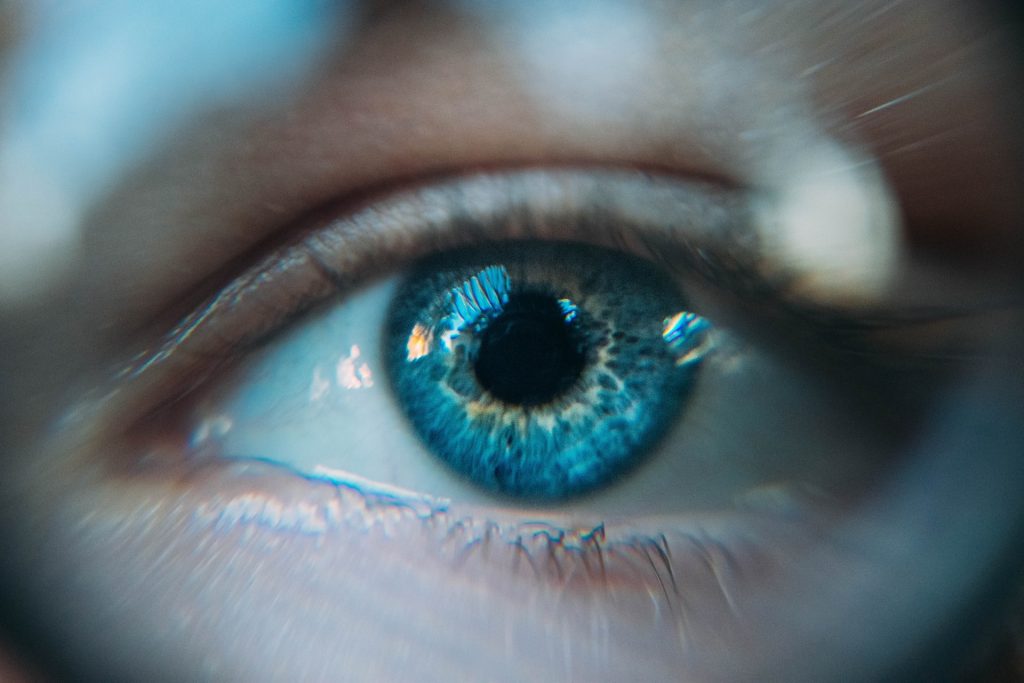
Eye
Source: Daniil Kuzelev on Unsplash

University of College London researchers have used gene therapy to partially restore the function of cone receptors in two children with achromatopsia, a rare genetic disorder which can cause partial or complete colourblindness.

The findings, published in Brain, suggest that treatment activates previously dormant communication links between the retina and the brain, thanks to the developing adolescent brain’s plastic nature.

The academically-led study has been running alongside a phase 1/2 clinical trial in children with achromatopsia, using a new way to test whether the treatment is changing the neural pathways specific to the cones.

Achromatopsia is caused by disease-causing variants to one of a few genes. As it affect the cones in the retina, are responsible for colour vision, people with achromatopsia are completely colourblind, while they also have very poor vision and photophobia. Their cone cells do not send signals to the brain, but many remain present, so researchers have been seeking to activate the dormant cells.

Lead author Dr Tessa Dekker said: “Our study is the first to directly confirm widespread speculation that gene therapy offered to children and adolescents can successfully activate the dormant cone photoreceptor pathways and evoke visual signals never previously experienced by these patients.

“We are demonstrating the potential of leveraging the plasticity of our brains, which may be particularly able to adapt to treatment effects when people are young.”

The study involved four young people with achromatopsia aged 10 to 15 years old.

The two trials, which each target a different gene implicated in achromatopsia, are testing gene therapies with the primary aim of establishing that the treatment is safe, while also testing for improved vision. Their results have not yet been fully compiled so the overall effectiveness of the treatments remains to be determined.

The accompanying academic study used a novel functional magnetic resonance imaging (fMRI) mapping approach to separate emerging post-treatment cone signals from existing rod-driven signals in patients, allowing the researchers to pinpoint any changes in visual function, after treatment, directly to the targeted cone photoreceptor system. They employed a ‘silent substitution’ technique using pairs of lights to selectively stimulate cones or rods. The researchers also had to adapt their methods to accommodate eye movements due to nystagmus, another symptom of achromatopsia. The results were compared to tests involving nine untreated patients and 28 volunteers with normal vision.

Each of the four children was treated with gene therapy in one eye, enabling doctors to compare the treatment’s effectiveness with the untreated eye.

For two of the four children, there was strong evidence for cone-mediated signals in the brain’s visual cortex coming from the treated eye, six to 14 months after treatment. Before the treatment, the patients showed no evidence of cone function on any tests. After treatment, their measures closely resembled those from normal sighted study participants.

The study participants also completed a test to distinguish between different levels of contrast. This showed there was a difference in cone-supported vision in the treated eyes in the same two children.

The researchers say they cannot confirm whether the treatment was ineffective in the other two study participants, or if there may have been treatment effects that were not picked up by the tests they used, or if effects are delayed.

Co-lead author Dr Michel Michaelides (UCL Institute of Ophthalmology and Moorfields Eye Hospital), who is also co-investigator on both clinical trials, said: “In our trials, we are testing whether providing gene therapy early in life may be most effective while the neural circuits are still developing. Our findings demonstrate unprecedented neural plasticity, offering hope that treatments could enable visual functions using signalling pathways that have been dormant for years.

“We are still analysing the results from our two clinical trials, to see whether this gene therapy can effectively improve everyday vision for people with achromatopsia. We hope that with positive results, and with further clinical trials, we could greatly improve the sight of people with inherited retinal diseases.”

Dr Dekker added: “We believe that incorporating these new tests into future clinical trials could accelerate the testing of ocular gene therapies for a range of conditions, by offering unparalleled sensitivity to treatment effects on neural processing, while also providing new and detailed insight into when and why these therapies work best.”

One of the study participants commented: “Seeing changes to my vision has been very exciting, so I’m keen to see if there are any more changes and where this treatment as a whole might lead in the future.

“It’s actually quite difficult to imagine what or just how many impacts a big improvement in my vision could have, since I’ve grown up with and become accustomed to low vision, and have adapted and overcome challenges (with a lot of support from those around me) throughout my life.”

Source: University College London

Illusion Tricks Pupils into Dilating

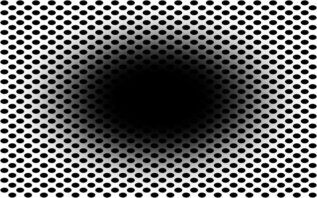
Looking at this image, do you perceive that the central black hole is expanding, as if you’re moving into a dark environment, or falling into a hole? If so, you’re not alone: a new study published in Frontiers in Human Neuroscience shows that this ‘expanding hole’ illusion, which is new to science, is perceived by approximately 86% of people.

The study’s first author, Professor Bruno Laeng at the University of Oslo, explained: “The ‘expanding hole’ is a highly dynamic illusion: The circular smear or shadow gradient of the central black hole evokes a marked impression of optic flow, as if the observer were heading forward into a hole or tunnel.”

Optical illusions aren’t simple curiosities: researchers study them to better understand the complex processes our visual system uses to anticipate and make sense of the visual world.

In the new study, Prof Laeng and colleagues demonstrated that the ‘expanding hole’ illusion deceives the brain so well that it even prompts a dilation reflex of the pupils to let in more light, just as if the observer was entering a dark area.

“Here we show based on the new ‘expanding hole’ illusion that that the pupil reacts to how we perceive light – even if this ‘light’ is imaginary like in the illusion – and not just to the amount of light energy that actually enters the eye. The illusion of the expanding hole prompts a corresponding dilation of the pupil, as it would happen if darkness really increased,” said Prof Laeng.

Prof Laeng and colleagues explored how the colour of the hole (besides black: blue, cyan, green, magenta, red, yellow, or white) and of the surrounding dots affect how strongly we mentally and physiologically react to the illusion. On a screen they presented variations of the “expanding hole” image to 50 women and men with normal vision, asking them to rate subjectively how strongly they perceived the illusion. While participants gazed at the image, the researchers measured their eye movements and their pupils’ unconscious constrictions and dilations. As controls, the participants were shown “scrambled” versions of the expanding hole image, with equal luminance and colours, but without any pattern.

The illusion appeared most effective when the hole was black. Fourteen percent of participants didn’t perceive any illusory expansion when the hole was black, while 20% didn’t if the hole was in color. Among those who did perceive an expansion, the subjective strength of the illusion differed markedly.

The researchers also found that black holes promoted strong reflex dilations of the participants’ pupils, while coloured holes prompted pupil to constriction. For black holes, but not for coloured holes, the stronger participants rated their perception of the illusion, the more their pupil diameter tended to change.

Minority not susceptible

Just why a minority seem unsusceptible to the “expanding hole” illusion is still unclear. It is also not known whether other vertebrate species, or even nonvertebrate animals with camera eyes such as octopuses, might perceive the same illusion as we do.

“Our results show that pupils’ dilation or contraction reflex is not a closed-loop mechanism, like a photocell opening a door, impervious to any other information than the actual amount of light stimulating the photoreceptor. Rather, the eye adjusts to perceived and even imagined light, not simply to physical energy. Future studies could reveal other types of physiological or bodily changes that can ‘throw light’ onto how illusions work,” concluded Prof Laeng.

Source: Frontiers

Anti-HIV Drugs may Combat Macular Degeneration

New research has shown that anti-HIV drugs may fight macular degeneration – overturning a preconception about DNA in the process.

Macular degeneration is the leading cause of blindness in developed countries. Even though HIV does not cause dry macular degeneration, the drugs prevented the loss of vision.

“We are extremely excited that the reduced risk was reproduced in all the databases, each with millions of patients,” said Jayakrishna Ambati, MD, a leading macular degeneration researcher at the University of Virginia School of Medicine. “This finding provides real hope in developing the first treatment for this blinding disease.”

A Big Data Archeology review of four health insurance databases showed that Nucleoside Reverse Transcriptase Inhibitors (NRTIs), a commonly used HIV treatment, reduced the incidence of dry macular degeneration by 40%. The records spanned two decades and covered over 100 million patients. The drugs had also previously been shown to possibly prevent diabetes.

The finding also comes with the discovery that DNA can be produced inside the cytoplasm. Alu DNA (found exclusively in primates), which makes up 10% of the human genome, is transposable and can insert itself into other places on the genome. It was long considered “junk” DNA, but are now believed to have important functions, such as allowing for multiple expressions of proteins from a single Alu element. Since it cannot replicate itself, Alu DNA requires a transposon called L1 to accomplish this, which was now reported to allow the production of Alu DNA outside the chromosome. The buildup of Alu DNA in cells contributes to macular degeneration, by killing off cells that support the retina.
The researchers are urging further investigation into NRTIs or safer derivatives known as Kamuvudines, both of which block a key inflammatory pathway, can be useful in preventing vision loss from dry macular degeneration.

“A clinical trial of these inflammasome-inhibiting drugs is now warranted,” said Ambati. “It’s also fascinating how uncovering the intricate biology of genetics and combining it with big data archeology can propel insights into new medicines.”

Source: Medical Xpress

Journal Information: Shinichi Fukuda el al., “Cytoplasmic synthesis of endogenous Alu complementary DNA via reverse transcription and implications in age-related macular degeneration,” PNAS (2021). www.pnas.org/cgi/doi/10.1073/pnas.202275111