Tag: viruses

A New Virus-like Entity has Just been Discovered – ‘Obelisks’ Explained

Photo by National Cancer Institute on Unsplash

Ed Feil, University of Bath

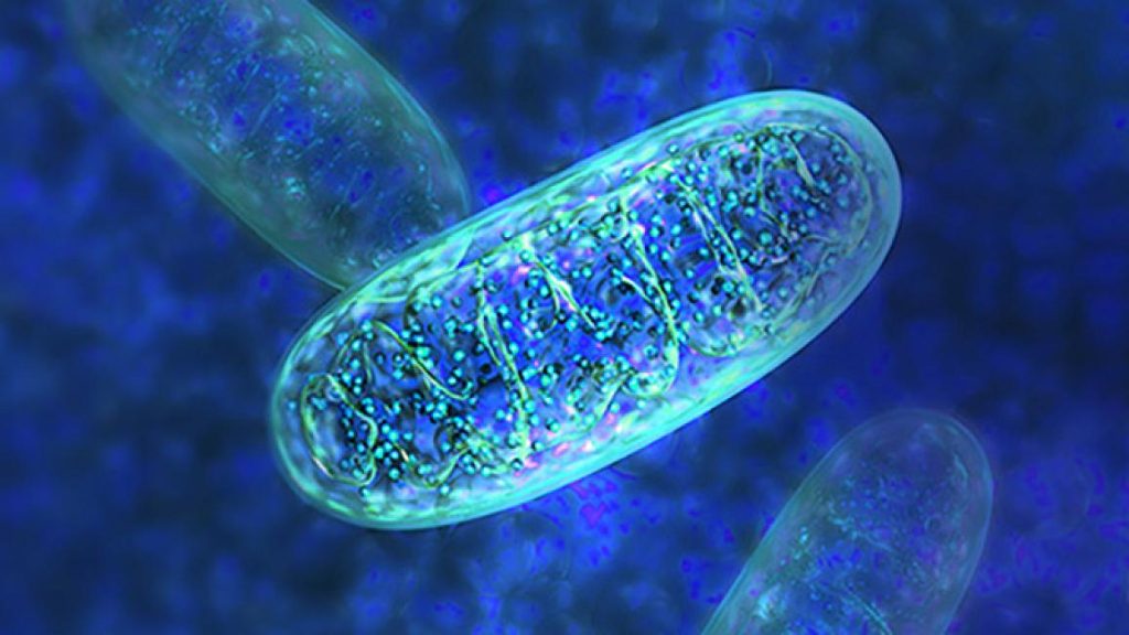
Biological entities called obelisks have been hiding – in large numbers – inside the human mouth and gut. These microscopic entities, which were recently discovered by a team at Stanford University, are circular bits of genetic material that contain one or two genes and self-organise into a rod-like shape.

Although the study is still in preprint form, meaning that it has not been peer-reviewed, it has already been extensively written about, including in two heavyweight journals: Nature and Science.

Let’s delve deeper into the strange world of very tiny “lifeforms”.

In biology, as in physics, things can get weirder and the rules fuzzier as we move through smaller and smaller scales.

Viruses, being unable to replicate without the help of a host, can most generously be considered to be on the edge of what constitutes life. Yet the estimated 10 nonillion (one followed by 31 zeroes) individual viruses on the planet can be found in every conceivable habitat and, through infecting and manipulating their hosts, have probably affected the evolutionary trajectories of all life.

Peering even further down into the world of minuscule biological entities, are the viroids – tiny scraps of genetic material (DNA-like molecules known as RNA) that cannot make proteins and, unlike viruses, don’t have a protective shell to encase their genome.

Viroids are examples of ribozymes: RNA molecules that may be a distant echo of the very first self-replicating genetic elements from which cellular life emerged.

Viroids can self-cleave (chop up) and re-ligate (stick back together) their genome as part of the replication cycle. And, despite their simplicity, they can cause serious disease in flowering plants.

Between a virus and a viroid – perhaps

The new preprint describes “viroid-like colonists of human microbiomes”. If “viroid-like” sounds non-committal, that is entirely deliberate. The newly discovered biological entity falls somewhere between viruses and viroids.

In fact, the name obelisks was proposed not only because of their shape, but also to provide wiggle room in case they turn out to be more like RNA plasmids (a different type of genetic element that resides inside bacteria) than either viruses or viroids.

Like viroids, obelisks have a circular single-stranded RNA genome and no protein coat but, like viruses, their genomes contain genes that are predicted to code for proteins.

All obelisks so far described encode a single major protein known as obulin, and many encode a second, smaller obulin.

Obulins bear no evolutionary resemblance, or “homology”, to any other protein found, and there are few clues as to their function.

By analysing existing datasets taken from the gut and mouth of humans as well as other diverse sources, the Stanford team found almost 30,000 distinct obelisk types.

These obelisk genomes have been previously overlooked because they are so dissimilar to anything described previously. The Stanford team found them using a clever bespoke method for searching databases for single-stranded circular RNA molecules to fish out any viroid-like elements.

It is clear from their results that obelisks are not rare. The researchers found them in datasets spanning the globe and in diverse niches.

These elements were detected in around 7% of microbiome datasets from the human gut and 50% of datasets from the mouth. However, whether these datasets provide a true representation of the prevalence and distribution of obelisks is unclear.

Different obelisk types were found in different body sites and in different donors. Long-term data revealed that people can harbour a single obelisk type for around a year.

Obelisks probably rely on microbial host cells to replicate, including those that live inside humans to replicate. Bacteria or fungi are likely hosts, but it is not known which exact species harbour these elements.

However, the researchers provide a critical lead through the analysis by providing strong evidence that a common bacterial component of dental plaque, Streptococcus sanguinis, plays host to a specific obelisk type.

We might have to rethink the gut microbiome.
Credit Darryl Leja National Human Genome Research Institute National Institutes Of Health

Friend or foe?

As S sanguinis is easy to grow and experiment on in the laboratory, this will provide a valuable model for understanding the fundamentals of obelisk biology.

This is critical, as nothing is known about the broader evolutionary and ecological significance of obelisks. They may be parasitic and harm host cells, or they may be beneficial.

Hosts may have evolved elaborate defence mechanisms against obelisks, or else actively recruit them to gain some unsuspected advantage. If obelisks change or upset the human microbiome, this may in turn have implications for human health – they may even have therapeutic potential.

Alternatively, obelisks may cause neither harm nor benefit to their microbial host, or to humans. Instead, they may simply exist as stealthy evolutionary passengers, silently and endlessly replicating, like the original “selfish gene”.

Ed Feil, Professor of Microbial Evolution at The Milner Centre for Evolution, University of Bath

This article is republished from The Conversation under a Creative Commons license. Read the original article.

Normal Breathing Can Transport Viruses Over 2 Metres

Researchers have demonstrated that normal breathing can transport viruses in saliva droplets up a distance of up to 2.2 metres in 90 seconds.

The World Health Organization and the Centers for Disease Control recommend social distancing to prevent the spread of COVID. The distances are estimated from various studies, but there is a need for further research into how viruses are transported from one person to another. 
Previous studies considered aerosol transport after coughing or sneezing, while this study focused on normal human breathing, using computer simulations with a more realistic model than prior studies. A normal breath produces periodic jet flows that contain saliva droplets, but those jets’ velocity is less than a tenth that of a cough or sneeze.

Wearing a face mask greatly reduces the distance which these droplets can travel. Saliva droplets restricted by a mask had travelled only 0.72 metres after two minutes, far short of the distance of 1.8 metres suggested by the CDC.

The investigators found even normal breathing produces a complex field of vortices that can move saliva droplets away from the person’s mouth. The role of these vortices has not previously been understood.

Study author, Ali Khosronejad, American Institute of Physics said: “Our results show that normal breathing without a facial mask generates periodic trailing jets and leading circular vortex rings that propagate forward and interact with the vortical flow structures produced in prior breathing cycles.”

This complex vorticity field can enable the transport of aerosol droplets over long distances despite the slow speeds. A face mask serves to dissipate the kinetic energy of the jet produced by an exhaled breath, thereby disrupting the vortices and limiting the travel of virus-laden droplets.

The researchers also took into account evaporation of the saliva droplets. With no mask, they found the saliva droplets near the front of the plume of exhaled breath had partially evaporated, reaching a size of only one-tenth of a micrometre. In stagnant indoor air, it would take days for droplets this small to settle to the ground.

Masks partially redirect the exhaled breath downward, significantly restricting forward motion of the plume, so the risk of suspended droplets remaining in the air is substantially reduced.

“To simplify the breathing process, we did not consider the flow of air-saliva mixture through the nose and solely accounted for the flow through the mouth,” Khosronejad said. “In future studies, we will explore the effect of normal breathing via both the nose and mouth.”

Source: News-Medical.Net

Journal reference: Khosronejad, A., et al. (2021) A computational study of expiratory particle transport and vortex dynamics during breathing with and without face masks. Physics of Fluids. doi.org/10.1063/5.0054204.

‘Nanotraps’ Capture COVID Virus and Prevent Infection

Researchers have developed an entirely new treatment for COVID: ‘Nanotraps’ that capture the viruses inside the body, allowing the immune systems to destroy them

The “Nanotraps” mimick the human cells the virus normally attaches to, and bind it to their surface, keeping the virus from reaching other cells and target it for destruction by the immune system. It is possible that Nanotraps could be used on SARS-CoV-2 variants, and could be administered as a nasal spray.

“Since the pandemic began, our research team has been developing this new way to treat COVID-19,” said Assistant Professor Jun Huang, whose lab led the research. “We have done rigorous testing to prove that these Nanotraps work, and we are excited about their potential.”

Postdoc Min Chen and graduate student Jill Rosenberg targeted the spike mechanism that SARS-CoV-2 uses to lock onto ACE2 proteins on human cells.

To create a trap that would bind to the virus in the same way, they designed nanoparticles with a high density of ACE2 proteins on their surface. Other nanoparticles were designed with neutralising antibodies on their surfaces.

ACE2 proteins and neutralising antibodies have both been used in COVID treatments, but by mounting them onto nanoparticles, a much more effective and robust means for trapping the virus was created.

The nanoparticles are smaller than cells, 500 nanometres in diameter, allowing them to reach deep inside tissue and trap the virus.

No evidence of toxicity was seen in tests with mice, and they then tested the Nanotraps against a non-replicating virus called a pseudovirus in human lung cells in tissue culture plates and saw that they completely prevented viral entry into the cells.

 When the nanoparticle binds to the virus (about 10 minutes after injection), it chemically signalled macrophages to engulf and destroy the nanoparticle and the attached virus. Macrophages normally engulf nanoparticles, so this merely sped up the process.

Testing the Nanotraps on a pair of donated lungs kept alive with a ventilator, they found that they completely prevented infection.

They also collaborated with researchers at Argonne National Laboratory to test the Nanotraps with a live virus (rather than a pseudovirus) in an in vitro system. They found a 10 times better performance than with neutralising antibodies or ACE2 inhibitor.

The researchers plan further tests, including live virus and its variants.

“That’s what is so powerful about this Nanotrap,” Rosenberg said. “It’s easily modulated. We can switch out different antibodies or proteins or target different immune cells, based on what we need with new variants.”

Storage is simple, as the Nanotraps can be kept in a standard freezer, and administration is simple, using a nasal spray. The researchers said it is also possible to serve as a vaccine by optimisation of the Nanotrap formulation.

Source: Phys.Org

Journal information: Min Chen et al, Nanotraps for the containment and clearance of SARS-CoV-2, Matter (2021). DOI: 10.1016/j.matt.2021.04.005

Predicting the Next Viral Pandemic

A group of experts has argued that trying to survey all of the viruses in the animal kingdom is a futile effort, and that we should rather focus on those most likely to cross over at the interface of humans and animals.

The observation that most of the viruses that cause human disease come from other animals has led some researchers to attempt “zoonotic risk prediction” to second-guess the next virus to cause a global pandemic. 
Zoonotic viruses, those that cross over from animal species into humans, have caused epidemics and pandemics in humans for centuries. This is exactly what is occurring today with the COVID pandemic: SARS-CoV-2—the coronavirus that causes the disease—emerged from an animal species, albeit which one is not yet known.

An essay published April 20th in the open access journal PLOS Biology, led by Dr Michelle Wille at the University of Sydney, Australia with co-authors Jemma Geoghegan and Edward Holmes outlines the great challenges in zoonotic risk prediction.

The authors argue that these zoonotic risk predictions are of limited value, and will not be able to predict which virus will cause the next great pandemic. Instead, they reason, the human-animal interface should be the target for intensive viral surveillance.

A key question is whether it is possible to predict which animal or which virus group will most likely cause the next pandemic. This has led to “zoonotic risk prediction,” in which researchers attempt to determine which virus families and host groups are most likely to carry potential zoonotic and/or pandemic viruses.

Dr Wille and her colleagues identified several key problems with zoonotic risk prediction attempts.

Firstly, they’re based on very small data sets. Despite decades of work, less than 0.001% of all viruses have likely been identified, even from the mammalian species from which the next pandemic virus is expected to emerge.

Second, these data are also already highly biased in favour of those the most infectious viruses  of humans or agricultural animals, or are already known to be zoonotic. Most animals have in fact not been surveyed for viruses, and that viruses evolve so quickly that any such surveys will soon be out of date and therefore be of limited value.

The authors instead argue that a new approach is needed, not trying to futilely survey all the viruses in the wild but instead undertaking extensive sampling at the animal-human interface. This would enable the detection of novel viruses as soon as they appear in humans. This kind of enhanced surveillance could help us forestall the next pandemic.

Source: Phys.Org

Journal information: Wille M, Geoghegan JL, Holmes EC (2021) How accurately can we assess zoonotic risk? PLoS Biol 19(4): e3001135. doi.org/10.1371/journal.pbio.3001135