Tag: traumatic brain injury

Head Injury Doubles Long-term Mortality Risk

Photo by John Simmons on Unsplash

Adults who suffered any head injury during a 30-year study period had two times the rate of mortality than those who did not have any head injury, and mortality rates among those with moderate or severe head injuries were nearly three times higher, according to new research published in JAMA Neurology.

Head injury can be attributed to a number of causes, from motor vehicle crashes, unintentional falls, or sports injuries. Furthermore, head injury has been linked with a number of long-term health conditions, including disability, late-onset epilepsy, dementia, and stroke.

Previous studies have shown increased short-term mortality among hospitalised patients with head injuries. This longitudinal study evaluated 30 years of data from over 13 000 community-dwelling participants (ie not hospitalised or in nursing homes) to determine if head injury has an impact on mortality rates in adults over the long term. Of these, 18.4% reported one or more head injuries during the study period, and of those who suffered a head injury, 12.4% were recorded as moderate or severe. The median period of time between a head injury and death was 4.7 years.

Death from all causes was recorded in 64.6% of those individuals who suffered a head injury, and in 54.6% of those without any head injury. Accounting for participant characteristics, investigators found that the mortality rate from all-causes among participants with a head injury was 2.21 times the mortality rate among those with no head injury. Further, the mortality rate among those with more severe head injuries was 2.87 times the mortality rate among those with no head injury.

“Our data reveals that head injury is associated with increased mortality rates even long-term. This is particularly the case for individuals with multiple or severe head injuries,” explained the study’s lead author, Holly Elser, MD, PhD, MPH a Neurology resident at Penn. “This highlights the importance of safety measures, like wearing helmets and seatbelts, to prevent head injuries.”

Investigators also evaluated the data for specific causes of death among all participants. Overall, the most common causes of death were cancers, cardiovascular disease, and neurologic disorders (which include dementia, epilepsy, and stroke). Among individuals with head injuries, deaths caused by neurologic disorders and unintentional injury or trauma (like falls) occurred more frequently.

When investigators evaluated specific neurologic causes of death among participants with head injury, they found that nearly two-thirds of neurologic causes of death were attributed to neurodegenerative diseases, like Alzheimer’s and Parkinson’s disease. These diseases composed a greater proportion of overall deaths among individuals with head injury (14.2%) versus those without (6.6%). Further research into this association is recommended.

Source: University of Pennsylvania School of Medicine

e-Scooter Injuries among Children on The Increase

Photo by Kelly Sikkema on Unsplash

Standing electric scooters, typically termed e-scooters, have been increasing in popularity over the past decade. According to a new research abstract presented during the 2022 American Academy of Pediatrics National Conference & Exhibition, e-scooter injuries among children are becoming much more common and increasingly severe.

The authors found hundreds of e-scooter injuries in the US between 2011-2020. The rate of hospital admittance for patients increased from fewer than 1 out of every 20 e-scooter injuries in 2011 to 1 out of every 8 requiring admittance into a hospital for care in 2020.

“The number of annual e-scooter injuries has increased from 2011 to 2020, likely due in some part to the rise in popularity of rideshare e-scooter apps,” said lead author Harrison Hayward, MD, Emergency Medicine fellow at Children’s National Hospital. “Our study has characterised the spectrum of injuries that occur in children, which helps emergency room doctors prepare for taking care of them and helps parents and families to practice better safety.”

Researchers examined a national database of paediatric e-scooter injuries that were seen in emergency departments at over 100 US hospitals from 2011–2020 to find out what kinds of injuries children were sustaining and if any trends existed. Over 10% of all patients had a head injury, including a concussion, skull fractures, and internal bleeding. The most common injuries were arm fractures (27%), followed by minor abrasions (22%) and lacerations needing stitches (17%). The average age was 11.1 years and 59% of patients were male. Admittance to a hospital rose from 4.2% in 2011 to 12.9% in 2020.

“Parents whose children are riding e-scooters need to know how best to be safe. To that end, helmets are a must, since over 10% of the reported cases were head injuries,” said Dr Hayward. “Children should absolutely be wearing helmets while riding an e-scooter. Research has broadly demonstrated that helmets save lives for bicycle riders, and we should think similarly about e-scooters.”

Source: American Academy of Pediatrics

Behavioural Problems in Kids after Traumatic Brain Injuries

Boy hanging from tree
Photo by Annie Spratt on Pexels

Kids who experience a traumatic brain injury (TBI), even a mild one, have more emotional and behavioural problems than kids who do not, according to a study published in NeuroImage.

“These hits to the head are hard to study because much of it depends on recall of an injury since the impacts do not all require a visit to a doctor,” said study first author Daniel Lopez, a PhD candidate at Del Monte Institute for Neuroscience. “But being able to analyse longitudinal data from a large cohort and ask important questions like this gives us valuable information into how a TBI, even a mild one, impacts a developing brain.”

Researchers used MRI and behavioural data collected from thousands of children who participated in the Adolescence Brain Cognitive Development (ABCD) Study. They revealed children with a mild TBI experienced a 15-percent increased risk of an emotional or behavioural problem. The risk was the highest in children around ten years old. Researchers found that children who had a significant hit to the head but did not meet diagnostic criteria for a mild TBI also had an increased risk of these behavioural and emotional problems.

The University of Rochester Medical Center is one of 21 research sites collecting data for the National Institutes of Health ABCD Study. Since 2017, 340 children have been part of the 10-year study that is following 11 750 children through early adulthood. It looks at how biological development, behaviours, and experiences impact brain maturation and other aspects of their lives, including academic achievement, social development, and overall health.

Researchers hope future ABCD Study data will better reveal the impact these head hits have on mental health and psychiatric problems. “We know some of the brain regions associated with increased risk of mental health problems are impacted during a TBI,” said Ed Freedman, PhD, associate professor of Neuroscience and co-principal investigator of the ABCD Study at the University of Rochester. Freedman also led this study. “With more time and data, we hope to gain a better understanding of the long-term impact of even a mild TBI.”

Source: University of Rochester Medical Center

Repeated Concussions can Result in Skull Thickening

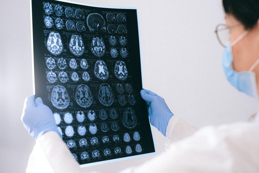
MRI images of the brain
Photo by Anna Shvets on Pexels

Published in the journal Scientific Reports, a study led by Associate Professor Bridgette Semple from Monash University, found that repeated concussions resulted in thicker, denser bones in the skull.

Although bones are considered a mostly structural component of the human body, bones are in fact active living tissues that can respond to applied mechanical forces. For example, martial arts training, with its kicks, punches and throws, has been shown to increase bone mineral density in the arms, legs and spines of practitioners.

At present, it is unclear whether this thickening of the skull is beneficial or detrimental: theoretically, a thicker skull is a stronger skull, suggesting that this may be the bone’s attempt to protect the brain from subsequent impacts.

“This is a bit of a conundrum,” Assoc Prof Semple said. “As we know, repeated concussions can have negative consequences for brain structure and function. Regardless, concussion is never a good thing.”

The team hopes that the microstructural skull alterations caused by concussion are now considered by researchers in the field to better understand how concussions affect the whole body.

A form of mild traumatic brain injury, concussion have been linked to long-term neurological consequences if they happen repetition.

While most studies focus on its effect on the brain and its function, they largely ignore the overlying skull bones.

Study collaborator Professor Melinda Fitzgerald, from Curtin University and the Perron Institute in Western Australia, has previously shown that repeated concussive impacts lead to subtle problems with memory, and evidence of brain damage.

In this new study, high-resolution neuroimaging and tissue staining techniques were used in a pre-clinical animal model, and revealed an increase in bone thickness and density, in close proximity to the site of injury.

“We have been ignoring the potential influence of the skull in how concussive impacts can affect the brain,” Associate Professor Semple said. “These new findings highlight that the skull may be an important factor that affects the consequences of repeated concussions for individuals.”

Future studies are planned, with collaborator and bone expert Professor Natalie Sims from St Vincent’s Institute of Medical Research in Melbourne, to understand if a thickened skull resulting from repeated concussions alters the transmission of impact force through the skull and into the vulnerable brain tissue underneath.

Source: Monash University

A Tangled Web of Brain Damage from Concussions in Children

Boy hanging from tree
Photo by Annie Spratt on Pexels

Concussion may cause different types of brain damage which lead to similar symptoms in children, according to research published in eLife. A new way of studying concussions could help inform the development of future treatments.

While most children fully recover after a concussion, some will have lasting symptoms. The findings help explain the complex relationships that exist between symptoms and the damage caused by the injury.

The researchers found that certain combinations of brain damage were associated with specific symptoms such as attention difficulties. Other symptoms, such as sleep problems, occurred in children with multiple types of injuries. For example, damage to areas of the brain that are essential for controlling sleep and wakefulness could cause challenges with sleeping, as could damage to brain regions that control mood.

The brain’s white matter holds clues

To do this, they examined how damage to the brain resulting from concussion affected its structural connection network, known as white matter. They then used statistical modelling techniques to see how these changes related to 19 different symptoms reported by the children or their caregivers.

Analysing symptoms may advance treatment

“Despite decades of research, no new treatment targets and therapies for concussions have been identified in recent years,” said lead author Guido Guberman, a Vanier Scholar and MDCM Candidate at McGill University. “This is likely because damage to the brain caused by concussions, and the symptoms that result from it, can vary widely across individuals. In our study, we wanted to explore the relationships that exist between the symptoms of concussion and the nature of the injury in more detail.”

Guberman and his colleagues analysed data collected from 306 children, aged nine to 10 years old, who had previously had a concussion. The children were all participants in the Adolescent Brain Cognitive Development (ABCD) Study.

“The methods used in our study provide a novel way of conceptualising and studying concussions,” says senior author Maxime Descoteaux, a Professor of Computer Science at Université de Sherbrooke. “Once our results are validated and better understood, they could be used to explore potential new treatment targets for individual patients. More broadly, it would be interesting to see if our methods could also be used to gather new insights on neurological diseases that likewise cause varied symptoms among patients.”

Source: McGill University

Long-term Symptoms of Concussion Tied to Vestibular Nerve Damage

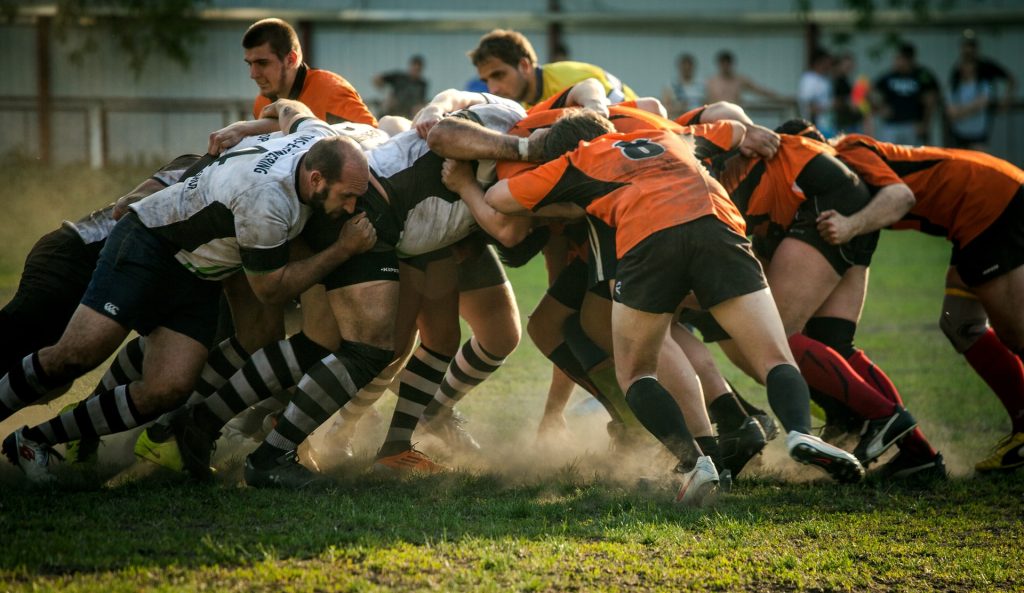
Rugby players
Photo by Olga Guryanova

A clinical study from Lund University in Sweden has shown that long-term problems such as dizziness and difficulty focusing after a concussion originate in an injury to the vestibular nerve. The researchers published their findings in the Journal of Neurotrauma.

Concussion resulting from a blow to the head is a hazard in many sports. In American football, where players who have suffered from repeated concussions have developed dementia, severe depression and cognitive impairment.

Concussion usually has only temporary symptoms, but an increasing number of athletes experience long-term problems that make it difficult to work, go to school or play sports. The symptoms are aggravated by activity or impressions and include headaches, depression, anxiety, nausea, difficulty focusing and problems with balance.

“It has been unclear what causes the symptoms, and it is difficult for healthcare professionals to help these athletes. We wanted to investigate this further to find out what really causes the symptoms,” said Professor Niklas Marklund, one of the study’s researchers.

The study included 21 healthy athletes without previous trauma to the head, and 21 athletes who all suffered from sports-related concussions and who had experienced persisting symptoms for more than six months. The researchers used a 7-Tesla MRI, to study the athletes’ brains to understand more about what caused the symptoms. They discovered impaired function of the balance organs in the inner ear of 13 athletes in the group with long-term problems. In the group of healthy athletes three people had similar findings.

“The test results show that the injury is located to the vestibular nerve, which is connected to the semicircular canals in a cavity inside the skull, and which is directly adjacent to the cochlea in the ear. These injuries lead to the inward nerve impulses not working properly, and the brain therefore does not receive important information about body movements and sensory impressions required to maintain a good balance,” said Anna Gard, doctoral student at Lund University and first author of the study.

Concussion often results from the head rotating too fast.

“We have not examined athletes with short-term problems after blows to the head, so we cannot say anything about them. This study applies to athletes with prolonged symptoms after concussion. The rotation of the head that occurs in connection with a concussion could lead to a stretch of the vestibular nerve, which then leads to impaired function. Now that we have more knowledge about where the problems are located, it is easier to find possible therapies that could help these athletes,” concluded Prof Marklund.

Source: Lund University

Over 70% of TBI Patients Report More Symptoms a Year On

Source: Pixabay

More than 70% of patients with traumatic brain injury (TBI) reported at least one problematic symptom at one year of follow-up that was new or worse than before injury according to a study published in the Journal of Neurotrauma. Half of the patients reported three or more such symptoms after a year.

Joan Machamer, from the University of Washington, Harborview Medical Center, and colleagues representing the TRACK-TBI Investigators, compared the frequency and persistence of symptoms in patients with TBI to two control groups: patients with orthopedic trauma and friend controls. The groups were evaluated at 2 weeks, and 3, 6, and 12 months after injury.

Physical symptoms such as headache, fatigue, and dizziness tended to occur earlier, according to the researchers, with cognitive symptoms becoming dominant later. While physical symptoms declined noticeably over time, cognitive symptoms remained more constant over time.

“Clinicians should inquire about symptoms in patients who have had a TBI, reassure them that experiencing symptoms is common, and direct them to seek treatment for symptoms that are disrupting their lives,” the researchers said.

Commenting on the study, David L. Brody, MD, PhD, Editor-in-Chief of Journal of Neurotrauma noted that is remarkable for several reasons. “First, it is among the largest studies of its type, with over 2,000 participants. Second, it confirms what many of us who practice brain injury medicine have observed for many years– a wide variety of symptoms can be very persistent and very troubling to our patients even after so-called ‘mild’ TBI. Third, the investigators used the right controls; people with orthopedic injury as well as friends of the patients with TBI. Symptoms in patients with TBI were substantially more common and more severe than in both control groups.”

Source: Mary Ann Liebert, Inc.

Study Uncovers Mechanism Behind Visual Impairment in Traumatic Brain Injury

A healthy neuron.
A healthy neuron. Credit: NIH

Traumatic brain injury can lead to long-term visual impairment, which researchers have found is caused by a dramatic drop in the number of neurons in the visual cortex. Their findings were published in Communications Biology.

Traumatic brain injury (TBI) is associated with mechanical brain damage and a wide range of neuronal abnormalities.  Injuries to the posterior occipital cortex are common in humans, and can result in visual impairment. Up to 75% of current or former soldiers live with permanent visual dysfunction or cortical blindness. 

The human brain possesses surprising neuroplasticity, which allows other areas of the brain to take over the functions of a damaged area.

Such neuroplasticity is also characteristic of the sensory areas of the visual cortex, which is final component of the visual pathway, responsible for receiving and processing visual impressions. The primary visual cortex (V1) is reached by the nerve fibres of the optic radiation, which carry nerve impulses from the retinas of both eyes.

Until now, scientists knew little about the effects of TBI on long-term visual circuit function. Using mice, a team of researchers examined how neurons respond to visual stimuli two weeks and three months after mild injury to the primary visual cortex (V1). V1 neurons normally show sensitivity to different features of a visual stimulus, such as colour or direction of movement. The preprocessed data is transmitted to subsequent areas of the visual cortex. This study showed that although the primary visual cortex remained largely intact after the brain injury, there was a 35% reduction in the number of neurons. This loss largely affected inhibitory neurons rather than excitatory neurons, which inhibit or stimulate action in the target cells, respectively.

After TBI, fewer than half of the isolated neurons were sensitive to visual stimuli (32% at two weeks after injury; 49% at three months after the event), compared with 90% of V1 cells in the control group. Up to a threefold decrease in neuronal activity was seen after the brain injury, and the cells themselves had worse spatial orientation. The overall results mean that even minor, superficial brain injuries cause long-term impairment in the way visual stimuli are perceived, persisting several months after the event.

Such a deeper understanding of the functional impairments in damaged visual cortex could provide a basis for developing circuit-level therapies for visual cortex damage.

Source: Institute of Physical Chemistry of the Polish Academy of Sciences

Good Outcomes for Severe Brain Injury Still Possible

Photo by Anna Shvets from Pexels

A new study adds to the growing body of evidence that decisions regarding moderate-to-severe traumatic brain injury (TBI) should not be made too soon after the injury, as a good prognosis can still emerge.

Researchers followed 484 patients with moderate-to-severe TBI and found that among the patients in a vegetative state, one quarter “regained orientation” — awareness of who, when and where they were —  within 12 months of their injury.

“Withdrawal of life-sustaining treatment based on early prediction of poor outcome accounts for most deaths in patients hospitalised with severe TBI,” said senior author Geoffrey Manley, MD, PhD,  noting that 64 of the 92 fatalities in the study occurred within two weeks of injury. Dr Manley is professor and vice chair of neurological surgery at UCSF and chief of neurosurgery at Zuckerberg San Francisco General Hospital.

“TBI is a life-changing event that can produce significant, lasting disability, and there are cases when it is very clear early on that a patient will not recover,” he said. “But results from this study show a significant proportion of our participants experienced major improvements in life functioning, with many regaining independence between two weeks and 12 months after injury.”

The patients in the study were enrolled by the brain injury research initiative TRACK-TBI, of which Dr Manley is the principal investigator. All patients were 17 and older and had presented to hospitals with level 1 trauma centers within 24 hours of injury. Their exams met criteria for either moderate TBI or severe TBI. The causes were falls, assault and primarily crashes involving a motor vehicle.

The patients, whose average ages were 35 in the severe TBI group (78 percent males) and 38 in the moderate TBI group (80 percent males), were assessed using the Glasgow Outcomes Scale Extended (GOSE), which ranges from 1 for death to 8 for “upper good recovery” and resumption of normal life. Impairment was also categorised with the Disability Rating Scale (DRS).

At two weeks post-injury, 93 percent of the severe TBI group and 79 percent of the moderate TBI group had moderate-to-severe disability, according to the DRS, and 80 percent had GOSE scores from 2 to 3, meaning they required assistance in basic everyday functioning.

But by 12 months, half of the severe TBI group and three-quarters of the moderate TBI group had GOSE scores of at least 4, indicating they could function independently at home for at least eight hours per day. Moreover, 19 percent of the severe TBI group had no disability, according to the DRS, and a further 14 percent had only mild injury, the researchers noted.

Most surprising were the findings for the 62 surviving patients who had been in a vegetative state. By the 12-month mark all patients had recovered consciousness and 1 in 4 had regained orientation. All but one survivor in this group recovered at least basic communication ability.

“These patients made the cut for favorable outcome,” said co-first author, Joseph Giacino, PhD, of Spaulding Rehabilitation Hospital, Massachusetts General Hospital and Harvard Medical School. “Their GOSE scores were 4 or higher, which meant they could be at home unsupervised for at least eight hours a day, since they were able to take care of basic needs, such as eating and toileting.”

In prior work, a significant percentage of patients with grave impairments had been shown to achieve favorable functionality after many months or years. This study coincided with the recommendation in 2018 from the American Academy of Neurology that in the first 28 days after injury, clinicians should refrain from telling families that a patient’s prognosis is beyond hope.

“While a substantial proportion of patients die or suffer lasting disability, our study adds to growing evidence that severe acute impairment does not portend uniformly poor long-term outcome,” said Manley, who is also affiliated with the UCSF Weill Institute for Neurosciences. “Even those patients in a vegetative state – an outcome viewed as dire – may improve, since this is a dynamic condition that evolves over the first year.”

Source: University of California, San Francisco

Journal information:JAMA Neurology (2021). DOI: 10.1001/jamaneurol.2021.2043

CT Scans Improve Outcomes for Concussion Patients

Photo by MART PRODUCTION from Pexels

A study found that CT scans for concussion patients provide crucial information on their risk for long-term impairment and their potential to make a complete recovery, and points to the need for more follow-up.

In the UC San Francisco-led study, researchers examined CT scans of 1935 patients, aged 17 and over, whose neurological exams met criteria for concussion, or mild traumatic brain injury (TBI). Outcomes for moderate and severe TBI have been linked to CT imaging features, but this may be the first time this link has been identified in patients with concussion. This contradicts previous research which had found no prognostic significance of specific types of CT abnormalities.

“Radiologists who routinely read trauma scans know intuitively that patterns of intracranial injury on CT are not random,” said first author Esther Yuh, MD, PhD, of the UCSF Department of Radiology and Biomedical Imaging. “We showed there are patterns of injury, that some of these are associated with worse outcome than others, and that they provide a window into mechanisms of injury that is reproducible across large studies.”

The study was published online in JAMA Neurology.

“Although concussions are referred to as mild traumatic brain injuries, there is nothing mild about some concussions,” explained senior author Geoffrey Manley, MD, PhD, professor and vice chair of neurological surgery at UCSF and chief of neurosurgery at Zuckerberg San Francisco General Hospital. “Patients with concussion may suffer from prolonged headache, poor sleep and impaired concentration, and they are at higher risk of self-medicating with drugs and alcohol. Concussion can also contribute to depression and anxiety, and increase the risk for suicide. We need to view concussion not as an event but as a disease requiring physician follow-up after a patient is discharged from the hospital.”

The participants were enrolled by the brain injury research initiative TRACK-TBI, of which Manley is the principal investigator. To enrich the number of so-called complicated concussions, the researchers drew exclusively from patients who had been seen at hospitals with level 1 trauma centres. This meant 37 percent of study participants had a positive CT, significantly more than the 9 percent of positive CTs from patients in US emergency departments.

The most common patterns of injury, affecting more than half of CT-positive patients, were combinations of subarachnoid haemorrhage (SAH), subdural haematoma (SDH), and/or contusion, which may be caused by injuries such as falls from standing. About 7 percent had intraventricular haemorrhage (IVH) or petechial haemorrhage, caused by head rotation as in some sporting, scooter and automobile accidents; and 5 percent had epidural haematoma (EDH), often seen in sports injuries such as being hit with a baseball.

Average age of the patients was 41 and 66 percent were male. They were followed-up at two weeks, and at three-, six- and 12 months following injury. Patients in the SAH/SDH/contusion group failed to make a complete recovery at 12 months post-injury and had a range of outcome impairments, from mild to more severe.

Patients in the IVH/petechial haemorrhage group tended toward more severe impairments, in the lower-moderate disability range, a level potentially affecting multiple areas of function, such as employment, social and leisure activities, up to 12 months post-injury. Patients with EDH fared significantly better and demonstrated complete recovery by their six-month assessment.

Results from CENTER-TBI, a parallel brain injury research group that had enrolled 2594 participants at European trauma centres. validated the findings. “The confirmation of the findings in an independent cohort confirms the fidelity of our results,” said Manley, adding that patients with EDH were one exception, with incomplete recovery lingering for months longer than those patients followed by TRACK-TBI. However, more severe outcomes were not seen at any point in either study.

The researchers noted that even among concussion patients with positive CT scans, only 39 percent get follow-up care, which should be routine. They also cautioned that their findings are not a call for increased CT use, which has radiation dose concerns and is restricted to known or suspected concussions.

Indeed, a recently approved rapid hand-held blood test may reduce the amount of CT scans. Manley found this test was more sensitive than CT in detecting concussion. The blood test measures biomarkers associated with TBI, which were nearly 52 times higher in MRI-identified concussion patients than in healthy participants.

In addition to challenging the belief that CT features in concussion are not relevant, the researchers are also challenging the idea that concussion is “what the patient brings to the injury,” said Manley, who is also affiliated with the UCSF Weill Institute for Neurosciences. “In moderate and severe TBI, it is anecdotally taught that outcome is determined by ‘what the injury brings to the patient,’ while concussion is determined by baseline characteristics like age, sex and years of education. While the study confirms the importance of these characteristics, we show that in some concussion cases, poor outcomes are also attributed to ‘what the injury brings to the patient.'”

Source: University of California, San Francisco

Journal information: Yuh EL et al., Pathological computed tomography features associated with adverse outcomes after mild traumatic brain injury, JAMA Neurology, July 19, 2021.