Tag: traumatic brain injury

Head Trauma may Activate Latent Viruses, Leading to Neurodegeneration

Source: CC0

In sports, the connection between head injuries and neurodegenerative diseases such as chronic traumatic encephalopathy, Alzheimer’s disease, and Parkinson’s disease is now well recognised.

Researchers at Tufts University and Oxford University have now uncovered mechanisms that may connect the dots between trauma events and the emergence of disease. They point to latent viruses lurking in most of our brains that may be activated by the jolt, leading to inflammation and accumulating damage that can occur over the ensuing months and years. 

The results suggest the use of antiviral drugs as potential early preventive treatments post-head injury. The findings are published in a study in Science Signaling.

The microbiome aids in digestion, immune system development, and protection against harmful pathogens. 

But the microbiome also includes dozens of viruses that swarm within our bodies at any given time. Some of these can be potentially harmful, but simply lie dormant within our cells. Herpes simplex virus 1 (HSV-1), found in over 80% of people, and varicella-zoster virus, found in 95% of people, are known to make their way into the brain and sleep within our neurons and glial cells.

Dana Cairns, GBS12, research associate in the Department of Biomedical Engineering and lead author of the study, had found evidence in earlier studies suggesting that activation of HSV-1 from its dormant state triggers the signature symptoms of Alzheimer’s disease in lab models of brain tissue: amyloid plaques, neuronal loss, inflammations, and diminished neural network functionality.

“In that study, another virus – varicella – created the inflammatory conditions that activated HSV-1,” said Cairns. “We thought, what would happen if we subjected the brain tissue model to a physical disruption, something akin to a concussion? Would HSV-1 wake up and start the process of neurodegeneration?”

The link between HSV-1 and Alzheimer’s disease was first suggested by co-author Ruth Itzhaki, visiting professorial fellow at Oxford University, who more than 30 years ago identified the virus in a high proportion of brains from the elderly population. Her subsequent studies suggested that the virus can be reactivated in the brain from a latent state by events such as stress or immunosuppression, ultimately leading to neuronal damage.

Blows to Brain-like Tissue

In the current study, the researchers used a lab model that reconstructs the environment of the brain to better understand how concussions may set off the first stages of virus reactivation and neurodegeneration.

The brain tissue model consists of a 6mm-wide donut-shaped sponge-like material made of silk protein and collagen suffused with neural stem cells, which are then coaxed into mature neurons, growing axons and dendrite extensions and forming a network. Glial cells also emerge from the stem cells to help mimic the brain environment and nurture the neurons.

The neurons communicate with each other through their extensions similarly to how they would communicate in a brain. And just like cells in the brain, they can carry within them the DNA of dormant HSV-1 virus.  

After enclosing the brain-like tissue in a cylinder and giving it a sudden jolt atop a piston, mimicking a concussion, Cairns examined the tissue under the microscope over time. Some of the tissue models had neurons with HSV-1, and some were virus-free. 

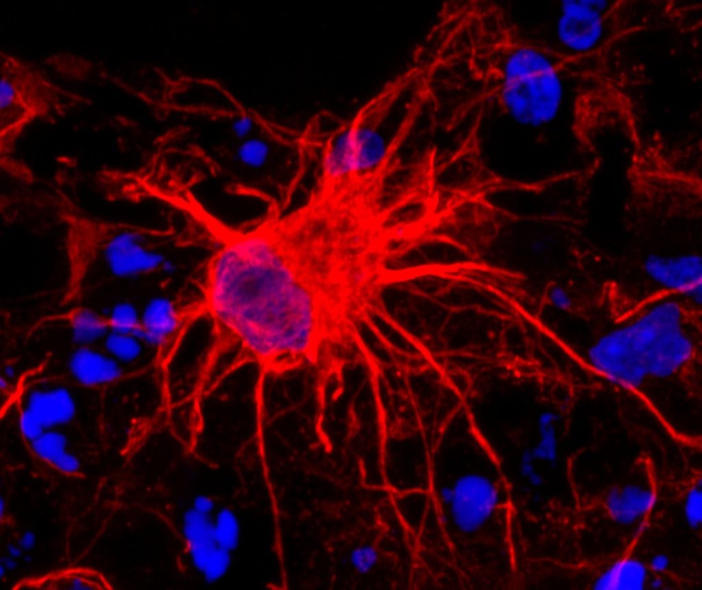
Following the controlled blows, she observed that the infected cells showed re-activation of the virus, and shortly after that the signature markers of Alzheimer’s disease, including amyloid plaques, p-tau (a protein that creates fiber-like “tangles” in the brain), inflammation, dying neurons, and a proliferation of glial cells called gliosis.

More strikes with the pistons on the tissue models mimicking repetitive head injuries led to the same reactions, which were even more severe. Meanwhile, the cells without HSV-1 showed some gliosis, but none of the other markers of Alzheimer’s disease.

The results were a strong indicator that athletes suffering concussions could be triggering reactivation of latent infections in the brain that can lead to Alzheimer’s disease. Epidemiological studies have shown that multiple blows to the head can lead to doubling or even greater chances of having a neurodegenerative condition months or years down the line.
 
“This opens the question as to whether antiviral drugs or anti-inflammatory agents might be useful as early preventive treatments after head trauma to stop HSV-1 activation in its tracks, and lower the risk of Alzheimer’s disease,” said Cairns.

The problem goes far beyond the concerns for athletes. Traumatic brain injury is one of the most common causes of disability and death in adults, affecting about 69 million people worldwide each year, at an economic cost estimated at $400 billion annually.

“The brain tissue model takes us to another level in investigating these connections between injury, infection, and Alzheimer’s disease,” said David Kaplan, Stern Family Endowed Professor of Engineering at Tufts.

“We can re-create normal tissue environments that look like the inside of a brain, track viruses, plaques, proteins, genetic activity, inflammation and even measure the level of signalling between neurons,” he said. “There is a lot of epidemiological evidence about environmental and other links to the risk of Alzheimer’s. The tissue model will help us put that information on a mechanistic footing and provide a starting point for testing new drugs.”

Source: Tufts University

Heads up – School Rugby and Head Injuries Explained

The rugby season is kicking off in schools across South Africa and players, parents, coaches and referees are preparing for exciting, yet physically demanding matches. In many sports, injuries are an unfortunate, common occurrence. Rugby, inherently a contact sport, also carries the inevitable risk of head injuries, ranging from minor concussions to severe Traumatic Brain Injuries (TBIs).

The importance of early detection

The early detection of head injuries is essential for effective treatment and preventing further complications. In many cases, the symptoms of a concussion or TBIs may not be immediately apparent and athletes may continue playing which can lead to further damage.

Accurate diagnosis and management of head injuries require a combination of clinical evaluation and advanced imaging techniques. Dr Hofmeyr Viljoen, radiologist at SCP Radiology talks about the nature of these injuries, the critical role radiology plays in diagnosing and managing them and what preventative measures can be taken.

Understanding head injuries in rugby

Dr Viljoen explains that there are several types of head injuries common in rugby. ‘The most frequent is concussion, a mild traumatic brain injury occurring when the brain is jolted inside the skull from an impact or violent movement. Concussions can be mild or lead to significant short and long-term issues. Occasionally, with more severe injuries we see skull fractures, contusions and haemorrhage surrounding the brain. These require urgent diagnosis and management.’

Recognising the symptoms

He emphasises awareness of concussion symptoms, including headaches, dizziness, nausea, confusion, memory problems, sensitivity to light and difficulty concentrating. ‘Immediate recognition is vital,’ he explains. ‘A player with any of these symptoms must be removed from play immediately to prevent further injury.’

The role of radiology

Radiology plays an essential part in accurately diagnosing the extent of head injuries. According to Dr Viljoen, Computed Tomography (CT) scans are always the first imaging method used in emergency settings. Although patients with concussion typically do not have significant imaging findings, it is crucial to image those patients with severe concussion or atypical symptoms. ‘CT scans rapidly detect serious issues like fractures, brain swelling and bleeding, providing crucial information for urgent treatment decisions,’ he explains.

Magnetic Resonance Imaging (MRI) is used in situations requiring more detailed evaluation, particularly when concussion symptoms persist or worsen. ‘MRI excels in identifying subtle injuries, such as microbleeds and brain swelling, often missed by CT scans,’ says Dr Viljoen. Unlike CT scans, MRI does not use radiation, making it a safer option for repeated assessments over time.

Advanced imaging methods

Emerging imaging techniques, such as Diffusion Tensor Imaging (DTI), show promise for better understanding and management of head injuries, especially the subtle effects of concussions. ‘DTI helps identify damage to the brain’s white matter, potentially guiding return-to-play decisions and treatment strategies,’ notes Dr Viljoen.

Understanding possible complications – Second Impact Syndrome (SIS)

SIS is a rare but extremely serious condition that occurs when a person sustains a second concussion before fully recovering from an initial concussion. This second injury doesn’t have to be severe to trigger SIS – it can even be minor – but it causes rapid and severe brain swelling (cerebral oedema).

The brain’s ability to regulate its blood flow and pressure is compromised following the initial concussion, making it vulnerable to catastrophic swelling after a subsequent impact. Symptoms can escalate quickly, often within minutes, including loss of consciousness, severe headache, dilated pupils, respiratory failure and even death. Young athletes are especially vulnerable to SIS. Due to its rapid progression and severity, SIS is considered a medical emergency requiring immediate intervention.

Preventing SIS involves strictly adhering to concussion management protocols, ensuring full recovery after any head injury and carefully monitoring symptoms before returning to sports or high-risk activities.

Addressing Chronic Traumatic Encephalopathy (CTE)

Dr Viljoen says CTE is a long-term degenerative brain condition linked to repeated head impacts. ‘CTE is challenging because currently, it can only be definitively diagnosed after death.  However, ongoing research aims to develop methods to detect CTE in living patients, potentially using advanced imaging techniques like Positron Emission Tomography (PET).’ Most research is focused on advancing non-invasive methods to see what is happening inside the brain of a living person and to track it over time.

Common causes of head injuries in rugby

  • These primarily arise from the high-impact nature of the sport, with tackling identified as a significant risk factor. Tackling, particularly when performed incorrectly or at a dangerous height, frequently leads to head trauma. Young players are especially vulnerable as their tackling techniques may not yet be fully developed, increasing the likelihood of injury. Teaching safe and correct tackling methods early is a way to mitigate these risks
  • Rugby’s dynamic gameplay often results in players being brought down forcefully or falling awkwardly. Even with protective gear, the impact of the head striking the playing surface can lead to concussions or more severe trauma
  • Due to the speed and intensity of the game, unintended impacts between players are inevitable. These include clashes of heads or impacts from knees and elbows, which can result in injuries ranging from mild concussions to more severe brain injuries. Preventative strategies and safer playing practices can reduce these risks

Prevention remains critical

Dr Viljoen emphasises the importance of proper training: ‘Educating young players on safe tackling techniques and enforcing protective protocols significantly reduces injury risks. Protective gear like headguards can minimise superficial injuries, though it does not prevent concussions.’

He also stresses the importance of concussion protocols. ‘Coaches at schools and clubs must rigorously apply concussion management strategies, ensuring players are adequately assessed and cleared by medical professionals before returning to the field.’ Under-reporting in schoolboy ruby often occurs because the player either wants to stay in the game and/or doesn’t recognise the symptoms of concussion.

Dr Viljoen concludes, ‘Rugby is a fantastic sport for building teamwork and resilience but player safety must always come first. Through awareness, timely medical intervention and proper preventative strategies, we can significantly reduce the risk and severity of head injuries, allowing young athletes to safely enjoy the game they love.’

Promising Findings in Testing Nasal Spray for TBI Treatment

Source: CC0

A new study led by researchers at Mass General Brigham suggests a nasal spray developed to target neuroinflammation could one day be an effective treatment for traumatic brain injury (TBI). By studying the effects of the nasal anti-CD3 in a mouse model of TBI, researchers found the spray could reduce damage to the central nervous system and behavioural deficits, suggesting a potential therapeutic approach for TBI and other acute forms of brain injury. The results are published in Nature Neuroscience.

“Traumatic brain injury is a leading cause of death and disability – including cognitive decline – and chronic inflammation is one of the key reasons,” said lead author Saef Izzy, MD, FNCS, FAAN, a neurologist and head of the Immunology of Brain Injury Program at Brigham and Women’s Hospital, a founding member of the Mass General Brigham healthcare system. “Currently, there is no treatment to prevent the long-term effects of traumatic brain injury.”

The study examines the monoclonal antibody Foralumab, made by Tiziana, which has been tested in clinical trials for patients with multiple sclerosisAlzheimer’s disease, and other conditions.

“This opens up a whole new area of research and treatment in traumatic brain injury, something that’s almost impossible to treat,” said senior author Howard Weiner, MD, co-director of the Ann Romney Center for Neurologic Diseases at Brigham and Women’s Hospital. “It also means this could work in intracerebral hemorrhage and other stroke patients with brain injury.”

Multiple experiments were done in mouse models with moderate-to-severe traumatic brain injury to explore the communication between regulatory cells induced by the nasal treatment and the microglial immune cells in the brain. Over time, researchers were able to identify how they modulate immune response.

“Modulating the neuroinflammatory response correlated with improved neurological outcomes, including less anxiety, cognitive decline, and improved motor skills,” Izzy said.

In addition to assessing the effects of the treatment, the research team was able to learn about immune response over time and compare the immune responses and effects of TBI in the mice.

The next step in the research is to translate the findings from preclinical models to human patients.

“Our patients with traumatic brain injury still don’t have an effective therapeutic to improve their outcomes, so this is a very promising and exciting time to move forward with something that’s backed up with solid science and get it to patients’ bedsides,” said Izzy.

Once in the clinical setting, Weiner said the hope is this treatment could be used on a variety of traumatic brain injury patients, including football players with repetitive concussions. 

“We envision giving a nasal spray right there on the sidelines,” said Weiner. “It isn’t something we can do yet, but we see the potential.”

Source: Mass General Brigham

Men More Than Three Times as Likely to Die From a Brain Injury, New Study Shows

Photo by Anna Shvets

A new analysis of mortality data reveals the disproportionate impact of traumatic brain injuries (TBI) on older adults, males and certain racial and ethnic groups. The study, published in the peer-reviewed journal Brain Injury, provides a comprehensive analysis of TBI-related deaths across different population groups across the US in 2021.

The findings indicate that suicides remain the most common cause of TBI-related deaths, followed by unintentional falls, and specific groups are disproportionately affected by these tragedies.

Men, in particular, were found to be most likely to die from a TBI – more than three times the rate of women (30.5 versus 9.4). The reasons observed were multifactorial and could reflect differences in injury severity following a fall or motor vehicle crash, to the interaction of sex and age – with TBI outcomes in men worsening with age, while postmenopausal women fare better than men of similar age.

“While anyone is at risk for getting a TBI, some groups have a higher chance than others of dying from one. We identified specific populations who are most affected. In addition to men, older adults are especially at risk, with unintentional falls being a major cause of TBI-related death. American Indian or Alaska Native people also have higher rates of these fatal injuries,” says lead author Alexis Peterson PhD, of the National Center for Injury Prevention and Control at the Centers for Disease Control and Prevention.

“These findings highlight the importance of tailored prevention strategies to reach groups who may be at higher risk and the role healthcare providers can play in reducing TBI-related deaths through early intervention and culturally sensitive care.”

TBI remains a leading cause of injury-related death in the US In 2020, TBIs were associated with around a quarter of all injury-related deaths.

Using data from the National Vital Statistics System, the new analysis identified 69 473 TBI-related deaths among US residents during 2021. The age-adjusted TBI-related mortality rate was 19.5 per 100 000, representing an 8.8% increase from 2020.

Through statistical modeling, the researchers examined the simultaneous effect of multiple factors such as geographic region, sex, race and ethnicity, and age, on TBI-related mortality.

Key findings include:

  • Older adults (75+) had the highest rates of TBI-related deaths, with unintentional falls being the most common cause in this age group.
  • Non-Hispanic American Indian/Alaska Native individuals experienced the highest TBI-related death rate (31.5) compared to other racial and ethnic groups.
  • There were 37,635 TBI-related deaths categorised as unintentional injuries (ie, motor vehicle crashes, unintentional falls, unintentionally struck by or against an object, other).
  • 30,801 were categorized as intentional injuries (ie, all mechanisms of suicide and homicide).
  • Children aged from birth to 17 years accounted for around 4% of TBI-related deaths (2,977).

The authors emphasise the critical role of healthcare providers in preventing TBI-related deaths, particularly with groups at higher risk. “By assessing patients who may be at higher risk for TBI, especially due to falls or mental health challenges, healthcare providers can make timely referrals and recommend culturally tailored interventions to prevent further injury or death,” says Dr Peterson.

Public health efforts should focus on addressing the underlying causes of TBI-related deaths, such as unintentional falls and mental health crises, to help prevent further loss of life. “TBIs remain a significant public health concern, especially among older adults, men, and certain racial and ethnic groups,” says Peterson.  “CDC has proven resources that healthcare providers can use to not only reduce health disparities that increase the risk for TBI but also improve care for anyone affected by a TBI.”

The authors note the COVID-19 pandemic could have influenced TBI-related death trends in 2021. They also acknowledge several limitations of this analysis, including potential misclassification or incomplete documentation of causes on death certificates, which may lead to inaccuracies in estimating TBI-related deaths.

Source: Taylor & Francis Group

Evidence Builds for Near Infrared Treatment of TBI

Coup and contrecoup brain injury. Credit: Scientific Animations CC4.0

Birmingham scientists have shown light therapy delivered transcranially can aid tissue repair after mild traumatic brain injury (mTBI). Their research, published in Bioengineering & Translational Medicine, indicates that this novel method could result in a new treatment option in an area of medicine that currently has few, if any, treatment options.

Traumatic brain injury (mTBI) results when the initial trauma of head injury is magnified by a complex set of inflammatory changes that occur in the brain. These secondary processes, which take place from minutes to hours after head injury, can dramatically worsen outcomes for patients.

The method invented by scientists at the University of Birmingham, UK and patented by University of Birmingham Enterprise aims to protect against this secondary damage, and stimulate faster, and better recovery for patients.

We want to develop this method into a medical device that can be used to enhance recovery for patients with traumatic brain or spinal cord injury, with the aim of improving outcomes for patients.

Professor Zubair Ahmed, College of Medicine & Health

In the study, the Birmingham team, comprising researchers Professor Zubair Ahmed, Professor Will Palin, Dr Mohammed Hadis and surgeons Mr Andrew Stevens and Mr David Davies, examined the effect of two wavelengths of near infrared light (660nm and 810nm) on recovery following injury.

The study in preclinical models used daily two-minute bursts of infrared light, delivered by a laser, for three days post-injury.

The findings showed significant reductions in the activation of astrocytes and microglial cells, which are heavily implicated in the inflammatory processes in the brain that follow head trauma, and significant reductions in biochemical markers of apoptosis (cell death).

At four weeks, there were significant improvements in performance in functional tests involving balance and cognitive function. The red light therapy also accelerated recovery compared to controls, with superior outcomes for light with a wavelength of 810nm.

The study builds on research published earlier this year which showed near infrared light delivered directly to the site of spinal cord injury both improves survival of nerve cells and stimulates new nerve cell growth.

Professor Ahmed, who led the study, said: “We want to develop this method into a medical device that can be used to enhance recovery for patients with traumatic brain or spinal cord injury, with the aim of improving outcomes for patients.”

The researchers are seeking commercial partners to co-develop the device and take it to market.

Source: University of Birmingham

Less Invasive Method for Measuring Intracranial Pressure After TBI

Coup and contrecoup brain injury. Credit: Scientific Animations CC4.0

Researchers at Johns Hopkins explored a potential alternative and less-invasive approach to evaluate intracranial pressure (ICP) in patients with serious neurological conditions. This research, using artificial intelligence (AI) to analyse routinely captured ICU data, was published in Computers in Biology and Medicine.

ICP is a physiological variable that can increase abnormally if one has severe traumatic brain injury, stroke or obstruction to the flow of cerebrospinal fluid. Symptoms of elevated ICP may include headaches, blurred vision, vomiting, changes in behaviour and decreased level of consciousness. It can be life-threatening, hence the need for ICP monitoring in selected patients who are at increased risk. But the current standard for ICP monitoring is highly invasive: it requires the placement of an external ventricular drain (EVD) or an intraparenchymal brain monitor (IPM) in the functional tissue in the brain consisting of neurons and glial cells by drilling through the skull.

“ICP is universally accepted as a critical vital sign – there is an imperative need to measure and treat ICP in patients with serious neurological disorders, yet the current standard for ICP measurement is invasive, risky, and resource-intensive. Here we explored a novel approach leveraging Artificial Intelligence which we believed could represent a viable noninvasive alternative ICP assessment method,” says senior author Robert Stevens, MD, MBA, associate professor of anaesthesiology and critical care medicine.

EVD procedures carry a number of risks including catheter misplacement, infection, and haemorrhaging at 15.3 %, 5.8 %, and 12.1 %, respectively, according to recent research. EVD and IPM procedures also require surgical expertise and specialised equipment that is not consistently available in many settings thus underscoring the need for an alternative method in examining and monitoring ICP in patients.

The Johns Hopkins team, a group that included faculty and students from the School of Medicine and Whiting School of Engineering, hypothesised that severe forms of brain injury, and elevations in ICP in particular, are associated with pathological changes in systemic cardiocirculatory function due, for example, to dysregulation of the central autonomic nervous system. This hypothesis suggests that extracranial physiological waveforms can be studied to better understand brain activity and ICP severity.

In this study, the Johns Hopkins team set out to explore the relationship between the ICP waveform and the three physiological waveforms that are routinely captured in the ICU: invasive arterial blood pressure (ABP), photoplethysmography (PPG) and electrocardiography (ECG). ABP, PPG and ECG data were used to train deep learning algorithms, resulting in a level of accuracy in determining ICP that rivals or exceeds other methodologies.

Overall study findings suggest a completely new, noninvasive alternative to monitor ICP in patients.

Stevens says, “with validation, physiology-based AI solutions, such as the one used here, could significantly expand the proportion of patients and health care settings in which ICP monitoring and management can be delivered.” 

Source: John Hopkins Medicine

Concussion is Associated with Iron Accumulation in Certain Brain Areas

Photo by Anna Shvets

People who suffer from headaches after experiencing concussions may also be more likely to have higher levels of iron in areas of the brain – a sign of injury to brain cells, according to a preliminary study presented at the American Academy of Neurology’s 76th Annual Meeting.

“These results suggest that iron accumulation in the brain can be used as a biomarker for concussion and post-traumatic headache, which could potentially help us understand the underlying processes that occur with these conditions,” said study author Simona Nikolova, PhD, of the Mayo Clinic in Phoenix, Arizona, and a member of the American Academy of Neurology.

The study involved 120 participants, 60 of whom who had post-traumatic headache (PTH) due to mild traumatic brain injury (mTBI), and 60 healthy controls. The injuries were due to a fall for 45% of the people, 30% were due to a motor vehicle accident and 12% were due to a fight. Other causes were the head hitting against or by an object and sports injuries. A total of 46% of the people had one mild traumatic brain injury in their lifetime, 17% had two, 16% had three, 5% had four and 16% had five or more mild traumatic brain injuries.

Participants underwent 3T brain magnetic resonance imaging (T2* maps). T2* differences were determined using age-matched paired t-tests. For the PTH group, scans were done an average of 25 days after injury. T2* correlations with headache frequency, number of lifetime mTBIs, time since most recent mTBI, and Sport Concussion Assessment Tool (SCAT) severity scale scores,

The researchers observed lower T2* values in PTH participants relative to HC in the right supramarginal area, left occipital, bilateral precuneus, right cuneus, right cerebellum, right temporal, bilateral caudate, genu of the corpus callosum, right anterior cingulate cortex and right rolandic operculum (p < 0.001).

Within PTH subjects, there were positive correlations with iron accumulation between lifetime mTBIs, the time since most recent mTBI and headache frequency in certain areas of the brain. For example, T2* levels in headache frequency with T2* in the posterior corona radiata, bilateral temporal, right frontal, bilateral supplemental motor area, left fusiform, right hippocampus, sagittal striatum, and left cerebellum were associated with headache frequency.

“Previous studies have shown that iron accumulation can affect how areas of the brain interact with each other,” Nikolova said. “This research may help us better understand how the brain responds and recovers from concussion.”

Nikolova said that using the indirect measure of iron burden also means that the change in that measure could be due to other factors such as haemorrhage or changes in tissue water rather than iron accumulation.

Source: American Academy of Neurology

Difference in Brain Structures may Explain Concussion Outcomes for Males and Females

Coup and contrecoup brain injury. Credit: Scientific Animations CC4.0

Important brain structures that are key for signalling in the brain are narrower and less dense in females, and more likely to be damaged by brain injuries, such as concussion. Long-term cognitive deficits occur when the signals between brain structures weaken due to the injury. These structural differences in male and female brains might explain why females are more prone to concussions and experience longer recovery from the injury than their male counterparts, according to a University of Pennsylvania-led preclinical study published in Acta Neuropathologica.

Each year, approximately 50 million individuals worldwide suffer a concussion, also referred to as mild traumatic brain injury (TBI). For more than 15% of individuals who suffer persisting cognitive dysfunction, which includes difficulty concentrating, learning and remembering new information, and making decisions.

Although males make up the majority of emergency department visits for concussion, this has been primarily attributed to their greater exposure to activities with a risk of head impacts compared to females. In contrast, it has recently been observed that female athletes have a higher rate of concussion and appear to have worse outcomes than their male counterparts participating in the same sport.

“Clinicians have observed for a long time that females suffer from concussion at higher rates than males in the same sports, and that they take longer to recover cognitive function, but couldn’t explain the underlying mechanisms of this phenomenon,” said senior author Douglas Smith, MD, a professor of Neurosurgery and director of Penn’s Center for Brain Injury and Repair. “The variances in brain structures of females and males not only illuminate why this disparity exists, but also exposes biomarkers, such as axon protein fragments, that can be measured in the blood to determine injury severity, monitor recovery, and eventually help identify and develop treatments that help patients repair these damaged structures and restore cognitive function.”

Axons connect neurons, allowing communication across the brain. These axons form bundles that make up white matter in the brain and play a large role in learning and communication between different brain regions. Axons are delicate structures and are vulnerable to damage from concussion.

Communication between axons in the brain is powered by sodium channels that serve as the brain’s electric grid. When axons are damaged, these sodium channels are also impaired, which causes loss of signaling in the brain. The loss of signaling causes the cognitive impairment experienced by individuals after concussion.

In this study, researchers used large animal models of concussion to identify differences in brains of males and females after a concussion. They found that females had a higher population of smaller axons, which researchers demonstrated are more vulnerable to injury. They also reported that in these models, females had greater loss of sodium channels after concussion.

“The differences in brain structure not only tell us a lot about how brain injury affects males and females differently but could offer insights in other brain conditions that impact axons, like Alzheimer’s and Parkinson’s disease,” said Smith. “If female brains are more vulnerable to damage from concussion, they might also be more vulnerable to neurodegeneration, and it’s worth further research to understand how sex influences the structure and functions of the brain.”

Source: University of Pennsylvania School of Medicine

Scientists Give Macrophages First-aid ‘Backpacks’ to Calm TBI Inflammation

Colourised electron micrograph image of a macrophage. Credit: NIH

Scientists have created a new treatment for traumatic brain injury (TBI). The new approach leverages macrophages, which can increase or decrease inflammation in response to infection and injury. The team attached “backpacks” containing anti-inflammatory molecules directly to the macrophages. These molecules kept the cells in an anti-inflammatory state when they arrived at the injury site in the brain, enabling them to reduce local inflammation and mitigate the damage caused. The research is reported in PNAS Nexus.

“Every year, millions of people suffer from a TBI, but there is currently no treatment beyond managing symptoms. We have applied our cellular backpack technology – which we previously used to improve macrophages’ inflammatory response to cancerous tumours – to deliver localised anti-inflammatory treatment in the brain, which helps mitigate the cascade of runaway inflammation that causes tissue damage and death in a human-relevant model,” said senior author Samir Mitragotri, PhD, in whose lab the research was performed.

Stopping a runaway inflammation train

There is currently no treatment for the damage caused to brain tissue during a traumatic brain injury (TBI), beyond managing a patient’s symptoms. One of the main drivers of TBI-caused damage is a runaway inflammatory cascade in the brain.

As cells die from the impact, they release a cocktail of pro-inflammatory cytokine molecules that attract immune cells to clean up the damage. But the same cytokine molecules can also disrupt the blood-brain barrier, which causes blood to leak into the brain. Blood accumulation in the brain causes swelling, impaired oxygen delivery, and increased inflammation, and creates a vicious cycle of bleeding and damage that drives even more cell death.

The Mitragotri lab saw an opportunity in this problem.

“It’s generally believed anti-inflammatory therapies can be effective for treating TBI, but so far, none of them have proven effective clinically. Our previous work with macrophages has shown us that we can use our backpack technology to effectively steer their behaviour when they arrive at the injury site. Since these cells are already active players in the body’s natural immune response to a TBI, we had a hunch we could augment that pre-existing biology to reduce the initial damage,” said co-first author Rick Liao, Ph.D., a Postdoctoral Fellow at the Wyss Institute and SEAS.

“Body, heal thyself”…with backpacks

Macrophages are very malleable cells and can “switch” between pro-inflammatory and anti-inflammatory states. While the team’s previous work in cancer had been focused on keeping macrophages in a pro-inflammatory state when they arrive at the inflammation-reducing microenvironment of a tumour, this new project would be trying to do the opposite: keep the macrophages “calm” in the inflammation-riddled setting of a brain injury.

To do so, they used a disc-shaped “backpack” they had previously designed to treat multiple sclerosis that contained layers of two anti-inflammatory molecules: dexamethasone, a steroid, and interleukin-4, a cytokine that encourages macrophages to adopt an anti-inflammatory state. They then incubated these microparticles with both human and pig macrophages in vitro and saw that the backpacks stably stuck to the cells without causing any negative effect. They also observed that application of their backpacks decreased the expression of pro-inflammatory biomarkers and increased the expression of anti-inflammatory biomarkers, retaining the pig macrophages in a healing state.

But to prove that this shift would work in the body, they had to test the backpack-bearing macrophages in vivo. They chose pigs as their model organism because their brains’ structures and responses to injury more closely mimic those of humans than mice.

“Probably our biggest challenge in this project was scaling up production to match what we needed to run the experiments. Our previous studies were done in rodents, which required about two million macrophages and four million backpacks administered per subject. For the porcine study, we needed 100 million macrophages and 200 million backpacks per subject – on the scale of what would be administered in humans – and lots of helping hands,” said co-first author Neha Kapate, PhD, a Postdoctoral Fellow at the Wyss Institute and SEAS.

Once they had generated enough backpack-wearing porcine macrophages, they infused them into the pigs’ bloodstreams four hours after a TBI. Seven days later, they analysed the animals’ brains. Pigs that had received the macrophage treatment showed a high concentration of the cells in the area immediately surrounding the injury site, their lesions were 56% smaller, and there was significantly less haemorrhaging than in untreated animals.

Local immune cells also displayed a lower amount of a pro-inflammatory activation marker called CD80, indicating that the macrophages had accomplished their damage control by reducing inflammation in the brain. Corroborating that data, the levels of two soluble biomarkers for inflammation in the blood and cerebrospinal fluid were lower in treated animals than in untreated animals. The macrophage treatment also did not cause any negative effects.

The team plans to conduct future studies that focus on elucidating exactly how their anti-inflammatory macrophage therapy affects the blood-brain barrier’s integrity to prevent bleeding, which could also hold promise for treating other conditions like hemorrhagic strokes.

“Macrophages’ susceptibility to their local environment has historically prevented scientists from taking full advantage of their immune-modulating capabilities. This impressive study describes a truly novel and potentially powerful macrophage-based therapy for treating the inflammation that is the root cause of so many human afflictions in an effective and non-invasive way that works with biology rather than against it,” said Wyss Founding Director Donald Ingber, MD, PhD.

Source: Wyss Institute for Biologically Inspired Engineering at Harvard

Key Protein Coordinates Healing in Brain Injuries

Image of an astrocyte, a subtype of glial cells. Glial cells are the most common cell in the brain. Credit: Pasca Lab, Stanford University NIH support from: NINDS, NIMH, NIGMS, NCATS

A new study published in PNAS Nexus provides a better understanding of how the brain responds to injuries. Researchers at the George Washington University discovered that a protein called Snail plays a key role in coordinating the response of brain cells after an injury.

The study shows that after an injury to the central nervous system (CNS), a group of localised cells start to produce Snail, a transcription factor or protein that has been implicated in the repair process. The GW researchers show that changing how much Snail is produced can significantly affect whether the injury starts to heal efficiently or whether there is additional damage.

“Our findings reveal the intricate ways the brain responds to injuries,” said senior author Robert Miller, the Vivian Gill Distinguished Research Professor and Vice Dean of the GW School of Medicine and Health Sciences.

“Snail appears to be a key player in coordinating these responses, opening up promising possibilities for treatments that can minimise damage and enhance recovery from neurological injuries.”

This study identified for the first time a special group of microglial-like cells that produce Snail. Microglial cells are found in the central nervous system. The researchers found that lowering the amount of Snail produced after an injury results in inflammation and increased cell death. During this process, the injury worsens and there are fewer connections or synapses between brain cells. In contrast, when Snail levels are increased the outcome of brain injury improves-suggesting this protein can help limit the spread of injury-induced damage.

The research raises questions about whether an experimental drug that affects Snail production could be used to limit the damage incurred after someone suffers a stroke or has been injured in an accident, Miller said.

Additional studies must be done to show that increasing Snail production could curtail injury or even promote healing of the brain.

Miller and his team also plan to study the regulation of Snail in diseases like multiple sclerosis, a disease resulting in damage to the myelin nerve sheath. If drugs targeting Snail could be used to stop that damage, many of the future symptoms of this disease could be eased, he says.

But researchers have years of work to do before new drugs targeting Snail can be tested in clinical trials. The payoff ultimately might be drugs that can lead to accelerated healing for stroke damage, head wounds and even neurodegenerative diseases like dementia.

Source: George Washington University