Tag: terahertz

Terahertz Biosensor can Accurately Detect Skin Cancer
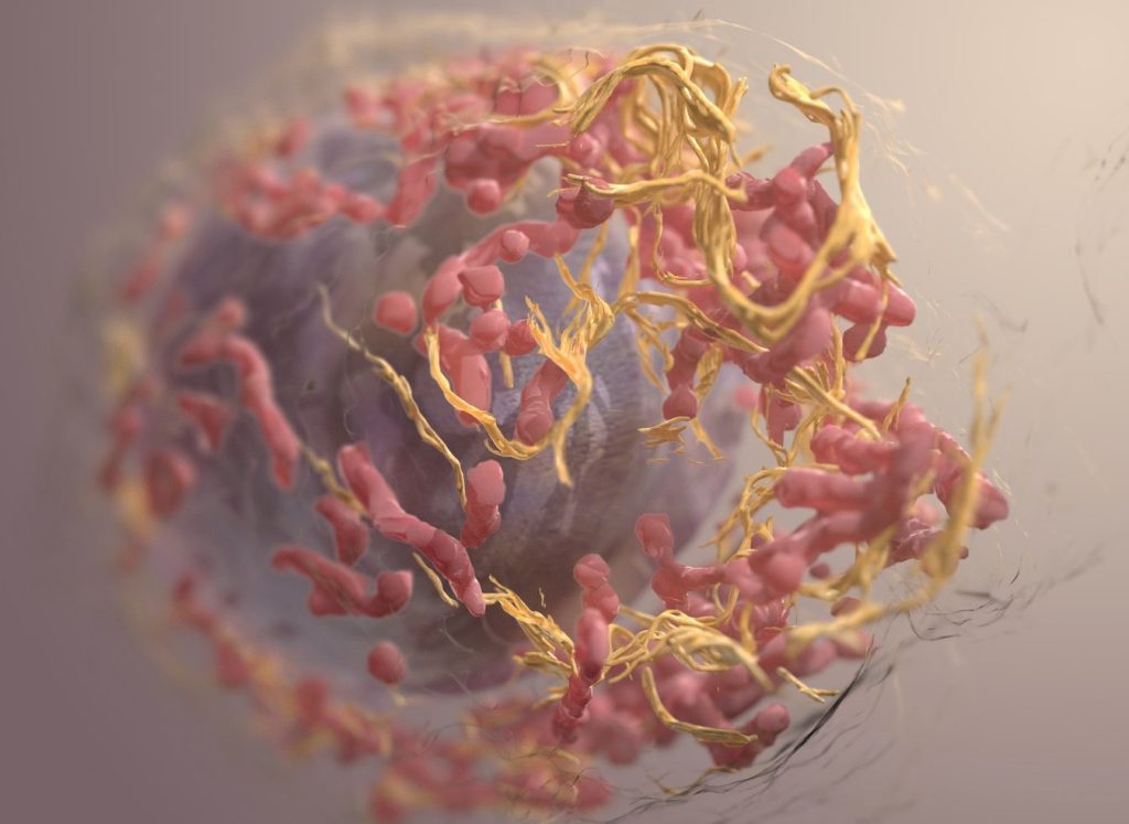
3D structure of a melanoma cell derived by ion abrasion scanning electron microscopy. Credit: Sriram Subramaniam/ National Cancer Institute

Researchers have developed a revolutionary biosensor using terahertz (THz) waves that can detect skin cancer with exceptional sensitivity, potentially paving the way for earlier and easier diagnoses. Published in the journal IEEE Transactions on Biomedical Engineering, the study presents a significant advancement in early cancer detection, thanks to a multidisciplinary collaboration of teams from Queen Mary University of London and the University of Glasgow.

“Traditional methods for detecting skin cancer often involve expensive, time-consuming, CT, PET scans and invasive higher frequencies technologies,” explains Dr Shohreh Nourinovin, Postdoctoral Research Associate at Queen Mary’s School of Electronic Engineering and Computer Science, and the study’s first author.

“Our biosensor offers a non-invasive and highly efficient solution, leveraging the unique properties of THz waves – a type of radiation with lower energy than X-rays, thus safe for humans – to detect subtle changes in cell characteristics.”

The key innovation lies in the biosensor’s design. Featuring tiny, asymmetric resonators on a flexible substrate, it can detect subtle changes in the properties of cells.

Unlike traditional methods that rely solely on refractive index, this device analyses a combination of parameters, including resonance frequency, transmission magnitude, and a value called “Full Width at Half Maximum” (FWHM). This comprehensive approach provides a richer picture of the tissue, allowing for more accurate differentiation between healthy and cancerous cells and to measure malignancy degree of the tissue.

In tests, the biosensor successfully differentiated between normal skin cells and basal cell carcinoma (BCC) cells, even at different concentrations. This ability to detect early-stage cancer holds immense potential for improving patient outcomes.

“The implications of this study extend far beyond skin cancer detection,” says Dr Nourinovin.

“This technology could be used for early detection of various cancers and other diseases, like Alzheimer’s, with potential applications in resource-limited settings due to its portability and affordability.”

Dr Nourinovin’s research journey wasn’t without its challenges.

Initially focusing on THz spectroscopy for cancer analysis, her project was temporarily halted due to the COVID pandemic. However, this setback led her to explore the potential of THz metasurfaces, a novel approach that sparked a new chapter in her research.

Source: Queen Mary University of London

A Handheld Terahertz Scanner Could Accurately Assess Burns

Ambulance
Photo by Camilo Jimenez on Unsplash

Researchers have developed a handheld terahertz (THz) wave imaging device to assess burns faster and more accurately than current methods. The new device uses neural network model that uses terahertz time-domain spectroscopy (THz-TDS) data for non-invasive burn assessment.

“It is important for healthcare professionals to accurately assess the depth of a burn to provide the most appropriate treatment,” said research team leader M. Hassan Arbab from Stony Brook University. “However, current methods of burn depth evaluation, which rely on visual and tactile examination, have been shown to be unreliable, with accuracy rates hovering around 60–75%. Our new approach could potentially improve the accuracy of burn severity assessments and aid in treatment planning.”

THz-TDS uses short pulses of terahertz radiation, which lies between infrared and microwave wavelengths, to probe a sample. It is being examined for assessing burn injuries because physical changes caused by a burn will produce alterations in the skin’s terahertz reflectivity.

In the journal Biomedical Optics Express, the researchers reported that their artificial neural network classification algorithm was able to accurately predict the ultimate healing outcome of in vivo burns in an animal study with 93% accuracy. Their method needs much less training data, potentially making it more practical to process big data sets obtained over large clinical trials.

“In 2018, approximately 416 000 patients were treated for burn injuries in emergency departments in the United States alone,” said Arbab. “Our research has the potential to significantly improve burn healing outcomes by guiding surgical treatment plans, which could have a major impact on reducing the length of hospital stays and number of surgical procedures for skin grafting while also improving rehabilitation after injury.”

Better burn assessment

Various technologies have been developed to improve burn assessment, but they haven’t been widely adopted in the clinic due to drawbacks such as long acquisition times, high costs and limited penetration depth and field of view. Although THz-TDS looks promising for burn assessment, early demonstrations were limited to point spectroscopy measurements, which don’t account for burn heterogeneity and spatial variations. THz spectroscopy setups also tend to be bulky and difficult to set up.

“To address these challenges, we developed the portable handheld spectral reflection (PHASR) scanner, a user-friendly device for fast hyperspectral imaging of in vivo burn injuries using THz-TDS,” said Arbab. The device allows for “rapid imaging of a 37 x 27 mm2 field of view in just a few seconds.”

Previously, the researchers used numerical methods to extract features from the THz-TDS images and machine learning techniques to estimate the severity grade of in vivo burn injuries using measurements from the PHASR scanner. However, this approach did not consider the physical dynamics and macroscopic changes of the dielectric permittivity of burned skin tissue. Dielectric permittivity describes how a material responds to an electric field, and the researchers used Debye theory to explain how biological material interacts with THz waves.

The researchers tested their method by using the PHASR scanner to obtain spectroscopic images of skin burns and measure the permittivity of the burns. The researchers used this data to create a neural network model based on labelled biopsies. The model estimated the severity of the burns with an average accuracy rate of 84.5% and predicted the outcome of the wound healing process with an accuracy rate of 93%.

The researchers note that clinical testing of both the technique and the handheld imaging device are needed before this technique could be integrated into the existing workflow of clinical burn assessment.

Source: Optica