Tag: T helper cells

T-helper Cells Near the Gut are Deliberately ‘Dysfunctional’
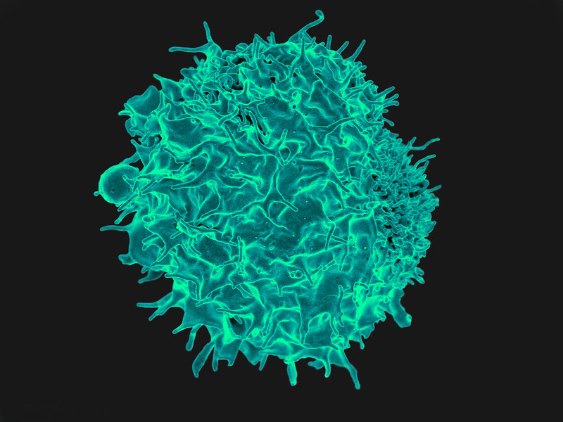
T cell
Scanning Electron Micrograph image of a human T cell. Credit: NIH/NIAID

A new study published in Nature has found that certain food proteins can cause T-helper cells in gut-associated lymphoid tissue to become dysfunctional in order for the immune system not to attack that particular food. Understanding how the process could be restarted could aid the development of food allergy treatments.

Led by Marc Jenkins, director of the University of Minnesota Medical School’s Center for Immunology, the research focused on why the immune system does not attack food in the way that it attacks other foreign entities like microbes.

“This study helps explain why our immune systems do not attack our food even though it is foreign to our bodies,” said Jenkins. “We found that ingested food proteins stimulate specific lymphocytes in a negative way. The cells become dysfunctional and eventually acquire the capacity to suppress other cells of the immune system.”

The gut associated-lymphoid tissue is a suppressive environment where lymphocytes that would normally generate inflammation undergo arrested development. This abortive response usually prevents dangerous immune reactions to food.

The research found that T-helper cells lack the inflammatory functions needed to cause gut pathology and yet the cells have the potential to produce regulatory T-cells that may suppress it. This means when people develop an intolerance or allergic reaction to certain foods, there may be a future capability to suppress that reaction by reintroducing dysfunctional lymphocytes.

Further research is needed to identify the mechanisms whereby food-specific lymphocytes become dysfunctional, knowledge which could be used to fight food allergies.

Source: University of Minnesota Medical School

Immune Cells Persist 6 Months after COVID Vaccination

Image of a syring for vaccination
Photo by Mika Baumeister on Unsplash

A recent study shows that T helper cells produced by people who received either of the two available messenger RNA (mRNA) vaccines for COVID persist six months after vaccination, at only slightly reduced levels from two weeks after vaccination. They are also at significantly higher levels than in unvaccinated individuals.

In the study, published in Clinical Infectious Diseases, the researchers also found that the T cells they studied recognise and help protect against the highly infectious delta variant of SARS-CoV-2.

“Previous research has suggested that humoral immune response – where the immune system circulates virus-neutralising antibodies – can drop off at six months after vaccination, whereas our study indicates that cellular immunity – where the immune system directly attacks infected cells – remains strong,” said Professor Joel Blankson, MD, PhD, study senior author. “The persistence of these vaccine-elicited T cells, along with the fact that they’re active against the delta variant, has important implications for guiding COVID vaccine development and determining the need for COVID boosters in the future.”

The researchers sampled blood from 15 study participants at three times: prior to vaccination, between seven and 14 days after their second Pfizer/BioNTech or Moderna vaccine dose, and six months after vaccination. The median age of the participants was 41 and none had evidence of prior SARS-CoV-2 infection.

CD4+ T lymphocytes are nicknamed helper T cells because they assist another type of immune system cell, the B lymphocyte (B cell), to respond to antigens on viruses such as SARS-CoV-2. Activated by the CD4+ T cells, immature B cells become either plasma cells that produce antibodies to mark infected cells for disposal from the body or memory cells that ‘remember’ the antigen’s biochemical structure for a faster response to future infections. Therefore, a CD4+ T cell response can serve as a measure of how well the immune system responds to a vaccine and yields humoral immunity.

The researchers found that the number of helper T cells recognising SARS-CoV-2 spike proteins was very low pre-vaccination, with a median of 2.7 spot-forming units (SFUs, the level of which is a measure of T cell frequency) per million peripheral blood mononuclear cells (PBMCs, identified as any blood cell with a round nucleus, including lymphocytes). Between 7 and 14 days after vaccination, the T cell frequency rose to a median of 237 SFUs per million PBMCs. At six months after vaccination, the level dropped slightly to a median of 122 SFUs per million PBMCs – a T cell frequency still significantly higher than before vaccination.

Six months after vaccination, the number of T cells recognising the delta variant spike protein was not significantly different from that of T cells attuned to the original virus strain’s protein.

“The robust expansion of T cells in response to stimulation with spike proteins is certainly indicated, supporting the need for more study to show booster shots do successfully increase the frequency of SARS-CoV-2-specific T cells circulating in the blood,” said Prof Blankson. “The added bonus is finding that this response also is likely strong for the delta variant.”

Source: John Hopkins Medicine