Tag: t cells

New T Cell ‘Rescue’ Therapy Promising for ARDS

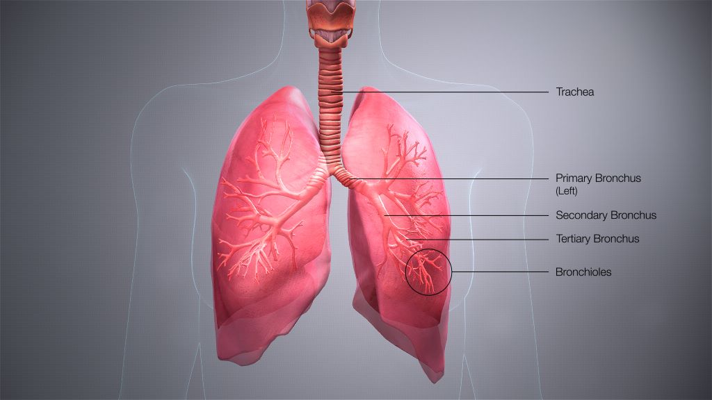
Credit: Scientific Animations CC4.0

Promising trial results indicate that a new type of cell therapy could improve the prognosis of those who are critically ill with acute respiratory distress syndrome (ARDS) resulting from severe COVID.

Published in the journal Nature Communications, Professor Justin Stebbing of Anglia Ruskin University (ARU) is the joint senior author of the new study investigating the use of agenT-797, MiNK Therapeutic’s allogeneic, unmodified invariant natural killer T (iNKT) cell therapy.

The iNKT cell therapy has the effect of rescuing exhausted T cells and prompting an anti-inflammatory cytokine response, potentially activating anti-viral immunity to help these patients fight infection as well as to reduce severe, pathogenic inflammation of the lung.

The new research was carried out at three medical centres and found that agenT-797, which is also under investigation in cancer trials, could be manufactured rapidly, had a tolerable safety profile, and appeared to have a positive effect on mortality among critically unwell Covid-19 ARDS patients receiving intensive care.

The exploratory trial included 20 mechanically ventilated patients with severe ARDS secondary to Covid-19. Of the 20 patients in the trial, 14 survived (70%) at 30 days (compared to a control group of 10%), and there was an 80% lower occurrence of bacterial pneumonia amongst those who received the highest dosage of agenT-797, compared to those who received fewer cells.

Twenty-one patients were treated overall (the main trial, plus one under compassionate use), which included five who were also receiving veno-venous extracorporeal membrane oxygenation (VV-ECMO), known as ‘the most aggressive salvage therapy’ for critically ill patients with ARDS.

In VV-ECMO, deoxygenated blood is pumped through a membrane lung and returned to the body via a cannula. This trial is believed to be the first immune cell therapy of any type to be used in critically unwell patients undergoing VV-ECMO.

Survival of the VV-ECMO cohort was 80% after 30 and 90 days, and 60% after 120 days. This compares favourably to overall survival of 51% for patients with Covid-19 who were treated with just VV-ECMO at the same institution, during the same timeframe.

Joint senior author Justin Stebbing, Professor of Biomedical Sciences at Anglia Ruskin University (ARU) in Cambridge, England, said: “During this small, exploratory study we observed that MiNK’s iNKT cell treatment, which is also being advanced for people with cancer, triggered an anti-inflammatory response in ARDS patients.

“Despite a poor prognosis, critically ill patients treated with this therapy showed favourable mortality rates and those treated at the highest dose also had reduced rates of pneumonia, underscoring the potential application of iNKT cells, and agenT-797 in particular, in treating viral diseases and infections more broadly.

Source: Anglia Ruskin University

New Therapy Eliminates ‘Problematic’ T Cells in Skin Autoimmune Diseases

Photo: CC0

In a groundbreaking study published in Science, researchers discovered distinct mechanisms controlling different types of immune cells, and found that, by precisely targeting these mechanisms, they could selectively eliminate ‘problematic cells’ and reshape the skin’s immune landscape.

The skin is packed with specialised immune cells that protect against infections and cancer and promote healing. These cells, called tissue-resident T cells or TRM cells, stay in place to fight infections and cancerous cells in the skin.

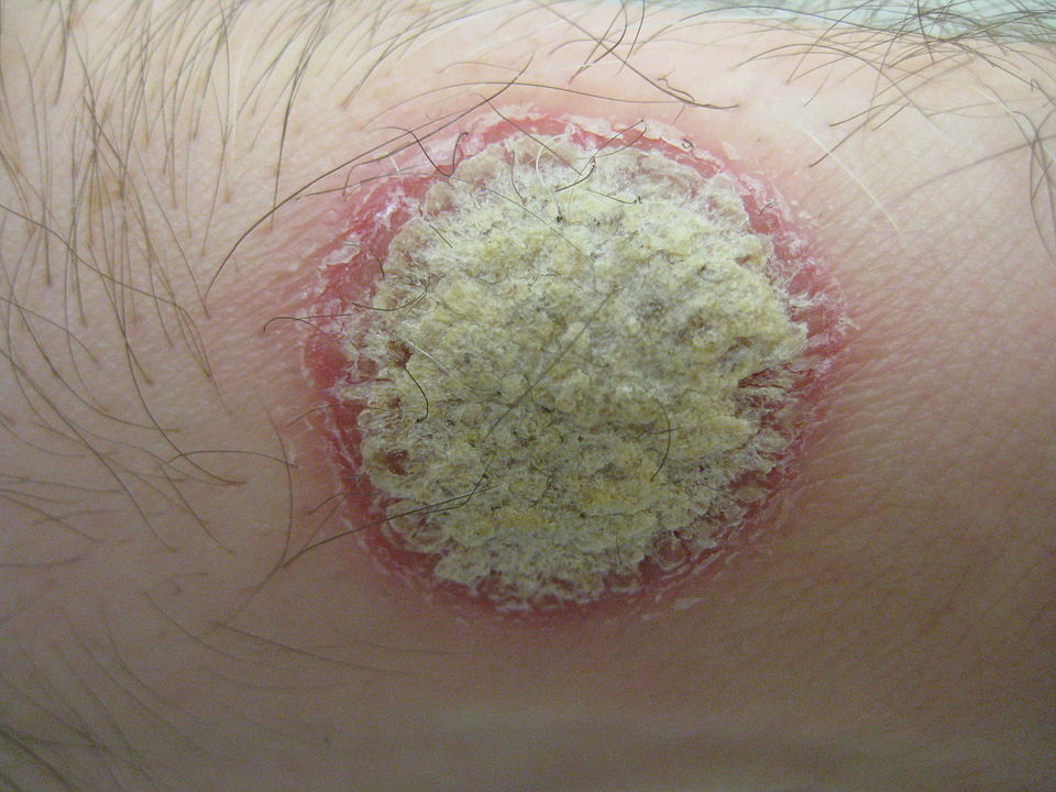
However, when not controlled properly, some of these skin TRM cells can contribute to autoimmune diseases, such as psoriasis and vitiligo.

Researchers, led by University of Melbourne’s Professor Laura Mackay, a Laboratory Head and Immunology Theme Leader at the Peter Doherty Institute of Infection and Immunity (Doherty Institute), found a way to redress this imbalance.

University of Melbourne’s Dr Simone Park, an Honorary Research Fellow and former Postdoctoral Fellow in the Mackay Lab at the Doherty Institute, and lead first author of the study, said that this research is the first to describe the unique elements that control various types of skin TRM cells in animal models, offering precise targets for potential treatment strategies.

“Specialised immune cells in our skin are diverse: many are critical to prevent infection and cancer, but others play a big role in mediating autoimmunity,” said Dr Park.

“We discovered key differences in how distinct types of skin T cells are regulated, allowing us to precisely edit the skin’s immune landscape in a targeted way.”

University of Melbourne’s Dr Susan Christo, Senior Research Officer in the Mackay Lab at the Doherty Institute and co-first author of the study, explained how these discoveries could advance efforts to treat skin disease.

“Most autoimmune therapies treat the symptoms of the disease rather than addressing the cause. Conventional treatments for skin disorders often impact all immune cells indiscriminately, meaning that we could also be wiping out our protective T cells,” said Dr Christo.

“Until now, we didn’t know how to pick apart ‘bad’ T cells in the skin from the ‘good’ protective ones. Through this research, we discovered new molecules that allow us to selectively remove disease-causing T cells in the skin.”

The research team harnessed this new knowledge to eliminate ‘problematic’ cells that can drive autoimmune disorders, while preserving the ‘good’ ones that are essential to maintain protective immunity.

University of Melbourne’s Professor Laura Mackay, senior author of the study, explained that these findings could pave the way for more precise and long-lasting therapies for skin disease.

“Skin conditions like psoriasis and vitiligo are difficult to treat long-term. The T cells driving disease are hard to remove, so patients often need life-long treatment. Our approach has the potential to revolutionise the way we treat these skin disorders, significantly improving outcomes for people dealing with challenging skin conditions,” said Professor Mackay.

With the study demonstrating successful removal of specific skin T cells in animal models, further research is necessary to validate the efficacy of these strategies in human subjects.

Dr Park hopes the study will inspire the development of new treatments for skin disease.

“These discoveries bring us one step closer to developing new drugs that durably prevent autoimmune skin disorders without compromising immune protection,” said Dr Park.

Source: The Peter Doherty Institute for Infection and Immunity

New Study Finds that Fatty Acid in Beef, Lamb and Dairy Boosts Cancer-fighting T Cells

Photo by Jose Ignacio Pompe on Unsplash

Trans-vaccenic acid (TVA), a long-chain fatty acid found in meat and dairy products from grazing animals such as cows and sheep, improves the ability of CD8+ T cells to infiltrate tumours and kill cancer cells, according to a new study by researchers from the University of Chicago.

The research, published in Nature, also shows that cancer patients with higher levels of TVA circulating in the blood responded better to immunotherapy, suggesting potential as a nutritional complement to conventional cancer therapy. Although trans fatty acids that are industrially produced are known to be harmful to health, natural ones such as TVA are linked to health benefits. But the researchers don’t envision prescribing diets packed with red meat and cheese – rather, TVA would be a supplement.

“There are many studies trying to decipher the link between diet and human health, and it’s very difficult to understand the underlying mechanisms because of the wide variety of foods people eat. But if we focus on just the nutrients and metabolites derived from food, we begin to see how they influence physiology and pathology,” said Jing Chen, PhD, professor of medicine at UChicago and one of the senior authors. “By focusing on nutrients that can activate T cell responses, we found one that actually enhances anti-tumour immunity by activating an important immune pathway.”

Searching for nutrients that activate immune cells

Chen’s lab focuses on understanding how metabolites, nutrients and other molecules circulating in the blood influence the development of cancer and response to cancer treatments. For the new study, they started with a database of around 700 known metabolites that come from food and assembled a ‘blood nutrient’ compound library consisting of 235 bioactive molecules derived from nutrients. They screened the compounds in this new library for their ability to influence anti-tumour immunity by activating CD8+ T cells, which are critical for killing cancerous or virally infected cells.

After the scientists evaluated the top six candidates in both human and mouse cells, they saw that TVA performed the best. TVA is the most abundant trans fatty acid present in human milk, but the body cannot produce it on its own. Only about 20% of TVA is broken down into other byproducts, leaving 80% circulating in the blood. “That means there must be something else it does, so we started working on it more,” Chen said.

Feeding mice a diet enriched with TVA significantly reduced the tumour growth potential of melanoma and colon cancer cells compared to mice fed a control diet. The TVA diet also enhanced the ability of CD8+ T cells to infiltrate tumours.

The team also performed a series of molecular and genetic analyses to understand how TVA was affecting the T cells. These included a new technique for monitoring transcription of single-stranded DNA called kethoxal-assisted single-stranded DNA sequencing, or KAS-seq, developed by Chuan He, PhD, professor of chemistry at UChicago and another senior author of the study. These additional assays, done by both the Chen and He labs, showed that TVA inactivates a receptor on the cell surface called GPR43 which is usually activated by short-chain fatty acids often produced by gut microbiota. TVA overpowers these short-chain fatty acids and activates a cellular signaling process known as the CREB pathway, which is involved in a variety of functions including cellular growth, survival, and differentiation. The team also showed that mouse models where the GPR43 receptor was exclusively removed from CD8+ T cells also lacked their improved tumour fighting ability.

Finally, working with other researchers, the team analysed blood samples taken from patients undergoing CAR-T cell immunotherapy treatment for lymphoma. They saw that patients with higher levels of TVA tended to respond to treatment better than those with lower levels. They also tested leukaemia cell lines and saw that TVA enhanced the ability of an immunotherapy drug to kill leukaemia cells.

TVA as a supplement

The study suggests that TVA could be used as a dietary supplement to help various T cell-based cancer treatments, although Chen points out that it is important to determine the optimised amount of the nutrient itself, not the food source. There is a growing body of evidence about the detrimental health effects of consuming too much red meat and dairy, so this study shouldn’t be taken as an excuse to eat more cheeseburgers and pizza; rather, it indicates that nutrient supplements such as TVA could be used to promote T cell activity. Chen thinks there may be other nutrients that can do the same.

“There is early data showing that other fatty acids from plants signal through a similar receptor, so we believe there is a high possibility that nutrients from plants can do the same thing by activating the CREB pathway as well,” he said.

‘The new research also highlights the promise of this ‘metabolomic’ approach to understanding how the building blocks of diet affect our health. Chen said his team hopes to build a comprehensive library of nutrients circulating in the blood to understand their impact on immunity and other biological processes like aging.

“After millions of years of evolution, there are only a couple hundred metabolites derived from food that end up circulating in the blood, so that means they could have some importance in our biology,” Chen said. “To see that a single nutrient like TVA has a very targeted mechanism on a targeted immune cell type, with a very profound physiological response at the whole organism level — I find that really amazing and intriguing.”

Source: University of Chicago

A Hard-to-treat Subtype of Asthma in Older Men

Credit: Pixabay CC0

Scientists have uncovered a group of T cells that may drive severe asthma, which gather in the lungs and seem most harmful in men who develop asthma in later life. The new research, published in MED, suggests asthma patients with these cells in their lungs may be more likely to have hard-to-treat, and potentially fatal, asthma attacks. These cells do not respond to the usual general therapy for asthma patients.

The scientists, from the University of Southampton and La Jolla Institute for Immunology (LJI), in California, uncovered these T cells, called ‘cytotoxic CD4+ tissue-resident memory T cells’, thanks to volunteers enrolled in the NHS clinic-based WATCH study. It follows hundreds of asthma patients of different ages, sexes, and disease severities. By following patients over many years, and analysing their immune cell populations, researchers are making new connections between asthma symptoms and immune cell activity.

“If you are male and you develop asthma after age 40, there’s a high chance this T cell population is in your lungs,” says LJI Research Assistant Professor Gregory Seumois, who co-led the study with LJI Professor Pandurangan Vijayanand.

“Once you understand the role of cells like these T cells better, you can start to develop treatments that target those cells,” says WATCH study director Dr Ramesh Kurukulaaratchy, Associate Professor at the University of Southampton and researcher at the NIHR Southampton Biomedical Research Centre.

Scientists now hope to learn more about these cells and their role in asthma development in order to develop personalised therapies for asthma patients.

How harmful T cells drive asthma

The ‘memory’ T cells help protect the body from viruses and bacteria it has encountered before, but the same T cell memory is a big problem for asthma patients. Their misguided T cells see harmless molecules, such as pollen, and produce a dangerous inflammatory response.

Men who developed asthma later in life had an overwhelming number of these potentially harmful T cells. Their lungs should have been home to a diverse bunch of CD4+ T cell types but, in this group, more than 65% of their cells were cytotoxic CD4+ tissue-resident memory T cells.

Personalised asthma treatments

Single-cell RNA sequencing by LJI scientists provides a ‘biomarker’ to help detect cytotoxic CD4+ tissue-resident memory T cells in more patients going forward.

Finding this biomarker represents a “paradigm shift” in asthma research, says Dr Kurukulaaratchy. Before now, scientists and clinicians separated asthma patients into just two groups: ‘T2 high’ and T2 low’. In a study published earlier this year, the research team showed the importance of drilling down to identify many more asthma patient subgroups; their analysis reveals that 93% of WATCH subjects with severe asthma were in the T2 high category.

Study co-author Professor Hasan Arshad, Chair in Allergy and Clinical Immunology at the University of Southampton, researcher at the NIHR Southampton Biomedical Research Centre, and Director of The David Hide Asthma and Allergy Research Centre, Isle of Wight says: “We have to think of severe asthma as having different subtypes, and the treatment has to be tailored according to these subtypes because one size does not fit all.”

The researchers now want to use sequencing tools and other techniques to discover additional biomarkers and asthma patient subtypes.

Source: University of Southampton

Contrary to Prior Belief, T Cells Even Protect the Cornea

Photo by Stormseeker on Unsplash

Researchers have discovered that the immune cells guarding the healthy human cornea from pathogens and inflammation are T cells, and dendritic cells, as previously thought. The discovery, published in PNAS, redefines current understanding of the immune cell landscape in the cornea of a healthy human eye. It builds on the team’s previous research in Cell Reports that showed that T cells protect the eye against virus infection in mice.

The collaborative research team jointly developed a new imaging technique as part of their investigation.

Research leader Professor Scott Mueller, from the Department of Microbiology and Immunology at the Doherty Institute explained that our knowledge of the various immune cell types in the human cornea is important for establishing the eye’s protective mechanisms against pathogens and disease.

“By combining our newly developed imaging technique with other advanced analytical approaches, we were able to discover that a significant number of cells at the surface of the healthy cornea are actually T cells,” said Professor Mueller.

“Until now, these cells were mistakenly classified as dendritic cells based on static imaging. This completely changes the current dogma in the field that only dendritic cells are present in the healthy cornea.”

The study’s first author, University of Melbourne’s Associate Professor Laura Downie said that being able to dynamically capture the cells’ normal behaviour, and in response to inflammation, provides unique understanding into the immune response in the eye.

“Using our non-invasive imaging approach, which we term Functional In Vivo Confocal Microscopy (Fun-IVCM), we have been able to see that these T cells move around quickly and interact with other cells and nerves in the outermost layer of the cornea. We also captured different cell dynamics in response to contact lens wear and in allergic eye disease, and quantified how these behaviours are modulated by drug treatments,” said Associate Professor Downie.

“These findings reshape our understanding of the distinct immune cell subsets in the human cornea, and how they respond to different stimuli. Using Fun-IVCM, we can achieve rapid, real-time insight into the cellular immune responses in living humans, in this accessible peripheral sensory tissue.”

Senior author Dr Holly Chinnery, also of the University of Melbourne, added that the new research will have major implications for the medical and immunology fields, including for patients and practitioners.

“Because this new technique involves non-invasive, time-lapse imaging of the human cornea, Fun-IVCM could be used in clinics directly to assess immune responses and ocular health. It could even be used for general immune system health,” said Dr Chinnery.

“Changes in T cells and behaviour could be used as a clinical biomarker of disease and assist with treatments.”

Source: The Peter Doherty Institute for Infection and Immunity

T Cell Monitoring may Help Prevent Type 1 Diabetes

A 3D map of the islet density routes throughout the healthy human pancreas. Source: Wikimedia CC0

Scripps Research scientists have shown that people at risk of developing type 1 diabetes could be identified by analysis of the T cells which drive the disease. The new approach, if validated in further studies, could be used to select suitable patients for a newly FDA-approved treatment that stops the autoimmune process, thereby making type 1 diabetes a preventable condition.

In the study, which appears in Science Translational Medicine, the researchers isolated T cells from mouse and human blood samples. By analysing the T cells that can cause type 1 diabetes, they were able to distinguish the at-risk patients who had active autoimmunity from those who had no significant autoimmunity – with 100% accuracy in a small sample.

“These findings represent a big step forward because they offer the possibility of catching this autoimmune process while there is still time to prevent or greatly delay diabetes,” says study senior author Luc Teyton, MD, PhD, professor in the Department of Immunology and Microbiology at Scripps Research.

The study’s first authors were graduate student Siddhartha Sharma and research assistants Josh Boyer and Xuqian Tan, all of the Teyton lab at the time of the study.

Type 1 diabetes usually occurs in childhood or early adulthood, in an autoimmune process that destroys the pancreas’s insulin-producing islet cells. The process can last years, with multiple starts and stops. Exactly how the process begins is not well understood, though it is known to involve genetic factors and may be triggered by routine viral infections.

In 2022, the US Food & Drug Administration approved an immune-suppressing therapy that can protect islet cells and at least delay diabetes onset by months to years if given in the early stages of autoimmunity. However, doctors have not had a good method for identifying people who could benefit from such treatment. They have traditionally examined levels of anti-islet antibodies in patient blood samples, but this antibody response has not been a very accurate measure of autoimmune progression.

“Anti-islet antibody levels are poorly predictive at the individual level, and type 1 diabetes is fundamentally a T cell-driven disease,” Teyton says.

In the study, Teyton and his team constructed protein complexes to mimic the mix of immune proteins and insulin fragments that CD4 T cells normally would recognise to initiate the autoimmune reaction. They used these constructs as bait to capture anti-insulin CD4 T cells in blood samples. They then analysed the gene activity within the captured T cells, and expression of proteins on the cells, to gauge their state of activation.

In this way, they were able to develop a classification algorithm that correctly identified which at-risk patients, in a set of nine, had ongoing anti-islet autoimmunity.

Teyton now hopes to validate the CD4 T cell-based approach with a long-term study in a larger cohort of participants, comparing this approach to the traditional approach of quantifying anti-islet antibodies.

Teyton and his colleagues also are working to make the process of isolating and analysing anti-islet T cells in blood samples more affordable and convenient, so that it can be used more easily in a clinical setting.

“If we can develop this into a useful method for identifying at-risk patients and tracking their autoimmunity status, we not only would have a way of getting the right people into treatment, but also would be able to monitor their disease progress and evaluate potential new preventive therapies,” Teyton says.

Source: Scripps Research Institute

Rare T Cell Could Guide Precision Treatment of Allergies

In a new Nature Immunology study, researchers sheds light on how a rare type of helper T cell, called Th9, can drive allergic disease, suggesting new precision medicine approaches to treating allergies in patients with high levels of Th9.

“Th9 cells are kind of like the black sheep of helper T cells,” said senior author Daniella Schwartz, MD, assistant professor of rheumatology at Pitt’s School of Medicine. “They need a perfect storm of occurrences to pop up, and they aren’t long-lived, which makes them hard to study. The other weird thing about Th9 cells is that they remain functional without seeing their antigen.”

T cells switch on when they encounter viruses, bacteria or other pathogens, causing them to ramp up production of inflammatory proteins called cytokines, which control a suite of immune responses via the JAK-STAT signalling pathway. The main “on” switch for T cells is when the T cell receptor recognises an antigen, a specific identifying feature of a threat. Beyond this specific form of activation, there’s also another type of switch known as bystander activation, which doesn’t involve the T cell receptor.

“Bystander activation usually requires other types of dangerous signals that indicate a threat,” said Schwartz. “What’s really unusual about Th9 cells is that they can be turned on even without these dangerous signals.”

To learn more about how Th9 cells are activated in allergic responses, the team measured the cytokine IL9, produced by Th9 cells, in T cells from patients with atopic dermatitis, and healthy volunteers. They found that Th9 cells from the allergy patients responded to bystander activation, but not those from healthy volunteers.

“This told us that there’s some sort of checkpoint that prevents non-specific activation of Th9 cells in healthy people,” explained Schwartz. “In allergy patients, we hypothesised that the checkpoint breaks down, so you’re getting production of the cytokine even without restimulating the cells with antigen.”

In most helper T cells, when antigen binds to T cell receptor, this highly specific recognition process causes DNA in the T cell’s nucleus to unwind like thread on a spool, opening up regions of DNA that encode the production of cytokines that unleash a suite of immune responses. When the threat is eliminated, there’s no more antigen to stimulate T cell receptors and the cells turn off. But the DNA structure remains open so that the cell is poised for a possible future encounter.

Schwartz and her team found that Th9 cells have a different type of regulation. These cells are activated by transcription factors called STAT5 and STAT6, which bind to the open region DNA around IL9 to activate the gene. Unusually, the DNA closes over time, shutting down production of IL9.

In healthy people, this opening and closing mechanism acts like a checkpoint to manage immune responses being on all the time. But when this checkpoint breaks down in allergy, the DNA remains open, keeping the IL9 gene switched on and driving allergic inflammation.

In a mouse model of allergic asthma driven by Th9, blocking JAK-STAT signaling with a drug called tofacitinib, which is approved for treating rheumatoid arthritis, atopic dermatitis and other inflammatory disorders, improved disease symptoms

Analysing data from allergic asthma patients, the researchers found that those with higher levels of Th9 cells had greater activation of STAT5 and STAT6-related genes. This finding supports the idea that Th9 could act as a biomarker to predict patients who are likely to respond to JAK inhibitors, pointing to new approaches for allergy precision medicine.

Source: University of Pittsburgh

Pseudomonas Aeruginosa Locks out Immune Cells

Pseudomonas
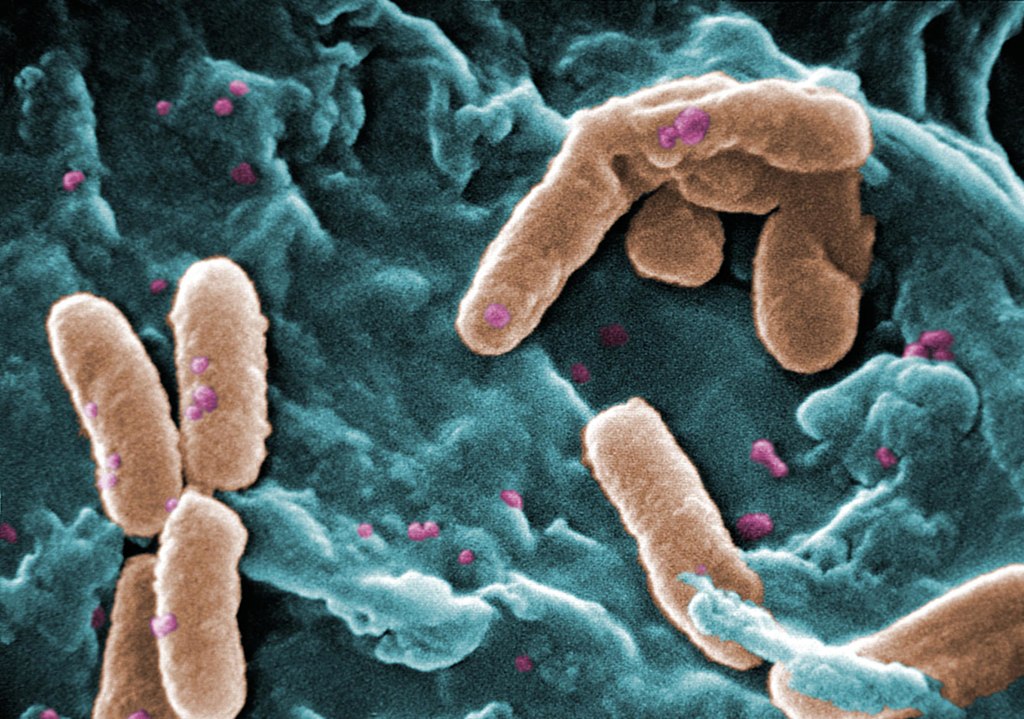
Scanning Electron Micrograph of Pseudomonas aeruginosa. Credit: CDC/Janice Carr

Pseudomonas aeruginosa bacteria are a common menace in hospital wards, causing life-threatening infections, and are often resistant to antibiotics. Researchers have discovered a mechanism that likely contributes to the severity of P. aeruginosa infections, which could also be a target for future treatments. The results were recently appeared in the journal EMBO Reports.

Many bacterial species use sugar-binding molecules called lectins to attach to and invade host cells. Lectins can also influence the immune response to bacterial infections. However, these functions have hardly been researched so far. A research consortium led by Prof Dr Winfried Römer at the University of Freiburg and Prof Dr Christopher G. Mueller at the CNRS/University of Strasbourg has investigated the effect of the lectin LecB from P. aeruginosa on the immune system. It found that isolated LecB can render immune cells ineffective: The cells are then no longer able to migrate through the body and trigger an immune response. The administration of a substance directed against LecB prevented this effect and led to the immune cells being able to move unhindered again.

LecB blockades immune cells

As soon as they perceive an infection, cells of the innate immune system migrate to a nearby lymph node, where they activate T and B cells, triggering a targeted immune response. LecB, according to the current study, prevents this migration. “We assume that LecB not only acts on the immune cells themselves in this process, but also has an unexpected effect on the cells lining the inside of the blood and lymph vessels,” Römer explains. “When LecB binds to these cells, it triggers extensive changes in them.” Indeed, the researchers observed that important structural molecules were relocated to the interior of the cells and degraded. At the same time, the cell skeleton became more rigid. “The cell layer thus becomes an impenetrable barrier for the immune cells,” Römer said.

An effective agent against LecB

Can this effect be prevented? To find out, the researchers tested a specific LecB inhibitor that resembles the sugar building blocks to which LecB otherwise binds. “The inhibitor prevented the changes in the cells, and T-cell activation was possible again,” Mueller said. The inhibitor was developed by Prof Dr Alexander Titz, who conducts research at the Helmholtz Institute for Pharmaceutical Research Saarland and Saarland University.

Further studies are needed to determine how clinically relevant the inhibition of the immune system by LecB is to the spread of P. aeruginosa infection and whether the LecB inhibitor has potential for therapeutic application. “The current results provide further evidence that lectins are a useful target for the development of new therapies, especially for antibiotic-resistant pathogens such as P. aeruginosa,” the authors conclude.

Source: University of Freiburg

Training Cells to Fight Both Chronic Inflammatory and Infectious Diseases

T cell
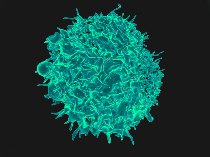
Scanning Electron Micrograph image of a human T cell. Credit: NIH/NIAID

Researchers from the University of Queensland have identified a pathway in cells that could be used to reprogram the body’s immune system to fight back against both chronic inflammatory and infectious diseases such as E. Coli.

Reporting their findings in the open-access journal PNAS, Dr Kaustav Das Gupta and Professor Matt Sweet found that a glucose-derived molecule in immune cells can both stop bacteria growing and dampen inflammatory responses.

According to Dr Das Gupta, the discovery is a critical step towards future therapeutics that train immune cells.

“The effects of this molecule called ribulose-5-phosphate on bacteria are striking – it can cooperate with other immune factors to stop disease-causing strains of the E. coli bacteria from growing,” Dr Das Gupta said.

“It also reprograms the immune system to switch off destructive inflammation, which contributes to both life-threatening infectious diseases such as sepsis as well as chronic inflammatory diseases like respiratory diseases, chronic liver disease, inflammatory bowel disease, rheumatoid arthritis, heart disease, stroke, diabetes and dementia.”

The research was carried out on a strain of E. coli bacteria, responsible for 80% of urinary tract infections and also a common cause of sepsis. Pre-clinical trials confirmed the role of this pathway in controlling bacterial infections.

Professor Sweet said that human cells were also used to demonstrate that ribulose-5-phosphate reduces the production of molecules that drive chronic inflammatory diseases.

“Host-directed therapies which train our immune systems to fight infections, will become increasingly important as more types of bacteria become resistant to known antibiotics,” Professor Sweet said.

“A bonus is that this strategy also switches off destructive inflammation, which gives it the potential to combat chronic disease.

“By boosting the immune pathway that generates ribulose-5-phosphate, we may be able to give the body the power to fight back against inflammatory and infectious diseases – not one, but two of the major global challenges for human health.”

Many current anti-inflammatory therapies target proteins on the outside of cells but because this pathway occurs inside cells, the researchers devised a new approach to target the pathway using mRNA technology.

Source: University of Queensland

Scientists Solve Epstein-Barr Virus Mystery

Photo by National Cancer Institute on Unsplash

Medical science has not yet been able to explain why the Epstein-Barr virus triggers infectious mononucleosis (IM) in some people with initial infections and not in others. But now researchers have identified a unusual T cell response to the virus as the cause, and as a potential target for the development of vaccines. The findings were recently published in the journal Blood.

T cells normally fight the proliferation of the Epstein-Barr virus (EBV) in humans as part of an antiviral immune response. In this response, certain EBV components (peptides) are presented to the T cells by a specific molecule (HLA-E), which is found on the surface of cells infected with EBV. This triggers a non-classical T-cell response that leads to the destruction of the infected cells. Due to a genetic variation (HLA-E*0103/0103), about one third of the population naturally has more HLA-E molecules on EBV-infected cells.

A recently published study has shown that the risk of developing IM following first-time infection with the Epstein-Barr virus depends strongly on this EBV-specific immune response.

“Our research revealed that people with the HLA-E*0103/0103 genetic variation have a lower risk of developing infectious mononucleosis than those who do not have the variation. Our experiments in the lab showed that this gene variation is associated with a highly pronounced EBV-specific -non-classical — immune response,” explained Hannes Vietzen from MedUni Vienna’s Center for Virology, the first author of the study.

Preventive and diagnostic possibilities

EBV is one of the most common viral infections in humans. On initial infection, the virus causes IM in some children and young adults; this disease is characterised by non-specific symptoms, such as fever, as well as exhaustion that in some cases can last for several months. Until now, it was unclear why a first-time EBV infection only leads to IM in a minority of people, while most do not present any symptoms whatsoever. The immune response that the researchers identified could also be a target for research into preventive measures: “This immune response was still measurable years after the initial EBV infection and generally provides long-lasting protection against reinfection with Epstein-Barr, so it might be worth focusing our attention on this mechanism with a view to developing new vaccines in future,” said Hannes Vietzen, looking ahead.

Another finding from the study could also open up new diagnostic options: “The combination of the unfavourable HLA-E genetic variation with certain EBV peptides also appears to play an important role in the development of EBV-associated lymphomas in transplant recipients,” Hannes Vietzen commented. “Analysis of the EBV strains found in these patients could be helpful in identifying high-risk patients at an early stage and treating them in good time.”

Source: Medical University of Vienna