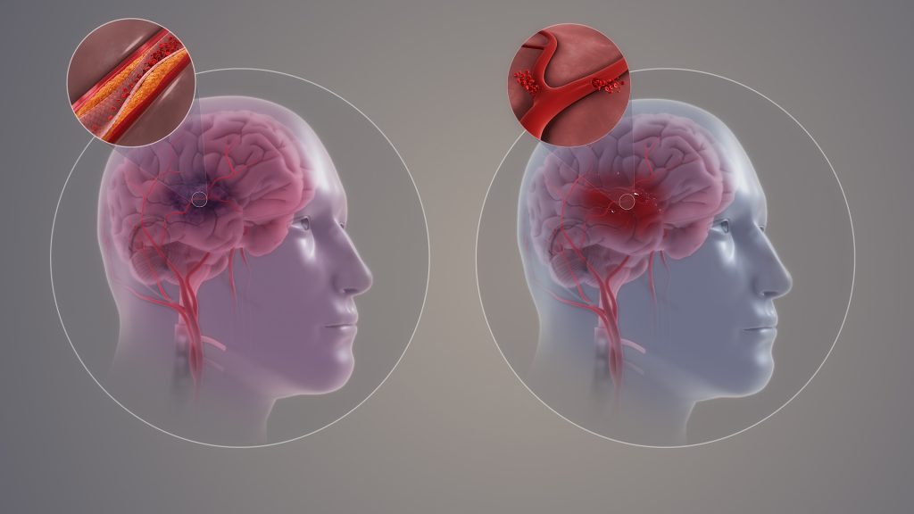
Data on 13 205 adults suggests an increased risk of later life stroke among people who had experienced parental divorce in childhood
People whose parents divorced during their childhood may be at a greater risk of stroke later in life, according to a new study published January 22, 2025 in the open-access journal PLOS One by Esme Fuller-Thomson of University of Toronto, Canada, and colleagues.
Each year, approximately 795 000 individuals in the U.S. have a stroke. Previous work has established many sociodemographic risk factors for stroke, as well as connections between adverse childhood events and stroke. In the new study, researchers looked specifically at the impact of childhood parental divorce among adults with no history of childhood abuse. They used data on 13 205 adults aged 65 and over from the 2022 Behavioral Risk Factor Surveillance System.
The study found that people who had experienced parental divorce before they were 18 years old had 1.61 times higher odds of having a stroke when compared to respondents who did not experience parental divorce (AOR=1.61, 95% CI=1.15-2.24). The association did not vary by sex, and remained even after controlling for known risk factors such as diabetes, depression, and small social support networks.
The current study was not designed to analyse the potential mechanism of this association, nor to prove causation. The conclusions may not be generalisable to younger generations, who have experienced overall higher rates of parental divorce. In addition, several potential confounding factors – including blood pressure, cholesterol, contraceptive use, age at parents’ divorce, and types of strokes – were not available in the data.
However, the authors say that their data supports an association between parental divorce during childhood and increased stroke risk, even in the absence of childhood abuse and other trauma.
Senior author Esme Fuller-Thomson adds: “It is extremely concerning that older adults who grew up in divorced families had 60% higher odds of stroke, even after excluding those who had been physically or sexually abused as children. The magnitude of the association between parental divorce and stroke was comparable to well-established risk factors for stroke such as male gender and having diabetes.”
Individuals with sickle cell disease are at a higher risk for stroke and resulting cognitive disability. But even in the absence of stroke, many such patients struggle with remembering, focusing, learning and problem solving, among other cognitive problems, with many facing challenges in school and in the workplace.
Now a multidisciplinary team of researchers and physicians at Washington University School of Medicine in St. Louis has published a study that helps explain how the illness might affect cognitive performance in sickle cell patients without a history of stroke. The study, appearing in JAMA Network Open, found such participants had brains that appeared older than expected for their age. Individuals experiencing economic deprivation, who struggle to meet basic needs, even in the absence of sickle cell disease, had more-aged appearing brains, the team also found.
“Our study explains how a chronic illness and low socioeconomic status can cause cognitive problems,” said Andria Ford, MD, a professor of neurology and chief of the section of stroke and cerebrovascular diseases at WashU Medicine and corresponding author on the study. “We found that such factors could impact brain development and/or aging, which ultimately affects the mental processes involved in thinking, remembering and problem solving, among others. Understanding the influence that sickle cell disease and economic deprivation have on brain structure may lead to treatments and preventive measures that potentially could preserve cognitive function.”
More than 200 young, Black adults with and without sickle cell disease, living in St. Louis and the surrounding region in eastern Missouri and southwestern Illinois, participated in brain MRI scans and cognitive tests. The researchers – including Yasheng Chen, DSc, an associate professor of neurology at WashU Medicine and senior author on the study – calculated each person’s brain age using a brain-age prediction tool that was developed using MRI brain scans from a diverse group of more than 14 000 healthy people of known ages. The estimated brain age was compared with the individual’s actual age.
The researchers found that participants with sickle cell disease had brains that appeared an average of 14 years older than their actual age. Sickle cell participants with older-looking brains also scored lower on cognitive tests.
The study also found that socioeconomic status correlates with brain age. On average, a seven-year gap was found between the brain age and the participants’ actual age in healthy individuals experiencing poverty. The more severe the economic deprivation, the older the brains of such study subjects appeared.
Healthy brains shrink as people age, while premature shrinking is characteristic of neurological illnesses such as Alzheimer’s disease. But a smaller brain that appears older can also result from stunted growth early in life. Sickle cell disease is congenital, chronically depriving the developing brain of oxygen and possibly affecting its growth from birth. Also, children exposed to long-term economic deprivation and poverty experience cognitive challenges that affect their academic performance, Ford explained.
As a part of the same study, the researchers are again performing cognitive tests and scanning the brains of the same healthy and sickle cell participants three years after their first scan to investigate if the older-looking brains aged prematurely, or if their development was stunted.
“A single brain scan helps measure the participants’ brain age only in that moment,” said Ford, who treats patients at Barnes-Jewish Hospital. “But multiple time points can help us understand if the brain is stable, initially capturing differences that were present since childhood, or prematurely aging and able to predict the trajectory of someone’s cognitive decline. Identifying who is at greatest risk for future cognitive disability with a single MRI scan can be a powerful tool for helping patients with neurological conditions.”
Updated clinical recommendations, including lifestyle changes, prevention strategies and treatment options, to reduce the risk of a first stroke outlined in a new guideline from the American Stroke Association
Healthy lifestyle behaviours, such as good nutrition, smoking cessation and being physically active, along with routine health screenings and managing risk factors for cardiovascular disease and stroke with medication, can help prevent individuals from having a first stroke. Screening for stroke risk and educating people on how to lower their chances of having a stroke ideally begin with their primary care professional and include evidence-based recommendations, according to a new clinical guideline from the American Stroke Association, and published in the journal Stroke.
“The most effective way to reduce the occurrence of a stroke and stroke-related death is to prevent the first stroke – referred to as primary prevention,” said Chair of the guideline writing group, Cheryl D. Bushnell, MD, MHS, FAHA, professor and vice chair of research in the department of neurology at Wake Forest University School of Medicine. “Some populations have an elevated risk of stroke, whether it be due to genetics, lifestyle, biological factors and/or social determinants of health, and in some cases, people do not receive appropriate screening to identify their risk.”
The “2024 Guideline for the Primary Prevention of Stroke” replaces the 2014 version and is a resource for clinicians in implementing a variety of prevention strategies for individuals with no prior history of stroke. The new guideline provides evidence-based recommendations for strategies to support brain health and prevent stroke throughout a person’s lifespan by improving healthy lifestyle behaviours and getting preventive care.
“This guideline is important because new discoveries have been made since the last update 10 years ago. Understanding which people are at increased risk of a first stroke and providing support to preserve heart and brain health can help prevent a first stroke,” said Bushnell.
Key stroke prevention recommendations include regular health screenings, identifying risk factors, lifestyle interventions and medications, when indicated.
Identifying and managing risk factors
Unidentified and unmanaged cardiovascular disease risk factors can cause damage to arteries, the brain and the heart years before cardiovascular disease and stroke occur. Primary care health professionals should promote brain health for patients through stroke prevention education, screenings and addressing risk factors from birth to old age.
Modifiable risk factors for stroke, such as high blood pressure, overweight and obesity, elevated cholesterol and elevated blood sugar, can be identified with physical exams and blood tests. These conditions should be addressed with healthy lifestyle and behavioural changes and may include medications for select patients. Antihypertensive medications to reduce blood pressure and statin medications to lower cholesterol can help to reduce the risk of first stroke in adults with increased cardiovascular disease risk and those receiving CVD care. A new recommendation is consideration of glucagon-like protein-1 (GLP-1) receptor agonist medications, which are FDA-approved to reduce the risk of cardiovascular disease in people with overweight or obesity and/or Type 2 diabetes.
Healthy lifestyle behaviours
The most common, treatable lifestyle behaviours that can help reduce stroke risk are detailed in the Association’s Life’s Essential 8 cardiovascular health metrics. They include healthy nutrition, regular physical activity, avoiding tobacco, healthy sleep and weight, controlling cholesterol, and managing blood pressure and blood sugar. The guideline recommends that adults with no prior cardiovascular disease, as well as those with increased risk, follow a Mediterranean dietary pattern. Mediterranean dietary programs have been shown to reduce the risk of stroke, especially when supplemented with nuts and olive oil.
Physical activity is also essential for stroke risk reduction and overall heart health. Physical activity can help to improve important health measures such as blood pressure, cholesterol, inflammatory markers, insulin resistance, endothelial function and weight. The guideline urges health care professionals to routinely screen patients for sedentary behaviour, a confirmed risk factor for stroke, and counsel them to engage in regular physical activity. The Association reinforces the U.S. Department of Health and Human Services Office of Disease Prevention and Health Promotion’s recommendation that adults get at least 150 minutes per week of moderate-intensity aerobic activity or 75 minutes per week of vigorous aerobic activity, or a combination of both, preferably spread throughout the week.
Health equity and stroke risk
New to the guideline is an emphasis on social determinants of health and the impact they have on stroke risk. Social determinants of health are non-medical factors, including education, economic stability, access to care, discrimination, structural racism and neighborhood factors (such as the lack of walkability, lower availability of healthy food and fewer health resources), that contribute to inequities in care and influence overall health. Health care professionals should ensure patient education is available for various educational and language levels, and advocate for their patients by choosing treatments and medications that are effective and affordable.
Health care professionals are also encouraged to connect patients to resources that help address health-related social needs such as food and housing insecurity, refer them to programs that support healthy lifestyle changes and direct them to support programs that may help defray health care costs including medication expenses.
New sex- and gender-specific recommendations
The guideline also includes some new gender- and sex-specific recommendations for women. Health professionals should screen for conditions that can increase a woman’s risk of stroke, including use of oral contraceptives, high blood pressure during pregnancy, other pregnancy complications such as premature birth, endometriosis, premature ovarian failure and early onset menopause. Treatment of elevated blood pressure during pregnancy and within six weeks of delivery is recommended to reduce the risk of maternal intracerebral haemorrhage.
Transgender women and gender-diverse individuals taking oestrogens for gender affirmation may also be at an increased risk of stroke. Evaluation and modification of any existing risk factors are needed to reduce the risk of stroke for these individuals.
“Implementing the recommendations in this guideline would make it possible to significantly reduce the risk of people having a first stroke. Most strategies that we recommend for preventing stroke will also help reduce the risk of dementia, another serious health condition related to vascular issues in the brain,” said Bushnell.
The writing group notes that writing recommendations focused on preventing a first stroke was challenging. There are limitations to some of the evidence that informed the guideline, including that many clinical trials enrolled adults who have already had a cardiovascular event that may include a stroke. The writing group also identified knowledge gaps to help inform topics for future research.
The guideline highlights the need for risk assessment in primary stroke prevention and includes the use of risk prediction tools to estimate risk for atherosclerotic cardiovascular disease so that patients receive timely prevention and treatment strategies. The Association has recently developed a new Predicting Risk of Cardiovascular Disease Events (PREVENT) risk calculator as a screening tool that can help inform preventive treatment decisions. The PREVENT calculator can estimate 10-year and 30-year stroke and heart disease risk in individuals starting at age 30 – a decade earlier than the Pooled Cohort Equations, another CVD risk calculator.
According to the American Stroke Association, learning the warning signs of stroke and preventative measures are the best way to avoid strokes and keep them from happening again. The abbreviation F.A.S.T. – for face drooping, arm weakness, speech difficulty, time to call 911 – is a useful tool to recognise the warning signs of stroke and when to call for help.
This guideline was prepared by the volunteer writing group on behalf of the American Stroke Association and is endorsed by the Preventive Cardiovascular Nurses Association and the Society for Vascular Surgery. The American College of Obstetricians and Gynecologists supports the clinical value of this document as an educational tool.
Since 1990, the American Stroke Association has translated scientific evidence into clinical practice guidelines with recommendations to improve cerebrovascular health. The “2024 Guideline for the Primary Prevention of Stroke” replaces the 2014 “Guidelines for the Primary Prevention of Stroke.” This updated guideline is intended to be a resource for clinicians to use to guide various prevention strategies for individuals with no history of stroke. The Association supports the development and publication of clinical practice guidelines without commercial support, and members volunteer their time to the writing and review efforts.
Initial prescriptions of benzodiazepines, a class of drugs used to treat anxiety and sleep problems after a stroke may include too many pills for adults ages 65 or older, finds new study in the Stroke journal
Although there has been a slight downward trend in the prescription of benzodiazepines (depressants that relieve anxiety, muscle spasms, produce sedation and reduce seizures) among older adults over the last decade, the rate of first-time prescriptions for these medications after an ischaemic stroke is still sizable, according to research published today in Stroke.
After a stroke, benzodiazepines may be used to calm anxiety and improve sleep, but also have a potential for abuse and addiction. When prescribed to older adults, these medications may increase the risk of falls and broken bones, as well as memory problems, confusion and other harmful effects.
Researchers reviewed data from Medicare claims in the US and analysed 10 years of first-time prescriptions for benzodiazepines among more than 120 000 people, ages 65 and older, who were hospitalised for ischaemic stroke. The rate of benzodiazepine prescriptions during the first three months after stroke were examined, and data were adjusted for race, sex and ethnicity. Then year-to-year prescription patterns were reviewed to identify the number of potentially excessive new benzodiazepine prescriptions given to stroke survivors.
“We reviewed stroke survivors at 90 days after a stroke because that window of time is critical for rehabilitation of motor, speech and cognitive function, as well as mental health. It’s often a very difficult time for patients who experience loss of mobility and independence. Benzodiazepines may inhibit recovery and rehabilitation,” said study co-author Julianne Brooks, MPH, a data analytics manager at the Center for Value-based Healthcare and Sciences at Massachusetts General Brigham in Boston. “For this older age group, guidelines recommend that benzodiazepine prescriptions should be avoided if possible. However, there may be cases where benzodiazepines are prescribed to be used as needed. For example, to treat breakthrough anxiety, a provider may prescribe a few pills and counsel the patient that the medication should only be used as needed. The increased risks of dependence, falls and other harmful effects should be discussed with the patient.”
The study found:
Within 90 days of stroke, 6127 (4.9%) people were started on a benzodiazepine for the first time.
Lorazepam (40%) and alprazolam (33%) were the most-prescribed benzodiazepine medications.
Three-quarters of the first-time benzodiazepine prescriptions were for a supply of over seven days, and more than half of the prescriptions were for a supply between 15 to 30 days.
Prescription rates were higher among women (5.5%) than men (3.8%).
Prescription fill rates were also higher in Hispanic adults (5.8%), though this group was limited by the small number of participants – 1.9% of the overall sample.
Overall, prescription rates were highest in the Southeast (5.1%) and lowest in the Midwest (4%) of the US. “The Southeast region is the stroke belt with a higher rate of strokes, so that could explain some differences in care in that region,” Brooks said.
There was an overall modest nationwide decline of initial prescriptions from 2013 to 2021 of 1.6%.
“We found a pattern of potential oversupply with these initial benzodiazepine prescriptions, which would be enough for patients to become long-term users or possibly addicted. The benzodiazepine prescriptions given under these circumstances may lead to dependence,” Brooks said. “Increased awareness and improved recommendations about the risks of these medications for older stroke survivors are needed.
“Although the overall prescription rate decreased slightly over 10 years, this prescription pattern is still a problem. It’s concerning because older adults are vulnerable to overprescribing and adverse outcomes. We know from previous studies that vulnerable and marginalized populations experience worse outcomes after stroke, so we want to understand the factors that may play a role so we can provide better care,” Brooks said.
The 2019 American Geriatrics Society Beers Criteria maintains a list of medications that health care professionals can reference to safely prescribe medications for adults older than 65. Beers criteria recommends avoiding benzodiazepines in all older adults due to the risk of cognitive impairment, delirium, falls, fractures and motor vehicle crashes.
“Other guidelines also suggest behavioural interventions such as cognitive behaviour therapy for insomnia, antidepressant medications for anxiety disorders and trying non-pharmaceutical interventions first,” Brooks said.
Researchers said more studies are needed to understand if there is a safe level for prescribing benzodiazepines that may be most appropriate for older adults. The main limitation was that this study used a large, national dataset that did not include information about why benzodiazepines were prescribed.
An analysis of data in the UK Biobank has found that COVID infection may increase the risk of myocardial infarction (MI), stroke and death from any cause for up to three years for people with and without cardiovascular disease, according to new research published in the American Heart Association’s peer-reviewed journal Arteriosclerosis, Thrombosis and Vascular Biology (ATVB).
“We found a long-term cardiovascular health risk associated with COVID, especially among people with more severe COVID cases that required hospitalisation,” said lead study author James Hilser, M.P.H., Ph.D.-candidate at the University of Southern California Keck School of Medicine in Los Angeles. “This increased risk of heart attack and stroke continued three years after COVID infection. Remarkably, in some cases, the increased risk was almost as high as having a known cardiovascular risk factor such as Type 2 diabetes or peripheral artery disease.”
Previous research has shown that COVID increases the risk of serious cardiovascular complications within the first month after infection. This study examined how long the increased risk lasted and whether it subsided after recovering from COVID infection.
Researchers reviewed health and genetic data in the UK Biobank for more than 10 000 adults, including approximately 8000 who had tested positive for SARS-CoV-2 from February 1 to December 31, 2020 and about 2000 who tested positive for the virus in a hospital setting in 2020. A group of more than 200,000 adults who had no history of COVID infection during the same time frame in the UK Biobank were also reviewed for comparison. None of the participants were vaccinated at the time of infection because COVID vaccines were not yet available in 2020.
The analysis found:
During the nearly 3-year follow-up period, the risk of heart attack, stroke and death was more than two times higher among adults who had COVID, and nearly four times greater among adults hospitalized with COVID, compared with the group with no history of COVID infection.
People hospitalized with COVID, without cardiovascular disease or without Type 2 diabetes, had a 21% greater risk of heart attack, stroke and death compared to people with cardiovascular disease and without COVID infection.
There was a significant genetic interaction among the non-O blood types and hospitalisation for COVID. People with severe COVID infections had an increased risk of heart attack and stroke, however, that risk was even higher in people who had non-O blood types (those with blood types A, B or AB).
The risk of heart attack and stroke was about 65% higher in adults with non-O blood types compared to those who had type O blood. A preliminary analysis did not show that Rh (positive or negative) blood type interacted with severe COVID, the authors noted.
“Worldwide, over a billion people have already experienced COVID infection. The findings reported are not a small effect in a small subgroup,” said co-senior study author Stanley Hazen, M.D., Ph.D., chair of cardiovascular and metabolic sciences in Cleveland Clinic’s Lerner Research Institute and co-section head of preventive cardiology. “The results included nearly a quarter million people and point to a finding of global health care importance that may translate into an explanation for a rise in cardiovascular disease around the world.”
Study details, background and design:
Health data was from the UK Biobank, a large-scale study of 503,325 adults living in the United Kingdom who were 40 to 69 years of age at enrollment between 2006 and 2010. The in-depth health and biomedical information was collected for participants registered in the UK National Health Service with a UK general practitioner (similar to a primary care physician in the U.S.).
This analysis included health data for 10,005 adults who tested positive for the COVID virus or were hospitalized with COVID between February 1, 2020, and December 31, 2020. An additional 217,730 peers enrolled in the UK Biobank who did not have COVID during the same time period were included. In the analysis, all participants were matched as closely as possible for demographics and similar health conditions.
Major adverse cardiovascular events (heart attack, stroke and all-cause death) were evaluated for long-term risk, through October 31, 2022, approximately 3 years later.
“This interesting paper is really two studies in one,” said Sandeep R. Das, M.D., M.P.H., MBA, FAHA, co-chair of the American Heart Association’s COVID-19 CVD Registry committee and director for quality and value in the cardiology division for UT Southwestern Medical Center in Dallas. “First, the authors show that having been hospitalized with COVID is a marker of increased cardiovascular risk, on par with having a pre-existing diagnosis of cardiovascular disease. Although proving direct cause and effect is very difficult to tease out in a study that only analyses past data collected for other purposes, this finding is important because it suggests a history of prior COVID hospitalization, even without a history of CVD, should be considered to initiate and possibly accelerate CVD prevention efforts. Whether severe COVID infection has a direct impact on the vascular system is an interesting area for study as well,” Das said.
“The second ‘study’ in this paper looks at the relationship between ABO blood type and COVID outcomes. They show that something located close to the genetic home of ABO blood type is associated with different degrees of susceptibility to COVID. This is really fascinating, and I look forward to seeing scientists tease out what the specific pathway may be.”
The study had several limitations, including that the data was from patients who had the original strain of the COVID virus before vaccines were widely available in 2021. Additionally, the researchers noted that UK Biobank information on medication use was not specific to the beginning of the pandemic in 2020 or the date that patients were infected with SARS-CoV-2. Also, because the majority of participants in the UK Biobank are white, additional research is needed to confirm that these results apply to people with diverse racial and ethnic backgrounds.
“The results of our study highlight the long-term cardiovascular effects of COVID infection. Given the increased risk of heart attack, stroke and death, the question is whether or not severe COVID should be considered as another risk factor for CVD, much like Type 2 diabetes or peripheral artery disease, where treatment focused on CVD prevention may be valuable,” said co-senior study author Hooman Allayee, Ph.D., a professor of population and public health sciences at the University of Southern California Keck School of Medicine in Los Angeles. “The results suggest that people with prior COVID infection may benefit from preventive care for cardiovascular disease.”
People who have had a stroke may be more likely to sleep too much or too little compared to those without prior stroke, according to a study published in Neurology®, the medical journal of the American Academy of Neurology. The study does not prove that stroke causes abnormal sleep; it only shows an association.
“Sleeping the right amount is considered essential for ideal brain and heart health,” said study author Sara Hassani, MD, of Duke University School of Medicine in Durham, North Carolina, and member of the American Academy of Neurology. “We know that abnormally long or short sleep after stroke can affect recovery and deteriorate quality of life, so these results should prompt us to screen for these issues and look at how we can help people improve their sleep habits.”
The study involved 39 559 participants, of whom 1572 had a stroke and 37 987 without stroke history. Every two years, participants were asked how much sleep they usually get at night on weekdays or workdays. Sleep duration was divided into three categories: short, less than six hours; normal, six to eight hours; and long, eight or more hours of sleep.
Researchers looked at how often participants had normal sleep, defined as six to eight hours. Normal sleep duration was less common for people who had a stroke than for those with no prior stroke for all age groups with 32% vs 54% for people age 18-44; 47% vs 55% for people age 45-64; and 45% vs. 54% for people over age 65.
After adjusting for factors that could affect sleep such as age, weight and high blood pressure, researchers found people who had a stroke were 54% more likely to report more than eight hours of sleep per night compared to those without stroke. Those with stroke were 50% more likely to get less than six hours of sleep per night when compared to those without stroke.
“In previous research, stroke has been linked to abnormal sleep, in particular sleep apnea,” said Hassani. “Conditions like insomnia and excessive sleepiness are common in stroke patients and may occur as a direct or indirect consequence of stroke itself. Future research should explore the links between stroke and duration of sleep and determine the effect of sleep duration on outcomes after stroke.”
One limitation of the study was that hours of sleep were self-reported, so participants may not have remembered accurately how much they slept.
Research conducted at the University of Gothenburg shows that daily physical activities, at work or in the home, are not sufficient to protect against stroke. Fortunately, the findings, published in JAMA Network Open, suggest that exercising in free time and using active modes of transport are associated with a decreased risk of stroke.
“Physical activity during leisure time and as transportation is becoming increasingly important now that many jobs and domestic activities are becoming more sedentary,” says lead author of the study Adam Viktorisson, researcher at Sahlgrenska Academy at the University of Gothenburg, Sweden.
Twenty year follow-up
The research study covers 3614 people from the region of Västra Götaland, 269 of whom suffered a stroke in the twenty years spanned by the study. Three months after the stroke, 120 of these had died or were dependent on help to carry out activities of daily living.
Physical activity data was gathered from surveys. Some participants were also given a pedometer to wear. Physical activity during leisure time or for transportation showed a link to the objective measurements from the pedometers, while physical activity at work did not.
Occupational physical activity not protective
The health benefits of physical activity are well known, but earlier studies tend to mainly focus on physical activity during leisure time. Research in recent years has shown that physical activity at work can instead have negative health impacts, increasing the risk of cardiovascular disease.
“How and when we carry out physical activity seems to play a crucial role in determining its health benefits. In our study leisure time and transport related physical activities were associated with a lower risk of stroke, whereas activities during work time or in the household were not” Adam Viktorisson points out.
“Physically demanding jobs are often linked to stress, little opportunity for recovery, air pollution and generally poorer socioeconomic conditions, which can counteract the positive effects of physical activity.”
Promote public health
The study used data from the INTERGENE cohort at the University of Gothenburg. Study participants were surveyed and data was collected from 2001 to 2004, consisting of both clinical and questionnaire data. The researchers hope that these results will bring greater awareness and lead to changes in public health policy to encourage physical activity in society.
“Encouraging people to be physically active in their daily lives, for example by walking, cycling and doing other types of exercise, can be an important strategy in reducing the number of strokes and improving the prognosis of people who suffer a stroke,” says Adam Viktorisson.
Why do new comorbidities arise because of ischaemic stroke? A study from Germany recently published in the journal Cell has discovered why this can happen – and ways in which it might be countered. The findings from the study show that the immune system is involved in damage to other organs, including the heart.
Besides the early mortality and morbidity resulting from the ischaemic brain injury itself, long-term morbidity after stroke is also due to the high prevalence of secondary comorbidities and complications, such as cognitive impairment and dementia, post-stroke depression, cardiac events, persistent vascular inflammation, and stroke-induced metabolic disturbances.
Liesz is the principal investigator of this new study. The researchers worked on the hypothesis that the high rate of comorbidities that develop after a stroke could have a common immunological cause. And they actually managed to find it: the origin of the dysfunctions in other parts of the body lies in the immunological memory of the blood-forming cells in bone marrow.
Using single-cell sequencing techniques, Liesz and his team demonstrated the presence of permanent proinflammatory changes in the transcriptome of certain immune cells (monocytes/macrophages) in several organs. In other words, certain gene segments are transcribed differently there after the stroke, which unbalances the proteome. These epigenetic modifications occur most frequently in the heart, where they can cause scarring and impair pumping function. “We managed to identify the protein IL-1b as the main culprit for the epigenetic modifications that affect immunological memory after a stroke,” says Liesz.
Promising therapeutic approaches on the horizon
The researchers demonstrated in a mouse model the connection between modified blood formation in bone marrow through overexpressed IL-1b and cardiac dysfunctions. Moreover, they showed that blocking IL-1b and inhibiting migration of the proinflammatory cells to the heart both successfully prevented cardiac problems after a stroke.
“These findings are hugely significant, as they open up the promise of effective therapeutic approaches for the prevention of secondary cardiac conditions after a stroke,” reckons Liesz.
The authors of the study believe that the epigenetic mechanisms they described for the reprogramming of the immune system in the brain-heart axis will create a new framework for explaining the development of various IL-1b-mediated comorbidities.
Research by West Virginia University has demonstrated that American Heart Association and American Stroke Association guidelines are effective at speeding up hospitals’ response times for stroke treatment and can be mastered even by members of ‘ad hoc‘ medical teams that assemble rapidly on the fly.
When a stroke patient arrives at an emergency room, specialists from across hospital departments – emergency medical services, neurologists, pharmacists, physicians, nurses, radiologists and technicians – rush to coordinate a team response. AHA and ASA guidelines put specific limits on how much time can optimally elapse between the onset of ischaemic stroke, in which blood flow to the brain is blocked, and subsequent events like arrival at the hospital and delivery of an infusion.
But experts have questioned whether the communication of those best practices helps medical teams that assemble temporarily and whose members don’t typically collaborate. In a Journal of Operations Management article, WVU associate professor Bernardo Quiroga and coauthors answer that question using data about more than 8000 patients who received stroke care at a large hospital between 2009 and 2017.
“‘Time is brain’ for stroke victims,” Quiroga explained. “Blocked blood flow to the brain kills almost two million neurons a minute, so your life or ability to walk or talk hinges on how quickly multiple professionals coordinate to restore blood flow. If you’re lucky, you’re treated within the first hour of symptom onset. Better yet, you receive a shot of Tissue Plasminogen Activator, which dissolves clots. TPA works better the earlier it’s given and usually isn’t effective after 4.5 hours.”
In 2010, the AHA and ASA launched Target: Stroke, a program that identifies stroke care best practices and standardises each step in the process. Participating hospitals reduced median treatment times from 79 minutes in 2009 to 51 minutes in 2017, but it wasn’t clear if that improvement was driven by adherence to best practices or by clinicians learning through repetition as they handled more stroke cases.
To figure that out, the researchers investigated whether repeated ‘learning by doing’ decreased the hospital’s stroke care time. Then, they evaluated whether deliberate, ‘induced’ learning and implementation of AHA/ASA best practices decreased the time further.
Learning through repetition worked. The more strokes the hospital treated, the faster it responded. For each doubling of cumulative stroke alerts, ‘door-to-needle time’ – the time to get patients from the hospital door to a TPA infusion – decreased by 10.2%.
Best practices also worked. Specifically, the researchers examined two best practices: the Helsinki Model protocol, which directs that EMS staff keep stroke patients on the stretcher for transport to the CT room rather than transferring them to ER beds; and the Rapid Administration of TPA protocol, which requires the pharmacist to be in the CT room with TPA before completion of the CT scan. Those protocols significantly reduced the hospital’s door-to-needle time beyond improvements from repetition-based learning.
According to Quiroga’s coauthor and former PhD student Brandon Lee, that matters because it demonstrates the efficacy of best practices and shows ad hoc teams learning guidelines and implementing them long-term.
However, Lee emphasised the importance of the presence of the hospital’s stroke advisory committee, which set targets, evaluated stroke teams’ performances and gave feedback.
Without similar “countermeasures to organisational forgetting,” Quiroga acknowledged that best practices aren’t always sustainable, especially on ad hoc teams.
“In the case of the best practice indicated by the Helsinki Model, compliance is difficult because the hospital needs to coordinate with multiple independent EMS systems. Some EMS providers may be reluctant to commit resources to extended time in the CT room, and EMS staff turnover may lead to forgetting,” Quiroga said.
Lee added, “Overall, because ad hoc teams are fluid, information sharing is harder. And when a group of people don’t know each other well, group learning slows. But although ad hoc teams learn more slowly, we determined they still learn.”
The research also assessed whether neurologists’ abilities to meet time goals were affected by their recent experiences treating prior stroke patients.
“As team leaders, neurologists can have an outsized influence on performance,” Quiroga said. “Because other members of the ad hoc team aren’t familiar with each other, they lean on their leader.”
But data showed stroke teams improving response times regardless of how many stroke cases the neurologist had treated individually or what the neurologist’s recent success rate was. Quiroga said that’s good news.
“The implication is that learning and sustaining best practices ensures an even quality of care for patients, regardless of individual neurologists’ experience levels.”
A new study by a global team of researchers has revealed that areas of age-related damage in the brain relate to motor outcomes after a stroke – a potentially under-recognised phenomenon in stroke research. The study was published in Neurology.
A stroke often leads to motor impairment, which is traditionally linked to the extent of damage to the corticospinal tract (CST), a crucial brain pathway for motor control. Signaling along the CST is involved in a variety of movements, including walking, reaching, and fine finger movements like writing and typing. However, stroke recovery outcomes aren’t fully predicted by damage to the CST, suggesting other factors are at play.
The new observational from the Enhancing Neuroimaging Genetics through Meta-Analysis (ENIGMA) Stroke Recovery working group. It examines how one such factor could be white matter hyperintensities (WMHs) – areas of age-related damage in the brain’s white matter, which represent vascular dysfunction and are known to impact cognitive functions. The goal of the ENIGMA Stroke Recovery working group is to understand how changes in the brain after stroke relate to functional outcomes and recovery. ENIGMA Stroke Recovery has data from over 2100 stroke patients collected across 65 research studies and 10 countries, comprising the most extensive multisite retrospective stroke data collaboration to date.
Study leader Sook-Lei Liew, PhD, said: “We are grateful for our many collaborators around the world who lead independent stroke research programs and who are willing to come together and enable large-scale investigations into these critical questions about the role of overall brain health in stroke recovery and rehabilitation.” Dr Liew is an associate professor at the Keck School of Medicine of USC.
The study analysed data from 223 stroke patients across four countries and found that larger WMH volumes were associated with more severe motor impairment after a stroke (e.g., difficulty moving or using their arm for daily tasks), independent of CST damage. WMHs are related to chronic hypertension, diabetes, high cholesterol, and smoking, among other factors and conditions, and have been strongly related to cognitive impairment, but not extensively studied in the context of motor impairment. Interestingly, the relationship between CST damage and motor impairment varied based on WMH severity. Patients with mild WMHs showed a typical relationship between CST damage and motor impairment, while patients with moderate to severe WMHs did not have this relationship. Instead, motor impairment was related to WMH volume, not CST damage.
These findings suggest that WMHs, indicative of cerebrovascular damage from a variety of sources, could provide additional context to understand an individual’s potential for recovery post-stroke. Therefore, assessing WMH volume could improve predictive models for stroke recovery.
“WMHs are related to overall cardiovascular and brain health as we age. By integrating assessments of age-related brain health, we may be better able to predict stroke recovery and tailor rehabilitation to individual needs. This personalised approach could open avenues to improve outcomes after stroke,” says lead author Jennifer K. Ferris, PhD, of Simon Fraser University.
The researchers’ next step is to pursue longitudinal studies to confirm their findings. This insight lays the groundwork for developing more accurate markers for recovery, which could transform post-stroke care and rehabilitation.