A novel drug holds promise for treating Duchenne muscular dystrophy (DMD), a rare genetic disorder that causes severe muscle degeneration.
McGill University researchers have discovered that an experimental compound called K884 can boost the natural repair abilities of muscle stem cells. Current treatments can slow muscle damage, but don’t address the root problem.
DMD affects about one in 5000 boys worldwide, often leading to wheelchair dependence by the teenage years and life-threatening complications in early adulthood.
“By strengthening muscle repair rather than just slowing degeneration, therapies that stimulate muscle stem cell function have the potential to improve quality of life for DMD patients. It may help restore muscle function and, ultimately, offer greater independence,” said senior author Natasha Chang, Assistant Professor in McGill’s Department of Biochemistry.
Building stronger muscles from stem cells
Biotechnology company Kanyr Pharma originally developed the drug for cancer and metabolic diseases, but it has not yet been approved for any specific use. This preclinical study marks the first time the drug has been tested in DMD cells.
The researchers put DMD-affected muscle stem cells from humans and mice under the microscope to see how they responded to the drug. They observed that experimental drug blocks specific enzymes, allowing muscle stem cells to develop into functional muscle tissue.
“What makes K884 particularly promising is its precision. It targets DMD-affected cells without affecting healthy muscle stem cells,” said Chang.
Unlike gene therapy, which targets specific genetic mutations and isn’t suitable for all patients, K884 works at the cellular level, restoring muscle repair regardless of the mutation causing the disease. This makes it a potential treatment option for all DMD patients, she added.
A new understanding of DMD
The findings, published in Life Science Alliance, add to a growing body of evidence that challenges previous assumptions about DMD’s root cause.
“This disease has historically been seen as a muscle problem caused by a missing protein called dystrophin,” said Chang. “But new research, including our own, shows that restoring stem cell function is just as critical for repairing muscle.”
The team plans to keep testing the drug, focusing on its safety and long-term effects, while also exploring other related compounds, some of which are already involved in early human trials.
Cell therapy has been explored as a new regenerative treatment for osteoarthritis, but the efficacy of stem cell transplantation from different sources for the treatment of knee osteoarthritis (KOA) remains controversial. A recent analysis of all relevant published studies indicates that stem cell transplantation from different sources is effective for treating knee osteoarthritis, the most prevalent chronic joint disease.
The review and meta-analysis, which is published in the Journal of Orthopaedic Research, included 16 studies involving 875 patients with knee osteoarthritis (441 in the stem cell transplantation group and 434 in the control group). Stem cell treatment was associated with significant reductions in patient-reported pain from the third month onwards. The most significant pain relief at different postoperative months came from fat-derived and umbilical cord–derived stem cells. A patient’s own fat-derived stem cells resulted in better pain alleviation compared with those from other donors. Also, a patient’s own fat-derived stem cells led to the most effective recovery of knee joint function.
“Stem cell transplantation proved safe and effective for knee osteoarthritis treatment,” the authors wrote. “Different sources stem cells have a good effect on alleviating knee joint pain, restoring knee joint function, and minimising patient trauma.”
In a study published in Nature Medicine, investigators explored the mesenchymal stem cells’ potential as a game-changing treatment option for knee osteoarthritis. This type of treatment seeks to regenerate damaged tissue, treating the problem directly instead of seeking only to relieve symptoms. However, the availability of robust data from well-designed randomised controlled trials has been limited, particularly in comparison to the gold-standard of treatment for knee osteoarthritis (OA), corticosteroid injections (CSI).
Characterised by extensive damage to joints and debilitating pain, knee OA affects millions of people worldwide is the most common cause of chronic knee pain and has long posed a substantial clinical and economic burden.
In spite of advances in diagnosis, medications, and short-term pain management solutions, the elusive goal of a disease-modifying OA drug has remained out of reach. In recent years though, the use of stem cell therapy has gained traction as a promising alternative to surgery and for improving patients’ quality of life.
The initial findings of this study describe a first-of-its-kind randomized clinical trial to identify the most effective source of cellular injections for knee OA. The research team compared three types of cellular preparations, including autologous bone marrow aspirate concentrate (BMAC), autologous stromal vascular fraction (SVF), and allogenic human umbilical cord tissue MSCs (UCT) against CSI. The primary outcome measures were the visual analogue scale (VAS) and the Knee Injury and Osteoarthritis Outcome Score (KOOS) for pain from baseline to one year. The question driving the research was whether cell therapies could outperform corticosteroids in the treatment of knee osteoarthritis at the one-year mark.
While the findings showed each group had a measurable improvement in pain and function, there was no significant advantage to using any of the tested cell products compared to the gold standard anti-inflammatory corticosteroid treatment at the 12-month follow-up regarding the change in VAS pain score from baseline. Similarly, the analysis of the KOOS pain score produced consistent results, with no significant differences between groups at the 12-month mark in the change in score from baseline.
“The study demonstrated no superiority of any cell therapy over corticosteroids in reducing pain intensity over the course of a year,” says Scott D. Boden, MD, director of the Emory Orthopaedics and Spine Center, and a senior author on the study. “While there is much enthusiasm about the regenerative capacity of stem cells, the findings call into question the comparative effectiveness of various injections for knee osteoarthritis and underscores the importance of a personalised approach in selecting the right treatment for each patient’s unique needs.”
The study’s extensive reach also extended to evaluating the safety of these procedures measuring every adverse reaction, ranging from mild joint discomfort and swelling to unrelated hospitalisations. Importantly, the study found no study-related serious adverse events or symptomatic knee infections across any of the treatment groups at any point during the follow-up.
According to Dr Boden, future papers from the ongoing analysis of our data will determine if certain subgroups of patients might preferentially benefit from one of these treatments more than another. The findings offer an important step forward in answering key questions about the comparative effectiveness of certain OA treatment options, but more in-depth analysis using MRIs and cellular analysis of each injectate will continue to help inform standards of care.
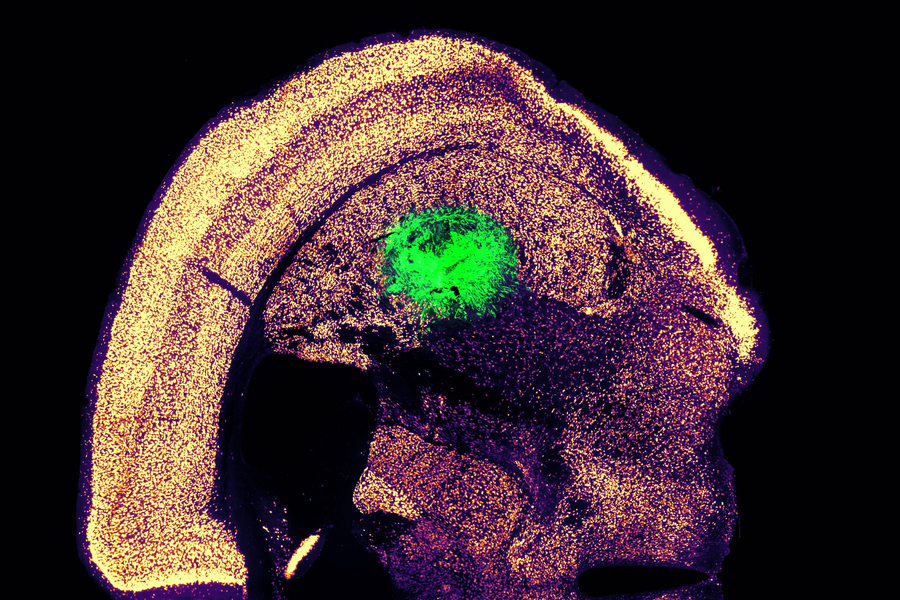
Researchers at the University of Oxford have produced an engineered tissue representing a simplified cerebral cortex by 3D printing human stem cells. The results, published in the journal Nature Communications, showed that, when implanted into mouse brain slices, the structures became integrated with the host tissue.
The breakthrough technique could lead to tailored repairs for brain injuries. The researchers demonstrated for the first time that neural cells can be 3D-printed to mimic the architecture of the cerebral cortex.
Brain injuries, including those caused by trauma, stroke and surgery for brain tumours, typically result in significant damage to the cerebral cortex. For example, each year, around 70 million people globally suffer from traumatic brain injury (TBI), with 5 million of these cases being severe or fatal. Currently, there are no effective treatments for severe brain injuries, leading to serious impacts on quality of life.
Tissue regenerative therapies, especially those in which patients are given implants derived from their own stem cells, could be a promising route to treat brain injuries in the future. Up to now, however, there has been no method to ensure that implanted stem cells mimic the architecture of the brain.
In this new study, the University of Oxford researchers fabricated a two-layered brain tissue by 3D printing human neural stem cells. When implanted into mouse brain slices, the cells showed convincing structural and functional integration with the host tissue.
Lead author Dr Yongcheng Jin (Department of Chemistry, University of Oxford) said: ‘This advance marks a significant step towards the fabrication of materials with the full structure and function of natural brain tissues. The work will provide a unique opportunity to explore the workings of the human cortex and, in the long term, it will offer hope to individuals who sustain brain injuries.’
The cortical structure was made from human induced pluripotent stem cells (hiPSCs), which have the potential to produce the cell types found in most human tissues. A key advantage of using hiPSCs for tissue repair is that they can be easily derived from cells harvested from patients themselves, and therefore would not trigger an immune response.
The hiPSCs were differentiated into neural progenitor cells for two different layers of the cerebral cortex, by using specific combinations of growth factors and chemicals. The cells were then suspended in solution to generate two ‘bioinks’, which were then printed to produce a two-layered structure. In culture, the printed tissues maintained their layered cellular architecture for weeks, as indicated by the expression of layer-specific biomarkers.
When the printed tissues were implanted into mouse brain slices, they showed strong integration, as demonstrated by the projection of neural processes and the migration of neurons across the implant-host boundary. The implanted cells also showed signalling activity, which correlated with that of the host cells. This indicates that the human and mouse cells were communicating with each other, demonstrating functional as well as structural integration.
The researchers now intend to further refine the droplet printing technique to create complex multi-layered cerebral cortex tissues that more realistically mimic the human brain’s architecture. Besides their potential for repairing brain injuries, these engineered tissues might be used in drug evaluation, studies of brain development, and to improve our understanding of the basis of cognition.
The new advance builds on the team’s decade-long track record in inventing and patenting 3D printing technologies for synthetic tissues and cultured cells.
Senior author Dr Linna Zhou (Department of Chemistry, University of Oxford) said: “Our droplet printing technique provides a means to engineer living 3D tissues with desired architectures, which brings us closer to the creation of personalised implantation treatments for brain injury.”
Senior author Associate Professor Francis Szele (Department of Physiology, Anatomy and Genetics, University of Oxford) added: “The use of living brain slices creates a powerful platform for interrogating the utility of 3D printing in brain repair. It is a natural bridge between studying 3D printed cortical column development in vitro and their integration into brains in animal models of injury.”
Senior author Professor Zoltán Molnár (Department of Physiology, Anatomy and Genetics, University of Oxford) said: “Human brain development is a delicate and elaborate process with a complex choreography. It would be naïve to think that we can recreate the entire cellular progression in the laboratory. Nonetheless, our 3D printing project demonstrates substantial progress in controlling the fates and arrangements of human iPSCs to form the basic functional units of the cerebral cortex.”
The vertebral bones that constitute the spine are derived from a distinct type of stem cell that secretes a protein favouring tumour metastases, according to a study led by researchers at Weill Cornell Medicine. The discovery, published in Nature, opens up a new line of research on spinal disorders and helps explain why solid tumours so often spread to the spine, and could lead to new orthopaedic and cancer treatments.
Vertebral bone was found to be derived from a stem cell that is different from other bone-making stem cells. Using bone-like “organoids” made from vertebral stem cells, they showed that the known tendency of tumours to spread to the spine rather than long bones is due largely to a protein called MFGE8, secreted by these stem cells.
“We suspect that many bone diseases preferentially involving the spine are attributable to the distinct properties of vertebral bone stem cells,” said study senior author Dr Matthew Greenblatt.
In recent years, Dr Greenblatt and other scientists have found that different types of bone are derived from different types of bone stem cells. Since vertebrae develop along a different pathway early in life, and also appear to have had a distinct evolutionary trajectory, Dr Greenblatt and his team hypothesised that a distinct vertebral stem cell probably exists.
The researchers started out by isolating what are broadly known as skeletal stem cells, which give rise to all bone and cartilage, from different bones in lab mice based on known surface protein markers of such cells. They then analysed gene activity in these cells to see if they could find a distinct pattern for the ones associated with vertebral bone.
This effort yielded two key findings. The first was a new and more accurate surface-marker-based definition of skeletal stem cells as a whole. This new definition excluded a set of cells that are not stem cells that had been included in the old stem cell definition, thus clouding some prior research in this area.
The second finding was that skeletal stem cells from different bones do indeed vary systematically in their gene activity. From this analysis, the team identified a distinct set of markers for vertebral stem cells, and confirmed these cells’ functional roles to form spinal bone in further experiments in mice and in lab-dish cell culture systems.
The researchers next investigated the phenomenon of the spine’s relative attraction for tumour metastases, including breast, prostate and lung tumours, compared to other types of bone. The traditional theory, dating to the 1940s, is that this “spinal tropism” relates to patterns of blood flow that preferentially convey metastases to the spine versus long bones. But when the researchers reproduced the spinal tropism phenomenon in animal models, they found evidence that blood flow isn’t the explanation, finding instead a clue pointing to vertebral stem cells as the possible culprits.
“We observed that the site of initial seeding of metastatic tumour cells was predominantly in an area of marrow where vertebral stem cells and their progeny cells would be located,” said study first author Dr Jun Sun, a postdoctoral researcher in the Greenblatt laboratory.
Subsequently, the team found that removing vertebral stem cells eliminated the difference in metastasis rates between spine bones and long bones. Ultimately, they determined that MFGE8, a protein secreted in higher amounts by vertebral compared to long bone stem cells, is a major contributor to spinal tropism. To confirm the relevance of the findings in humans, the team collaborated with investigators at Hospital for Special Surgery to identify the human counterparts of the mouse vertebral stem cells and characterise their properties.
The researchers are now exploring methods for blocking MFGE8 to reduce the risk of spinal metastasis in cancer patients. More generally, said Dr Greenblatt, they are studying how the distinctive properties of vertebral stem cells contribute to spinal disorders.
“There’s a subdiscipline in orthopaedics called spinal orthopaedics, and we think that most of the conditions in that clinical category have to do with this stem cell we’ve just identified,” Dr Greenblatt said.
Researchers have identified stem cells in the human thymus for the first time. These cells represent a potential new target to understand immune diseases and cancer and how to boost the immune system. Their reported their discovery in the journal Developmental Cell.
The thymus is a gland located in the front part of the chest, the place where thymocytes (the cells in the thymus) mature into T cells, specialised immune cells crucial to fighting disease. The thymus has a unique and complex 3D structure, including an epithelium (a lining of cells able to guide T cell maturation) that forms a mesh throughout the whole organ and around the thymocytes.
Owing to its relatively inaccessible location, comparatively recent discovery and the fact that it shrinks with age, the thymus has only been investigated for a short period of time compared to other organs. Until now, scientists believed it didn’t contain ‘true’ epithelial stem cells, but only progenitors arising in foetal development.
However, these findings from researchers at the Francis Crick Institute, show for the first time the presence of self-renewing stem cells, which give rise to the thymic epithelial cells instructing thymocytes to become T cells. This suggests the thymus plays an important, regenerative role beyond childhood, which could be exploited to boost the immune system.
In the course of their experiments, the researchers examined these stem cells based on the expression of specific proteins in the human thymus. They identified stem-cell niches (areas where stem cells are clustered) in two locations in the thymus: underneath the organ capsule, or outer layer, and around blood vessels in the medulla, the central part.
They demonstrated that thymic stem cells contribute to the environment by producing proteins of the extracellular matrix, which functions as their own support system.
By using state-of-the-art techniques to map gene expression in single cells and tissue sections, they found that these stem cells, named Polykeratin cells, express a variety of genes allowing them to give rise to many cell types not previously considered to have a common origin. They can develop into epithelial as well as muscle and neuroendocrine cells, highlighting the importance of the thymus in hormonal regulation.
The researchers isolated Polykeratin stem cells in a dish and were able to show that thymus stem cells can be extensively expanded. They demonstrated that all the complex cells in the thymus epithelium could be produced from a single stem cell, highlighting a remarkable and yet untapped regenerative potential.
Roberta Ragazzini, postdoctoral research associate at the Crick and UCL, and first author, said: “It’s paradoxical that stem cells in the thymus – an organ which reduces in size as we get older – regenerate just as much as those in the skin – an organ which replaces itself every three weeks. The fact that the stem cells give rise to so many different cell types hints at more fundamental functions of the thymus into adulthood.”
It’s understood that the thymus’ activity is tightly regulated in adults, providing enough immune support to fight infections but not overshooting to the degree of attacking the body’s own cells.
However, in certain people, the thymus isn’t working properly, or their immune system has reduced capacity. Today’s findings suggest it could be helpful in these cases to stimulate the stem cells to regrow the thymus and rejuvenate their immune system.
Paola Bonfanti, senior group leader of the Epithelial Stem Cell Biology and Regenerative Medicine Laboratory at the Crick, said: “This research is a pivotal shift in our understanding of why we have a thymus capable of regeneration. There are so many important implications of stimulating the thymus to produce more T cells, like helping the immune system respond to vaccinations in the elderly or improving the immune response to cancer.”
The researchers will next study thymic stem cell properties throughout life and how to manipulate them for potential therapies.
Cancer researchers have shown that immunotherapy after stem cell transplantation effectively combats neuroblastomas in children. Crucially, stem cells from a parent provide children with a new immune system that responds much better to immunotherapies. These results of an early clinical trial were published in the Journal of Clinical Oncology.
Tumours of the nervous system, neuroblastomas are associated with an unfavourable prognosis if the tumour is classified as a high-risk type. and particularly poor for patients in the relapsed stage. In this study by scientists at St. Anna Children’s Cancer Research Institute and the Eberhard Karls University of Tübingen, immunotherapy following stem cell transplantation is now associated with long-term survival in a substantial proportion of the patients. Compared to an earlier study the survival rate was increased.
“After the transplantation of stem cells from a parent, the patients are equipped with a new immune system. This enables a better immune response to the subsequent immunotherapy and clearly improves the outcome,” explains Prof Ruth Ladenstein, MD, co-first author.
Five-year survival exceeds 50%
“After a median follow-up of about eight years, we see that more than half of the study patients live five years or longer with their disease,” Prof Ladenstein reports (5-year overall survival: 53%). In comparison, the 5-year overall survival in an earlier study, in which stem cell transplantation was not followed by immunotherapy, was only 23%. Those patients who showed a complete or partial response to prior treatment had significantly better survival.
“In summary, immunotherapy with dinutuximab beta following transplantation of stem cells from matched family donors resulted in remarkable outcomes when patients had at least a partial response to prior treatment,” says Prof Ladenstein. “In our study, there were no unexpected side effects and the frequency of graft-versus-host-disease was low.”
Restoring natural killer cell potency
Dinutuximab beta is a monoclonal antibody that binds to a molecule, GD2, on the surface of tumour cells, marking them for destruction by natural killer cells. But prior chemotherapies may impair natural killer cells. “Therefore, a transplantation of intact natural killer cells from matched family donors seems reasonable before immunotherapy is administered. The transplanted, new natural killer cells are now able to target the tumour cells more efficiently – by means of an antibody-dependent reaction,” explains Prof Ladenstein.
According to the authors, further studies are needed to determine the individual components of the therapeutic approaches. Recently, conventional chemotherapy has also been combined with immunotherapy early in the treatment strategy, resulting in similarly improved response rates. The hope is that a renewed immune system through a healthy parent in combination with the described transplantation procedure could further increase survival rates: “Our approach could thus result in stronger and longer lasting tumour control. A randomised study would be necessary to scientifically substantiate the additional potential benefit of a new immune system in the context of relapse therapy,” Prof Ladenstein adds.
New research from Oregon Health & Science University is helping explain why at least five people have become HIV-free after receiving a stem cell transplant. The study’s insights may bring scientists closer to developing what they hope will become a widespread cure for HIV, hopefully without the need for costly techniques like stem cell therapy.
Published today in the journal Immunity, the OHSU-led study describes how two nonhuman primates were cured of the monkey form of HIV after receiving a stem cell transplant. It also reveals that two circumstances must co-exist for a cure to occur and documents the order in which HIV is cleared from the body – details that can inform efforts to make this cure applicable to more people.
“Five patients have already demonstrated that HIV can be cured,” said the study’s lead researcher, Jonah Sacha, PhD, OHSU professor.
“This study is helping us home in on the mechanisms involved in making that cure happen,” Sacha continued. “We hope our discoveries will help to make this cure work for anyone, and ideally through a single injection instead of a stem cell transplant.”
The first known case of HIV being cured through a stem cell transplant was reported in 2009. A man who was living with HIV was also diagnosed with acute myeloid leukemia, a type of cancer, and underwent a stem cell transplant in Berlin, Germany. Stem cell transplants, which are also called bone marrow transplants, are used to treat some forms of cancer. Known as the Berlin patient, he received donated stem cells from someone with a mutated CCR5 gene, which normally codes for a receptor on the surface of white blood cells that HIV uses to infect new cells. A CCR5 mutation makes it difficult for the virus to infect cells, and can make people resistant to HIV. Since the Berlin patient, four more people have been similarly cured.
This study was conducted with a species of nonhuman primate known as Mauritian cynomolgus macaques, which the research team previously demonstrated can successfully receive stem cell transplants. While all of the study’s eight subjects had HIV, four of them underwent a transplant with stem cells from HIV-negative donors, and the other half served as the study’s controls and went without transplants.
Of the four that received transplants, two were cured of HIV after successfully being treated for graft-versus-host disease, which is commonly associated with stem cell transplants.
Other researchers have tried to cure nonhuman primates of HIV using similar methods, but this study marks the first time that HIV-cured research animals have survived long term. Both remain alive and HIV-free today, about four years after transplantation. Sacha attributes their survival to exceptional care from Oregon National Primate Research Center veterinarians and the support of two study coauthors, OHSU clinicians who care for people who undergo stem cell transplants: Richard T. Maziarz, M.D., and Gabrielle Meyers, M.D.
“These results highlight the power of linking human clinical studies with pre-clinical macaque experiments to answer questions that would be almost impossible to do otherwise, as well as demonstrate a path forward to curing human disease,” said Maziarz, a professor of medicine in the OHSU School of Medicine and medical director of the adult blood and marrow stem cell transplant and cellular therapy programs in the OHSU Knight Cancer Institute.
The how behind the cure
Although Sacha said it was gratifying to confirm stem cell transplantation cured the nonhuman primates, he and his fellow scientists also wanted to understand how it worked. While evaluating samples from the subjects, the scientists determined there were two different, but equally important, ways they beat HIV.
First, the transplanted donor stem cells helped kill the recipients’ HIV-infected cells by recognizing them as foreign invaders and attacking them, similar to the process of graft-versus-leukaemia that can cure people of cancer.
Second, in the two subjects that were not cured, the virus managed to jump into the transplanted donor cells. A subsequent experiment verified that HIV was able to infect the donor cells while they were attacking HIV. This led the researchers to determine that stopping HIV from using the CCR5 receptor to infect donor cells is also needed for a cure to occur.
The researchers also discovered that HIV was cleared from the subjects’ bodies in a series of steps. First, the scientists saw that HIV was no longer detectable in blood circulating in their arms and legs. Next, they couldn’t find HIV in lymph nodes, or lumps of immune tissue that contain white blood cells and fight infection. Lymph nodes in the limbs were the first to be HIV-free, followed by lymph nodes in the abdomen.
The step-wise fashion by which the scientists observed HIV being cleared could help physicians as they evaluate the effectiveness of potential HIV cures. For example, clinicians could focus on analysing blood collected from both peripheral veins and lymph nodes. This knowledge may also help explain why some patients who have received transplants initially have appeared to be cured, but HIV was later detected. Sacha hypothesises that those patients may have had a small reservoir of HIV in their abdominal lymph nodes that enabled the virus to persist and spread again throughout the body.
Sacha and colleagues continue to study the two nonhuman primates cured of HIV. Next, they plan to dig deeper into their immune responses, including identifying all of the specific immune cells involved and which specific cells or molecules were targeted by the immune system.
New research published in the journal Immunity challenges the prevailing hypothesis for how donor stem cell grafts cause graft-versus-host disease (GVHD) and offers an alternative model that could guide development of novel therapies.
The study showed in a mouse model that GVHD, which often affects the skin, gut and liver, is maintained by donor T cells that seed those tissues soon after transplant and not by the continual recruitment of T cells from the blood as previously thought.
“This study changes the paradigm of how people think about GVHD,” said co-senior author Warren Shlomchik, MD, professor of medicine and immunology at the University of Pittsburgh School of Medicine. “It provides important mechanistic detail about what’s going on in the tissues affected by GVHD, which could ultimately inform the development of better therapeutics and lead to better outcomes for stem cell recipients.”
Allogeneic stem cell transplantation involves infusion of stem cells from a healthy donor’s blood or bone marrow to a recipient. While often lifesaving for patients with leukaemia and other blood disorders, the treatment also comes with a risk of developing GVHD, a life-threatening disease that occurs when donor alloreactive T cells attack the recipient’s healthy tissues.
According to a widely held theory, GVHD is maintained by T cells that continually migrate from secondary lymphoid organs throughout the body, including the spleen and lymph nodes, to affected tissues via the blood.
However, a different model posits that the disease is maintained locally by T cells in the tissues with little input from the blood. In the new study, Shlomchik, lead author Faruk Sacirbegovic, PhD, research assistant professor of surgery at Pitt, and their team investigated the two hypotheses for how GVHD is sustained in tissues.
The researchers developed a system to track alloreactive T cells in a mouse model of GVHD by labelling individual cells with unique tags to create different T cell “flavours.” By measuring the tags over time, they monitored where the T cells travelled and replicated.
The analysis showed that each tissue affected by GVHD had unique T cell populations with varying frequencies of each T cell flavour.
“This finding is strong evidence that the disease is locally maintained by T cells in each of the tissues,” explained Shlomchik. “If tissues were constantly getting T cells from circulating blood, then the frequencies of T cell flavors in each tissue should become more and more alike over time — but we didn’t see that.”
The team used mathematical models to predict that progenitor T cells seed out into recipient tissues early after transplant, differentiating there into disease-causing cells.
Next a series of experiments was conducted to confirm this prediction and identified these progenitors as T cells expressing a gene called Tcf7.
“We think that progenitor T cells are long-lived in target tissues and are critical for maintaining GVHD,” said co-senior author Thomas Höfer, PhD, professor of theoretical systems biology at the University of Heidelberg. “After the initial seeding phase, the disease is mostly sustained within the tissue itself without a lot of input from new T cells in the blood.”
Stem cell recipients are typically treated with immunosuppressants to prevent and treat GVHD. As these powerful drugs act systemically to suppress the immune system, they also lower immunity to infections and have other side effects.
According to the researchers, the study’s insights could eventually lead to new, targeted therapies for GVHD.
“Now that we know the identity of progenitor cells, we might be able to prevent them forming early post-transplant or target them directly after they’ve formed,” said Shlomchik. “The findings also suggest that treating GVHD in the tissues themselves would be effective – although targeting tissues beyond the skin remains a challenge.”
With better ways to minimise the risk of GVHD after stem cell transplantation, the procedure could become more widely used to treat a broader range of diseases, including blood disorders such as sickle cell anaemia and autoimmune diseases such as lupus and multiple sclerosis.
In a world first, researchers have launched a clinical trial of lab-grown red blood cells for transfusion into another person. These manufactured blood cells were grown from stem cells from donors, for transfusion into volunteers in the RESTORE randomised controlled clinical trial.
If our trial is successful, it will mean that patients who currently require regular long-term blood transfusions will need fewer transfusions in future, helping transform their care
Professor Cedric Ghevaert, chief investigator
If the technique is proven safe and effective, manufactured blood cells could in time revolutionise treatments for people with blood disorders such as sickle cell and rare blood types. It can be difficult to find enough well-matched donated blood for some people with these disorders.
To produce the lab-grown blood cells, stem cells are first magnetically extracted from a normal 470ml blood donation. These stem cells are then coaxed into becoming red blood cells. Over the three week process, an initial pool of about half a million stem cells generates 50 billion red blood cells.
Chief Investigator Professor Cedric Ghevaert, Professor in Transfusion Medicine and Consultant Haematologist at the University of Cambridge and NHS Blood and Transplant, said: “We hope our lab grown red blood cells will last longer than those that come from blood donors. If our trial, the first such in the world, is successful, it will mean that patients who currently require regular long-term blood transfusions will need fewer transfusions in future, helping transform their care.”
Professor Ashley Toye, Professor of Cell Biology at the University of Bristol and Director of the NIHR Blood and Transplant Unit in red cell products, said: “This challenging and exciting trial is a huge stepping stone for manufacturing blood from stem cells. This is the first-time lab grown blood from an allogeneic donor has been transfused and we are excited to see how well the cells perform at the end of the clinical trial.”
The trial is studying the lifespan of the lab grown cells compared with infusions of standard red blood cells from the same donor. The lab-grown blood cells are all fresh, so the trial team expect them to perform better than a similar transfusion of standard donated red cells, which contains cells of varying ages.
Additionally, if manufactured cells last longer in the body, patients who regularly need blood may not need transfusions as often. That would reduce iron overload from frequent blood transfusions, which can lead to serious complications.
The trial is the first step towards making lab grown red blood cells available as a future clinical product. For the foreseeable future, manufactured cells could only be used for a very small number of patients with very complex transfusions needs. NHSBT continues to rely on the generosity of donors.
Co-Chief Investigator Dr Rebecca Cardigan, Head of Component Development NHS Blood and Transplant and Affiliated Lecturer at the University of Cambridge, said: “It’s really fantastic that we are now able to grow enough red cells to medical grade to allow this trial to commence. We are really looking forward to seeing the results and whether they perform better than standard red cells.”
Thus far, two people have been transfused with the lab grown red cells. They are well and healthy, and were closely monitored with no untoward side effects were reported. The amount of lab grown cells being infused varies but is around 5-10mls.
Donors were recruited from NHSBT’s blood donor base. They donated blood to the trial and stem cells were separated out from their blood. These stem cells were then grown to produce red blood cells in a laboratory at NHS Blood and Transplant’s Advanced Therapies Unit in Bristol. The recipients of the blood were recruited from healthy members of the NIHR BioResource.
A minimum of 10 participants will receive two mini transfusions at least four months apart, one of standard donated red cells and one of lab grown red cells, to see if the young lab-made red blood cells last longer than cells made in the body.
Further trials are needed before clinical use, but this research marks a significant step in using lab grown red blood cells to improve treatment for patients with rare blood types or people with complex transfusion needs.