Tag: skull fusion

Why Breakdancing can Give You a Cone-shaped Head

Photo by Zac Ong on Unsplash

Adam Taylor, Lancaster University

For those of a certain age, Coneheads is an iconic 90s film. But for breakdancers, it seems, developing a cone-shaped head can be an occupational hazard.

According to a 2024 medical case report, a breakdancer who’d been performing for 19 years was treated for “headspin hole”, a condition also known as “breakdancer bulge” that’s unique to breakdancers. It entails a cone shaped mass developing on top of the scalp after repetitive head-spinning. Additional symptoms can include hair loss and sometimes pain around the lump.

Approximately 30% of breakdancers report hair loss and inflammation of their scalp from head-spinning. A headspin hole is caused by the body trying to protect itself. The repeated trauma from head-spinning causes the epicranial aponeurosis – a layer of connective tissue similar to a tendon, running from the back of your head to the front – to thicken along with the layer of fat under the skin on top of the head in an attempt to protect the bones of skull from injury.

The body causes a similar protective reaction to friction on the hands and feet, where callouses form to spread the pressure and protect the underlying tissues from damage. Everyday repetitive activities from holding smartphones or heavy weights through to poorly fitting shoes can result in callouses.

But a cone-shaped head isn’t the only injury to which breakdancers are prone, however. Common issues can include wrist, knee, hip, ankle, foot and elbow injuries, and moves such as the “windmill” and the “backspin” can cause bursitis – inflammation of the fluid filled sacs that protect the vertebrae of the spine. A headspin hole isn’t the worst injury you could sustain from breakdancing either. One dancer broke their neck but thankfully they were lucky enough not to have any major complications.

Others, such as Ukrainian breakdancer Anna Ponomarenko, have experienced pinched nerves that have left them paralysed. Ponomarenko recovered to represent her country in the Paris 2024 Olympics.

As with other sports, it’s unsurprising to hear that the use of protective equipment results in the reduction of injuries in breakdancing too.

But breakdancers aren’t the only ones to develop cone shaped heads.

Newborns

Some babies are born with a conical head after their pliable skull has been squeezed and squashed during the journey through the vaginal canal and the muscular contractions of mother’s uterus.

A misshapen head can also be caused by caput secundum, where fluid collects under the skin, above the skull bones. Usually, this condition resolves itself within a few days. Babies who’ve been delivered using a vacuum assisted cup (known as a Ventouse) – where the cup is applied to the top of the baby’s head to pull them out – can develop a similar fluid lump called a chignon.

Vacuum assisted delivery can also result in a more significant lump and bruising called a cephalohematoma, where blood vessels in the bones of the skull rupture. This is twice as common in boys than in girls and resolves within two weeks to six months.

If you’ve ever seen newborns wearing tiny hats in the first few hours of their life, then one of these conditions may be the reason.

Some children may also present with “cone-head” due to craniosynostosis, which occurs in about one in every 2000-2500 live births.

Newborn skulls are made up of lots of small bony plates that aren’t fused together, which enables babies’ brains to grow without restriction. Usually, once the brain reaches a slower growth pace that the bones can keep up with, the plates fuse together. In craniosynostosis, the plates fuse together too early creating differently shaped heads. Surgery can prevent brain growth restriction but is usually unnecessary if the child hasn’t been identified as having an shaped head by six months of age.

Adam Taylor, Professor and Director of the Clinical Anatomy Learning Centre, Lancaster University

This article is republished from The Conversation under a Creative Commons license. Read the original article.


Newly Discovered Bone Stem Cell Drives Premature Skull Fusion

Photo by Mathew Schwartz on Unsplash

Craniosynostosis, the premature fusion of the top of the skull in infants, is caused by an abnormal excess of a previously unknown type of bone-forming stem cell, according to a preclinical study published in Nature.

Occurring in one in 2500 babies, craniosynostosis arises from one of several possible gene mutations. By constricting brain growth, it can lead to abnormal brain development if not corrected surgically. In complex cases, multiple surgeries are needed.

Led by researchers at and led by researchers at Weill Cornell Medicine, the team focused on what happens in the skull of mice with one of the most common mutations found in human craniosynostosis. They found that the mutation drives premature skull fusion by inducing the abnormal proliferation of a type of bone-making stem cell, the DDR2+ stem cell, that had never been described before.

“We can now start to think about treating craniosynostosis not just with surgery but also by blocking this abnormal stem cell activity,” said study co-senior author Dr Matt Greenblatt, an associate professor of pathology and laboratory medicine at Weill Cornell Medicine and a pathologist at NewYork-Presbyterian/Weill Cornell Medical Center.

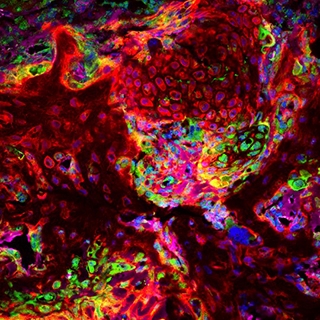
Histology image with stem cells labeled in red and skull region in green

A new stem cell driving disorders of premature skull fusion was transplanted (red), showing that it makes the cartilage seen at sites of skull fusion (green). Credit: Greenblatt lab.

In a study published in Nature in 2018, Dr Greenblatt, study co-senior author Dr Shawon Debnath and their colleagues, described the discovery of a type of bone-forming stem cell they called the CTSK+ stem cell. Because this type of cell is present in the top of the skull, or “calvarium,” in mice, they suspected that it has a role in causing craniosynostosis.

For the new study, they knocked out genes associated with craniosynostosis in CSTK+ stem cells in mice. They expected that the gene deletion somehow would induce these calvarial stem cells to go into bone-making overdrive. This new bone would fuse the flexible, fibrous material called sutures in the skull that normally allow it to expand in infants.

“We were surprised to find that, instead of the mutation in CTSK+ stem cells leading to these stem cells being activated to fuse the bony plates in the skull as we expected, mutations in the CTSK+ stem cells instead led to the depletion of these stem cells at the sutures – and the greater the depletion, the more complete the fusion of the sutures,” Dr Debnath said.

The unexpected finding led the team to hypothesise that another type of bone-forming stem cell was driving the abnormal suture fusion. After further experiments, and a detailed analysis of the cells present at fusing sutures, they identified the culprit: the DDR2+ stem cell, whose daughter cells make bone using a different process than that utilised by CTSK+ cells.

The team found that CTSK+ stem cells normally suppress the production of the DDR2+ stem cells. But the craniosynostosis gene mutation causes the CTSK+ stem cells to die off, allowing the DDR2+ cells to proliferate abnormally.

Collaborating with other researchers, they found the human versions of DDR2+ stem cells and CTSK+ stem cells in calvarial samples from craniosynostosis surgeries—underscoring the likely clinical relevance of their findings in mice.

The findings suggest that inappropriate DDR2+ stem cell proliferation in the calvarium, in infants with craniosynostosis-linked gene mutations, could be treated by suppressing this stem cell population, through mimicking the methods that CTSK+ stem cells normally use to prevent expansion of DDR2+stem cells. The researchers found that the CTSK+ stem cells achieve this suppression by secreting a growth factor protein called IGF-1, and possibly other regulatory proteins.

“We observed that we could partly prevent calvarial fusion by injecting IGF-1 over the calvarium,” said study first author Dr Seoyeon Bok, a postdoctoral researcher in the Greenblatt laboratory.

“I can imagine DDR2+ stem cell-suppressing drug treatments being used along with surgical management, essentially to limit the number of surgeries needed or enhance outcomes,” Dr. Greenblatt said.

In addition to treatment-oriented research, he and his colleagues now are looking for other bone-forming stem cell populations in the skull.

“This work has uncovered much more complexity in the skull than we ever imagined, and we suspect the complexity doesn’t end with these two stem cell types,” Dr Greenblatt said.

Source: Weill Cornell Medicine