Tag: sex differences

Female and Male Hearts may Respond Differently to Noradrenaline

Source: Wikimedia CC0

A new study published in Science Advances shows that female and male hearts respond differently to the stress hormone noradrenaline. The study in mice may have implications for human heart disorders like arrhythmias and heart failure and how different sexes respond to various drugs.

Using fluorescence imaging, the researchers were able to see in real time and in vivo how a mouse heart responds to hormones and neurotransmitters, including noradrenaline.

The results reveal that male and female mouse hearts respond uniformly at first after exposure to noradrenaline. However, some areas of the female heart return to normal more quickly than the male heart, producing differences in the heart’s electrical activity.

“The differences in electrical activity that we observed are called repolarisation in the female hearts. Repolarisation refers to how the heart resets between each heartbeat and is closely linked to some types of arrhythmias,” said Jessica L. Caldwell, first author of the study.

“We know that there are sex differences in the risk for certain types of arrhythmias. The study reveals a new factor that may contribute to different arrhythmia susceptibility between men and women,” Caldwell said.

Methods

The novel imaging system uses a genetically modified ‘CAMPER’ mouse to emit light during a very specific chemical reaction in the heart: cAMP binding.

The cAMP molecule (an abbreviation of cyclic adenosine 3′,5;-monophosphate) is an intermediate messenger that turns signals from hormones and neurotransmitters, including noradrenaline, into action from heart cells.

The light signals from the CAMPER mouse are transmitted by a biosensor that uses a fluorescence signal that can be picked up at high speed and high resolution by a new imaging system specially designed for hearts. This allows the researchers to record the heart’s reaction to noradrenaline in real time, along with changes in electrical activity.

This new imaging approach revealed the differences in the breakdown of cAMP in female and male mice and the associated differences in electrical activity.

Including female mice leads to discoveries

The researchers had not planned to study sex-based responses, according to Crystal M. Ripplinger, senior author of the study. But the researchers started seeing a pattern of different reactions, which led them to realise the differences were sex-based.

When Ripplinger started her lab at the UC Davis School of Medicine over a decade ago, she exclusively used male animals. That was the norm for most research at the time. But several years ago, she began including male and female animals in her studies.

“Sometimes the data between the two sexes is the same. But if the data start to show variation, the first thing we do is look at sex differences. Using both male and female mice has revealed clues into differences we would never have suspected. Researchers are realising you can’t extrapolate to both sexes from only studying one,” Ripplinger said.

She notes that with the current study, it’s not clear what the differences in cAMP and electrical activity may mean.

“The response in the female mice may be protective – or it may not. But simply documenting that there is a measurable difference in the response to a stress hormone is significant. We are hoping to learn more in future studies,” Ripplinger said.

Source: University of California – Davis Health

Sex and Age Influence the Circadian Rhythm

Photo by Malvestida on Unsplash

In a new study published in the journal Science, researchers exploring circadian molecular rhythms were able to uncover the organisation of gene expression rhythms in particular human tissues, and found that sex and age are involved, with females having a more regular pattern of rhythms.

In model organisms, analysing molecular rhythms is usually done using time-stamped measurements, but such data are not readily available in humans. To work around this, the researchers used existing measurements from a large cohort of post-mortem donors, combined with a novel computer algorithm that was designed to assign internal clock times to nearly one thousand donors.

“Interestingly, the data-science algorithm we developed turned out to resemble models from magnetic systems, which are well studied in statistical physics,” says study leader Felix Naef at Ecole Polytechnique Fédérale de Lausanne. Using this innovative approach, the researchers obtained the first comprehensive and accurate whole-organism view of 24-hour gene expression rhythms in 46 human tissues.

While the core clock machinery properties are conserved across the body and do not change significantly with sex and age, their analysis also revealed extensive programs of gene expression rhythms across major compartments of metabolism, stress response pathways and immune function, and these programs peaked twice a day.

In fact, the emerging whole-body organisation of circadian timing shows that rhythmic gene expression occurs as morning and evening waves, with the timing in the adrenal gland peaking first, while brain regions displayed much lower rhythmicity compared to metabolic tissues.

Dividing the donors by sex and age revealed a previously unknown richness of sex- and age- specific gene expression rhythms spread across biological functions. Strikingly, gene expression rhythms were sex-dimorphic (different in males and females) and more sustained in females, while rhythmic programs were generally reduced with age across the body.

Sex-dimorphic rhythms were particularly noticeable in the liver’s “xenobiotic detoxification,” the process by which liver breaks down harmful substances. Additionally, the study found that with age, the rhythm of gene expression decreases in the heart’s arteries, which may explain why older people are more susceptible to heart disease. This information could be useful in the field of “chronopharmacology,” which is the study of how a person’s internal clock affects the effectiveness and side effects of medication.

This study provides new insights into the complex interplay between our body clock, sex, and age. By understanding these rhythms, we might find new ways of diagnosing and treating pathologies such as sleep disorders and metabolic diseases.

Source: Ecole Polytechnique Fédérale de Lausanne

Why Many Lung Diseases are Sex-biased

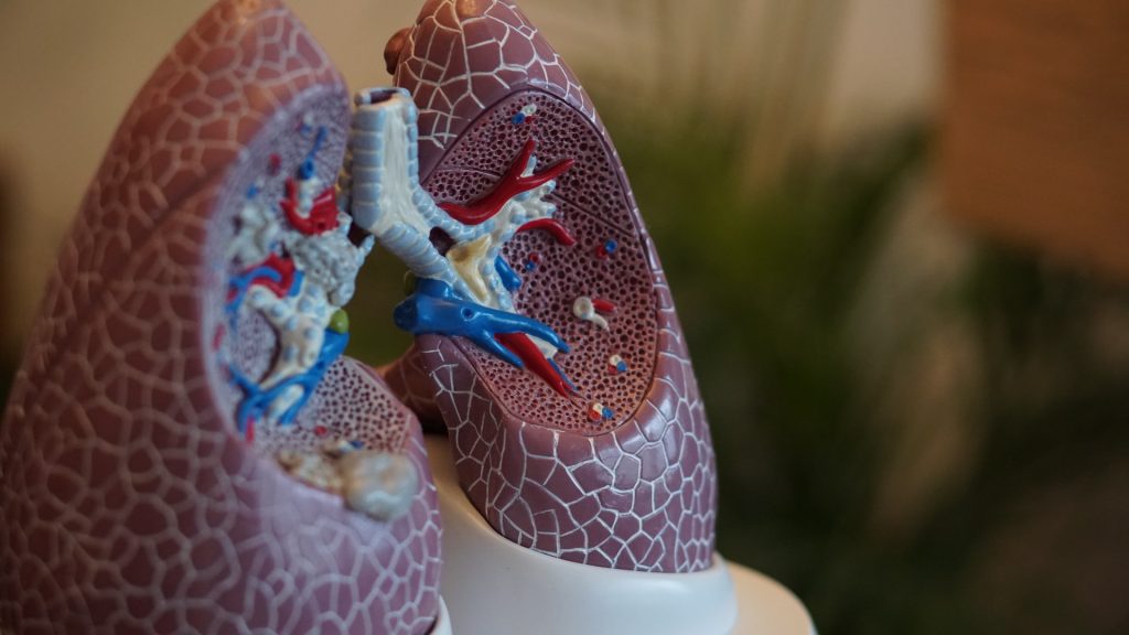
Anatomical model of lungs
Photo by Robina Weermeijer on Unsplash

By studying sex differences in expression across more than 2500 genes in mouse lung cells, researchers may have found an explanation for sex biases in the prevalence and severity of lung diseases, such as the greater severity of COVID for males. In particular, very high numbers of X-linked genes escape transcriptional silencing in lung alveolar type 2 (AT2s) cells, according to their study published in Stem Cell Reports.

“Our study is the first to compare male and female AT2 cells for gene expression, and our findings suggest that there are likely sex differences in lung repair following viral-induced injury,” says co-senior author Montserrat Anguera, an associate professor at the University of Pennsylvania.

Sex differences exist for many lung diseases, but the mechanistic basis for this remains unclear. “We started this project during the beginning of the pandemic, because we were curious about the sex bias with COVID disease, where more older men have increased morbidity, and wondered whether X-chromosome inactivation (XCI) might contribute to this sex bias,” Anguera says. “We realised that the SARS-CoV2 virus first encounters AT2 cells in the lung, and that the virus enters cells through the angiotensin-converting enzyme 2 (Ace2) receptor, which is located on the X chromosome.”

XCI is a process by which one of the copies of the X chromosome is inactivated in female mammals. The inactive X chromosome is silenced by being packaged into a transcriptionally inactive structure called heterochromatin. XCI prevents female mammals from having twice as many X-chromosome gene products as males, who only possess a single copy of the X chromosome.

In the new study, Anguera and co-senior author Andrew Vaughan, an assistant professor at the University of Pennsylvania, investigated XCI maintenance and sex-specific gene expression profiles using male and female AT2s. The results showed that approximately 68% of expressed X-linked genes in mouse AT2s escape XCI. These genes include Ace2, which serves as the entry point into cells for SARS-CoV-2, but is also involved in lung repair.

There were genome-wide expression differences between male and female AT2s, possibly contributing to sex differences in lung injury and repair in multiple settings. Taken together, the findings demonstrate that AT2 cells have the highest levels of XCI escape for mouse cells reported to date and support a renewed focus on AT2s as a potential contributor to sex-biased differences in lung disease.

In addition, the results showed that AT2 cells, similar to immune cells, do not strictly follow the classic rules of XCI. “We were surprised to find that female AT2 cells lack canonical epigenetic modifications that are typically enriched on the inactive X as a result of XCI. These include the long noncoding RNA Xist and heterochromatic histone modifications H3K27me3 and H2AK119-ubiquitin,” Anguera says. “Because the inactive X in female AT2 cells has fewer epigenetic marks, this enables more gene expression chromosome wide, including the Ace2 gene.”

For now, it remains an open question whether ACE2 escapes XCI in human AT2 cells. to The authors say this is a likely scenario because there are significantly higher numbers of XCI escape genes in human cells compared to mouse cells.

Moving forward, the authors plan to investigate how expression from the inactive X in AT2 cells is affected by SARS-CoV2 infections. They also will continue to study how expression from the X chromosome is regulated in other cell types that do not exhibit conventional XCI maintenance. “Our findings open the door to future work investigating the genetic and epigenetic basis, residing within the X chromosome, of sex differences in immune responses to inhaled viruses,” Anguera says.

Source: Science Daily

Why Obesity’s Health Impacts are Worse for Males

Toilet sign male and female
Photo by Tim Mossholder on Unsplash

A newly published study in iScience sheds light on the biological underpinnings in sex differences in obesity-related disease, with researchers observing “striking” differences in the cells that build blood vessels in the fatty tissue of male versus female mice.

Men are more likely than women to develop conditions associated with obesity such as cardiovascular disease, insulin resistance and diabetes, says study leader Professor Tara Haas at York University.

“People have used rodent models to study obesity, and the diseases that are associated with obesity – like diabetes – but they’ve typically always studied male rodents, because females are resistant to developing the same kinds of diseases,” says Haas. “We were really interested in exploring that difference because, to us, it spoke of something really fascinating happening in females that protects them.”

In earlier work, Haas and her team saw that when mice become obese, females grow a lot of new blood vessels to supply the expanding fat tissue with oxygen and nutrients, whereas males grow a lot less. For this study, Haas and her co-authors focused on differences in the endothelial cells that make up the building blocks of these blood vessels in fat tissue.

The team used software to help sift through thousands of genes to zero in on the ones that would be associated with blood vessel growth. They discovered that processes associated with the proliferation of new blood vessels were high in the female mice, whereas the males had a high level of processes associated with inflammation.

“It was very striking the extent of inflammation-associated processes that were prevalent in the males,” Haas recalls. “Other studies have shown that when endothelial cells have that kind of inflammatory response, they’re very dysfunctional, and they don’t respond to stimuli properly.”

York PhD student Alexandra Pislaru, who works in Haas’ lab and is a co-first author of the study, participated in this project as part of her dissertation.

“It is exciting to observe the continuing resilience that female endothelial cells display even when stressed by a long-term high-fat diet,” Pislaru says. “The findings from our study can help researchers to get a better understanding of why obesity manifests differently in men and women.”

The researchers also examined the behaviour of the endothelial cells when they were taken out of the body and studied in petri dishes.

“Even when we take them out of the body where they don’t have the circulating sex hormones or other kinds of factors, male and female endothelial cells still behave very differently from each other,” Haas explains.

Female endothelial cells replicated faster, while male endothelial cells displayed greater sensitivity to an inflammatory stimulus. By comparing with previously published data sets, the researchers found endothelial cells from aged male mice also displayed a more inflammatory profile compared to female cells.

“You can’t make the assumption that both sexes are going to respond to the same series of events the same way,” says Haas. “This isn’t just an obesity related issue – I think it’s a much broader conceptual problem that also encompasses healthy aging. One implication of our findings is that there will be situations where the treatment that is ideal for men is not going to be ideal for women and vice-versa.”

While humans and mice have different genes that may be turned up or down, Haas believes the general findings would likely apply and is interested studying the same cells in humans in future research.

Source: York University

Women with Autism have a Greater Mental Illness Risk

Mirror symbolising schizophrenia
Source: Vince Fleming on Unsplash

Autistic young men and women are more affected by psychiatric conditions and have an increased risk of hospitalisation as a result of their mental illness. Autistic women are particularly vulnerable, as shown in a study published in JAMA Psychiatry.

Autistic people have an increased risk of suffering from mental illness. Current data indicates that autistic women are more vulnerable than autistic men, but few studies have been able to establish that there are sex differences.

The researchers, from Karolinska Institutet, conducted a register-based cohort study with more than 1.3 million people in Sweden who were followed from the age of 16 to 24 between 2001 and 2013. Just over 20 000 of these were diagnosed with autism.

“We saw an increased risk of eleven different psychiatric conditions, including depression, anxiety disorders, self-harm and difficulty sleeping,” says Miriam Martini, a doctoral student in psychiatric epidemiology at Karolinska Institutet and first author of the study.  

High hospitalisation rates 

Something that Miriam Martini finds particularly worrying is that 32 out of 100 autistic women had been hospitalised as a result of their mental illness, compared with 19 out of 100 autistic men. For non-autistic people, the corresponding figure was less than five out of 100.   

The study focuses on young adults who are at a crucial time in their life when many mental health problems increase, while the transition to adulthood often means poorer access to care, says Miriam Martini.   

“Healthcare for young adults needs to be expanded, especially for autistic women, so that mental illness can be detected in time to avoid worsening of symptoms resulting in hospitalization,” says Miriam Martini.  

The reason why autistic women are more affected by mental illness than autistic men is not clear, but in the study, the researchers point to several possible factors. Previous research has shown that autistic women to a greater extent use compensatory behaviours to camouflage their autism, which may be due to the fact that women generally tend to adapt to the expectations of those around them. This delays diagnosis and the provision of assistance, which can negatively affect their mental health.  

Overlooked by the healthcare system 

Another possible explanation may be that it could be difficult to detect autism in women using diagnostic criteria.  

“It may be that autism manifests differently in women than in men, which means that women are not detected using today’s diagnostic criteria. This is something we need to do more research on,” says Miriam Martini.  

Source: Karolinska Institutet

Sex Differences in Post-stroke Biomarkers

Credit: American Heart Association

Although males and females are equally affected by stroke Since oestrogen and progesterone have known neuroprotective effects, it is important to compare the size, severity and biochemical composition of the brain tissue following stroke in female and male animal models. In a paper published in IBRO Neuroscience Reports, researchers have discovered that certain biomarkers were different in the brains of male and female mice.

Stroke neurosurgeon and research coordinator Prof Nicole Sylvain and her colleagues are looking at treatments for post-stroke recovery that help supplement these energy losses. Using the Canadian Light Source (CLS) at the University of Saskatchewan (USask), the team was able to identify energy biomarkers in the brain, which could eventually inform clinicians about the effects of potential stroke treatments on brain recovery after a stroke.

The group’s recent study examined post-stroke differences between male and female mice, and found that female mice have higher amounts of glycogen in their brains. Glycogen is a sugar-like substance that circulates in our blood and nourishes our cells. When the supply of glycogen is disrupted by stroke, the brain is severely impacted.

Most pre-clinical stroke research has been performed using male lab animals, with results usually generalised to both sexes. In clinical stoke cases, females have a higher incidence of ischaemic stroke and poorer outcomes, compared to males.

“We found that, for the most part, male data can be generalised for females, however, some of the metabolic markers we measured were actually different,” Sylvain said. “It’s really important to do the research on both sexes.”

It would be impossible for the team to detect the biomarkers without to the Mid-IR beamline.

“The only way to detect them in such an accurate way across the brain is with infrared imaging, so the CLS has been absolutely vital to our research.”

Source: University of Saskatchewan

Sex of Red Blood Cell Donors Does not Affect Recipient Survival

https://www.pexels.com/photo/a-close-up-shot-of-bags-of-blood-4531306/
Photo by Charlie-Helen Robinson on Pexels

A study published in JAMA Internal Medicine shows that, after taking haemoglobin levels into count, sex and previous pregnancy of blood donors do not affect the survival of patients receiving red blood cell transfusions. Differences in recipient survival depend rather on the haemoglobin quantity in the transfusion, the researchers found.

Female donor sex and previous pregnancy are established risk factors for transfusion-related acute lung injury following plasma and platelet transfusions, which is a leading cause of transfusion-related mortality.

Previous studies have produced conflicting results as to how donor sex affects the recipient’s survivability in the recipient following red blood cell transfusion. Some studies have indicated higher mortality in patients who have received red blood cells from women, in men who have received red blood cells from women who have been pregnant, and in sex-mismatched transfusions. Other studies, however, have not reported such correlations.

This question has now been further explored by researchers from Karolinska Institutet in a register study of almost 370 000 patients in Sweden who received a red blood cell transfusion for the first time between 2010 and 2018.

The aim of the study was to see how the sex and previous pregnancy status of the donor affects survival in the recipient within two years from transfusion. It also looked at how the risk of needing more transfusions differed between patients who received red blood cells from female and male donors. Blood from women on average contains less haemoglobin than blood from men, meaning that more transfusions might be required to obtain the desired level of haemoglobin in a recipient.

The study demonstrates that the median value for haemoglobin was lower in female blood donors (135g/L than male (149g/L) and that patients who received blood from a woman had a 12% higher risk of needing another transfusion within 24 hours than blood from a man. However, this sex difference was eliminated when adjusting for the donors’ haemoglobin levels, which the researchers say was an expected effect that had not been factored into previous studies.

“When we take into account the lower haemoglobin levels in blood from women, we see no difference in survival among patients who received a blood transfusion from women compared with from men, regardless of how many times the female donors had been pregnant and of the patients’ sex and age,” said the study’s first author Jingcheng Zhao, adjunct researcher at Karolinska Institutet. “Differences in haemoglobin levels are a source of error that previous studies have not taken into consideration and that might explain the conflicting results that has been seen previously.”

Data for the study was drawn from national population, health and blood donor registries. The study also shows that donor sex is naturally randomly distributed in the patient material since no regard is paid to the sex and previous pregnancies of the donors by the blood donor centres when supplying blood. According to the researchers, this means that more far-reaching conclusions be drawn.

Dr Zhao said this allows them to determine causality. “We’ll now continue developing methods for studying causal relationships in transfusion epidemiology using observational data, on things like donor characteristics and how blood is handled. There’s still much we don’t know about blood transfusion and its effects.”

One limitation is that it was not possible to separately study transfusions where the red blood cells had not undergone leukoreduction (the filtering out of white blood cells), since this procedure has been standard in Sweden since the 1990s. The researchers therefore add a caveat about generalizing the conclusions to erythrocyte concentrates that have not undergone leukoreduction, which, however, is relatively uncommon now.

Source: Karolinska Institutet

One in 500 Men Carry an Extra Sex Chromosome, Increasing Disease Risk

Photo by Cottonbro on Pexels

Around one in 500 men could be carrying an extra sex chromosome (X or Y), putting them at increased risk of diseases such as type 2 diabetes, atherosclerosis and thrombosis, according to a study published in Genetics in Medicine.

Researchers from the universities of Cambridge and Exeter analysed genetic data on 200 000 men aged 40 to 70 from UK Biobank. They found 356 men who carried either an extra X chromosome or an extra Y chromosome.

Some men have an extra X or Y chromosome – XXY or XYY, which is usually not obvious without a genetic test. Men with extra X chromosomes, a condition known as Klinefelter syndrome, are sometimes identified during investigations of delayed puberty and infertility; however, most are unaware that they have this condition. Men with an extra Y chromosome tend to be taller as boys and adults, but otherwise they have no distinctive physical features.

In today’s study, the researchers identified 213 men with an extra X chromosome and 143 men with an extra Y chromosome. As the participants in UK Biobank tend to be ‘healthier’ than the general population, this suggests that around one in 500 men may carry an extra X or Y chromosome.

Only a small minority of these men had a diagnosis of sex chromosome abnormality on their medical records or by self-report: fewer than one in four (23%) men with XXY and only one of the 143 XYY men (0.7%) had a known diagnosis.

By linking genetic data to routine health records, the team found that men with XXY have much higher chances of reproductive problems, including a three-fold higher risk of delayed puberty and a four-fold higher risk of being childless. These men also had significantly lower blood concentrations of testosterone. Men with XYY appeared to have a normal reproductive function.

Men with either XXY or XYY had higher risks of several other health conditions: a three-fold higher risk of developing type 2 diabetes, six-fold risk of venous thrombosis, three-fold risk of pulmonary embolism, and four-fold risk of chronic obstructive pulmonary disease (COPD).

It is unclear why an extra chromosome should increase the risk, said the researchers, or why the risks were so similar regardless of which sex chromosome was duplicated.

Yajie Zhao, a PhD student at the Medical Research Council (MRC) Epidemiology Unit at the University of Cambridge, the study’s first author, said: “Even though a significant number of men carry an extra sex chromosome, very few of them are likely to be aware of this. This extra chromosome means that they have substantially higher risks of a number of common metabolic, vascular, and respiratory diseases — diseases that may be preventable.”

Professor Ken Ong, also from the MRC Epidemiology Unit at Cambridge and joint senior author, added: “Genetic testing can detect chromosomal abnormalities fairly easily, so it might be helpful if XXY and XYY were more widely tested for in men who present to their doctor with a relevant health concern.

“We’d need more research to assess whether there is additional value in wider screening for unusual chromosomes in the general population, but this could potentially lead to early interventions to help them avoid the related diseases.”

Professor Anna Murray, at the University of Exeter, said: “Our study is important because it starts from the genetics and tells us about the potential health impacts of having an extra sex chromosome in an older population, without being biased by only testing men with certain features as has often been done in the past.”

Previous studies have found that around one in 1,000 females have an additional X chromosome, which can result in delayed language development and accelerated growth until puberty, as well as lower IQ levels compared to their peers.

Source: University of Cambridge

Sex-differentiation Genes Also Contribute to Disease Risks

Man and woman about to sprint
Source: Andrea Piacquadio on Pexels

Some physical traits that differ between sexes are known to be linked to certain single nucleotide polymorphisms (SNPs) outside the X and Y chromosomes. New research now suggests that many of these ‘sex-heterogenous’ SNPs also contribute to a person’s risk for a variety of diseases. Michela Traglia and colleagues at the University of California San Francisco presented their findings in PLOS Genetics.

Millions of SNPs are in each genome, with each SNP representing a difference in a certain DNA building block in a particular stretch of DNA. Many associations have been uncovered between certain SNPs and people’s distinct traits. Understanding SNPs has a number of applications, such as predicting individual treatment effectiveness or disease risks.

Traglia and colleagues previously found that SNPs associated with certain differences in physical traits between men and women, such as waist-hip ratio and basal metabolic rate, may also affect the biology of autism spectrum disorder and other complex diseases. Building on this work with two large genomic datasets, the identified an updated list of 2320 sex-heterogeneous SNPs.

Analysis of these SNPs revealed that they are also associated with a variety of health-related traits and diseases, some with strong sex bias and some without, including schizophrenia, type 2 diabetes, anorexia, heart failure, and ADHD.

These SNPs are located in stretches of DNA that are either within or near genes involved in skeletal and muscle development in a growing embryo. In addition, these SNPs appear to play a role in regulating gene expression and DNA methylation, which are fundamental processes by which a person’s DNA is translated into their distinct biology and traits.

Overall, the researchers conclude that the identified SNPs play a role in early-life biological processes shaping sex-distinct traits and which also affect health and disease risk later in life. More work is needed to understand the mechanisms behind these sex-heterogeneous SNPs.

“We found that genetic alleles with differing effects on measured physical traits in men and women also play an outsized role in health risks,” remarked study co-author Lauren Weiss. “We hope this work helps us to understand the genetic underpinnings of sexual dimorphism and its relationship with both early development and later disease risk.”

Source: EurekAlert!

Sex Differences in Nonalcoholic Fatty Liver Disease Explained

Toilet sign male and female
Photo by Tim Mossholder on Unsplash

Investigators may have discovered the reason why fewer women than men develop nonalcoholic fatty liver disease (NAFLD). They published their findings in Nature Communications.

One of the most common disorders globally, NAFLD is a leading cause of death worldwide. Its progressive form, ‘nonalcoholic steatohepatitis’ (NASH), affects about 30% of all NAFLD patients, and can lead to cirrhosis and liver cancer. Despite intensive research, the underlying mechanisms of NAFLD/NASH are still poorly understood and effective treatment is lacking as a result.

However, it is known that NAFLD/NASH is more common among men than women, especially premenopausal women. The reasons for this are still unclear, but evidence so far suggests that oestrogen plays a protective role. On the other hand, the protein formyl peptide receptor 2 (FPR2) is known to play an important role in mediating inflammatory responses in multiple organs. However, no study so far has determined its role in the liver. Could FPR2 be involved in the sex-related differences regarding NAFLD prevalence and severity?

Addressing this question, a research team led by Professor Youngmi Jung of Pusan National University, Korea, recently conducted a study using mice model, shedding light on the role of FPR2 in NAFLD/NASH and its relationship to the observed sex-based differences. This work is among the very few studies on NAFLD that relies on sex-balanced animal experiments rather than the more common male-only designs.

The researchers first found that Fpr2 was highly expressed in healthy livers of female mice. Furthermore, it was expressed differently in the livers of male and female mice that were fed a special NAFLD-inducing diet. Silencing the Fpr2 gene made the male and female mice equally vulnerable to NAFLD, suggesting that FPR2 has a protective effect on the liver.

Interestingly, the researchers also found that FPR2 production in the liver is mediated by oestrogen. Males supplemented with external oestrogen produced more Fpr2 and were more resistant to NAFLD, whereas females that had their ovaries removed exhibited reduced liver Fpr2 levels. “Taken together, our findings suggest that FPR2 is a potential therapeutic target for developing pharmacological agents to treat NAFLD/NASH,” says Prof Jung. “In addition, our results could help in the development of gender-based therapies for NASH.”

This unprecedented discovery of the female-specific production of FPR2 in the liver and its role in providing resistance against NAFLD/NASH will hopefully pave the way not only for novel treatments but also a more comprehensive and sex-aware approach when doing science. Prof Jung remarked on this: “Our research highlights the pressing need for designing and developing better sex-balanced animal experiments, considering that the sex-specific expression of FPR2 in the liver had been completely overlooked in previous studies.”

Source: Pusan National University