Tag: sex differences

How, When and Where: Sex Matters in Melanoma Development

Photo by Rfstudio on Pexels

Melanoma rates differ consistently between men and women in terms of the ages at which melanomas occur and the locations on the body where they occur. Over time, melanoma rates have increased in both men and women, but the trends differ by body site. A new study in the Journal of Investigative Dermatology, published by Elsevier, presents the findings from a large-scale, long-term melanoma data analysis investigating incidence trends by age, sex, and anatomic site.

Lead investigator David C. Whiteman, MBBS, PhD, Cancer Control Group, QIMR Berghofer Medical Research Institute, and Faculty of Medicine, The University of Queensland, Brisbane, Australia, explains, “There has been a general observation in numerous populations that melanomas appear to arise at different rates in men and women. We decided to investigate this observation rigorously and assess whether these differences have been constant through time or across generations by using large-scale data from population registries to investigate long-term melanoma trends in men and women.”

The research team analysed more than 40 years of melanoma data from Queensland, Australia, the USA, and Scotland. These three populations were chosen because historically they have had high (Queensland), moderate (USA), and low (Scotland) rates of melanoma. Over time, the rates of melanoma increased in all three populations, especially among women. In women in all populations, melanomas arise most commonly on the limbs, whereas in men, melanomas arise most commonly on the trunk and head and neck. In both sexes, there has been a steady increase in melanomas on the head and neck with increasing age.

Researchers found that in virtually all investigated populations, women experience higher rates of melanoma than men in early life (up to age ~45 years), but men develop melanomas at higher rates than women later in life (from ages ≥ 65 years). Furthermore, these sex-specific trends reflect complex patterns of incidence across body sites that vary consistently with age. Thus, in early life, women experience higher rates of lower limb melanomas than men, which persists into older ages. Also, on the upper limbs, women experience substantially higher rates than men from young ages until middle age (45–64 years), after which men experience higher rates. In contrast, on the head and neck and the trunk, melanomas occur at higher incidence in men than in women early in life. On all body sites, the rate at which melanoma incidence rises with age is much more rapid for men than for women.

The study confirms that men and women experience melanoma in different ways. While this is most likely driven by different patterns of sun exposure between men and women, there appear to be inherent differences in the ways in which melanomas develop at different body sites in women compared with men. Understanding the underlying biological differences could provide important clues about the etiology of this enigmatic cancer.

Source: Elsevier

Older Women more Vulnerable to Heat than Men, Researchers Find

Photo by Loren Joseph on Unsplash

As global climate change causes extreme heat waves to become more common around the world, epidemiological studies have shown that heat kills more women than men. Now, a new study by researchers at Penn State has found that older women are physiologically more vulnerable to high heat and humidity than older men, and that women between the ages of 40 and 64 are as vulnerable as men 65 years of age or older. This is the first study to determine this disparity exists due to physiological differences rather than from a preponderance of women at old age due to greater longevity.

Led by Olivia Leach, doctoral candidate in kinesiology at Penn State, and her adviser, W. Larry Kenney, professor of physiology and kinesiology at Penn State, the researchers demonstrated that middle-aged and older women were affected by heat at lower temperature/humidity combinations than middle-aged and older men. The results, published in the American Journal of Physiology-Regulatory, Integrative and Comparative Physiology, were somewhat unexpected, according to Leach, because there are no differences in heat vulnerability based on biological sex in adults younger than 30.

While the researchers did not directly compare middle-aged men to middle-aged women, the physiological responses of middle-aged women were similar to the responses of older men in the study, which demonstrated that middle-aged women are more vulnerable to heat than men of the same age.

“In addition to demonstrating that middle-aged and older women are at greater risk from extreme heat, we also identified what levels of heat and humidity are safe for women as they age,” Leach said. “This information is presented as a temperature/humidity curve based on a person’s age, and it can be useful for setting policies designed to keep people safe during a heat wave.”

The researchers tested the heat thresholds of 72 participants between 40 and 92 years of age in a specialized environmental chamber in Kenney’s laboratory. Before the experiment, participants swallowed a tiny device encased in a capsule that measured their core temperature throughout the experiment.

During the study, participants entered the specialised environmental chamber where they performed light physical activity to simulate the effort of minimal day-to-day tasks – the types of things people would need to do even during a heat wave. The researchers then gradually increased the temperature and/or humidity in the chamber until the participant’s body could no longer adequately cool itself, and their core temperature began to rise.

The study is part of the PSU HEAT, or Human Environmental Age Thresholds, project, led by Kenney. For five years, researchers in the PSU HEAT project have examined the levels of combined heat and humidity that humans can tolerate before their core temperatures begin to rise. When core temperatures rise, people become vulnerable to heat-related illnesses including heat exhaustion, heat stroke and even death.

“We’re not saying that people who experience a certain temperature will necessarily become sick or die,” Kenney said. “We are identifying the limits of livability – the thresholds where people can no longer continue their daily life unimpeded. Once people reach these temperatures, they need to take actions like seeking air conditioning to cool their bodies.”

Previous research by Kenney and others demonstrated that people become increasingly vulnerable to heat as they age, because their ability to efficiently sweat and pump blood to the skin – two primary cooling mechanisms – decreases. Sweat evaporation carries heat away from the body, while extra blood pumped to the skin dissipates heat to the environment and supports sweating.

To date, the PSU HEAT project has conducted more than 600 experiments on nearly 200 participants between ages 18 and 92, but the results of this experiment still yielded surprises, according to Leach.

“Among young adults, there is no difference in heat vulnerability between men and women,” Leach said. “Young people tend to be healthier, so any measurable health metric – from blood pressure to cholesterol – is more homogeneous among young people than it is among older people.”

As with other health measures, older adults have a wide range in their vulnerability to heat, Leach explained.

“We have examined many factors that might explain who faces the most risk in a heat wave,” Leach said. “We found that age and biological sex are the two most important factors that can predict whether a healthy adult would be at risk from high heat and humidity.”

While cardiovascular health and certain medications can affect a person’s sensitivity to heat, biological sex and age appear to be the two primary drivers of heat vulnerability among healthy people, the researchers said.

“Other factors – for example someone’s cardiovascular fitness or their body mass – have little impact on how vulnerable a person is to heat at rest or during light activity,” Leach continued. “Older women really are at greater risk from heat than other people. As governments and other social leaders prepare for extreme heat to become more common, the vulnerability of older women needs to factor into their planning.”

Source: Penn State

New Research Explains Differences in Men’s and Women’s Immune Systems

Photo by Daniil Onischenko on Unsplash

By analysing the immune system of female-to-male transgender individuals, Swedish researchers demonstrate the role of sex hormones in regulating the immune system. This newfound knowledge, published in Nature, explains differences between men and women, particularly in terms of immune signalling, and can be used to develop new immunological medications according to researchers.

Sex differences in the immune system are regulated both by genetics and by sex hormones. However, immunological comparisons between men and women can never fully distinguish the significance of genetic versus hormonal variations.

Now, three Swedish research groups led by Karolinska Institutet and Uppsala University has conducted a unique study analysing the regulation and adaptation of the immune system over time in 23 trans men who have undergone gender-affirming testosterone treatment, starting at the age of 18–37 years.

“We have followed individuals who were assigned female sex at birth and later received testosterone treatment in adulthood. Their genetic profile remains unchanged, while their hormone profile shifts entirely from typically female to male hormone levels,” says Petter Brodin, paediatrician and professor of paediatric immunology at the Department of Women’s and Children’s Health, Karolinska Institutet, who led the study together with Nils Landegren, assistant professor at Uppsala University, and Olle Kämpe, Professor at the Department of Medicine, Solna, Karolinska Institutet. “This unique change allows us, for the first time, to identify which parts of a person’s immune system are directly regulated by sex hormones rather than genetic sex differences.” 

The researchers can now demonstrate that increased testosterone levels and the accompanying reduction in oestrogen particularly affect the balance between two crucial immune signalling systems: antiviral interferon type 1 (IFN-1) and proinflammatory signals such as tumour necrosis factor alpha (TNFα).  

Specifically, they found that testosterone modulates a cross-regulated axis between type-I interferon and tumour necrosis factor. This is mediated by functional attenuation of type-I interferon responses in both plasmacytoid dendritic cells and monocytes. Conversely, testosterone potentiates monocyte responses leading to increased tumour necrosis factor, interleukin-6 and interleukin-15 production and downstream activation of nuclear factor kappa B-regulated genes and potentiation of interferon-γ responses, primarily in natural killer cells. 

The immune system changes throughout life

They also have a hypothesis about why the immune system needs to be dynamically regulated by hormones throughout life. 

“All individuals must be able to adjust their immune systems over the course of their lives to be optimally regulated for the conditions and challenges we face. During puberty and sexual maturation, new demands arise, and the immune system must be regulated differently to enable pregnancy in women and muscle growth in men,” says Petter Brodin. 

By regulating these key functions via sex hormones, this can be achieved, and in women, it is dynamically controlled even during a menstrual cycle,” he adds. 

The results of the study open an entirely new field of research, according to Nils Landegren. 

“The newfound knowledge will help us better influence people’s immune systems even without using sex hormones. For example, new drugs can be developed to impact these regulatory mechanisms and thus rebalance the immune response, especially for women with the autoimmune rheumatic disease SLE,” he explains. 

However, the results also have a more direct implications for transgender individuals. 

“This research is also of crucial for transgender individuals undergoing gender-affirming hormone therapy, and I believe that this group deserves significantly more scientific attention and follow-up to ensure their long-term health,” says Petter Brodin. 

Source: Karolinska Institutet

Oestrogen’s Protection against Fatty Liver Points to New Drug Treatment

Photo by The Creative Exchange on Unsplash

New research from Karolinska Institutet shows how oestrogen protects against MASLD, a fatty liver disease on the increase in the obesity epidemic. The study, published in Molecular Systems Biology, shows how a new drug under development could become a future treatment for fatty liver disease and liver cancer.

The global obesity epidemic has resulted in a dramatic increase in fatty liver, a disease in which fat that does not fit into fat cells is stored in liver cells instead.

Since last year, fatty liver due to obesity (and not excessive alcohol consumption) is known as MASLD (metabolic dysfunction-associated steatotic liver disease). According to previous research, as many as one in three adults are affected by some degree of MASLD, which in the worst cases can develop into cirrhosis and liver cancer.

Protection until menopause

However, the disease is very unevenly distributed between the sexes, with a large majority of affected individuals being men.

“Women have a natural protection until menopause due to the female sex hormone oestrogen,” explains study leader Claudia Kutter, senior researcher at the Department of Microbiology, Tumor and Cell Biology, Karolinska Institutet.

Although women’s protection has been known for some time, the mechanism behind the protective effect has been less clear. Now Claudia Kutter’s research team may have found the answer.

Through genetic analyses of mice of both sexes fed a high-fat diet, with some of the male mice also receiving oestrogen, the researchers were able to identify a key protein in the development of fatty liver. 

The protein, called TEAD1, was found to play an overall role in regulating how liver cells absorb fat. Blocking TEAD1 protected liver cells from the harmful accumulation of fat. Mice receiving oestrogen treatment had lower TEAD1 activity and less fat accumulation in the liver.

New drug under development

In the next step, the researchers tested blocking TEAD1 in human liver cells with the same result. The fact that this was possible at all, however, was a bit of luck.

“It turned out that a pharmaceutical company is developing an anti-cancer drug that blocks TEAD1, which allowed us to test our hypothesis,” says Claudia Kutter.

The fact that TEAD1 is also involved in cancer does not worry her, quite the contrary.

“Since the activity of TEAD proteins is elevated in cancer, blocking TEAD at an early stage can also be positive from a cancer point of view,” she says. “Patients suffering from liver cancer are currently diagnosed very late. If the patient is given this drug early in the process to protect against fatty liver, it can hopefully also prevent the development of liver cancer.”

The pharmaceutical company will now start clinical trials of the drug as a protection against fatty liver disease, while Claudia Kutter’s research team will continue researching further ways to tackle the disease.

“We want to focus on how to find the disease earlier and identifying new treatment targets,” she says. “Different approaches may be needed for different patients depending on their gender and hormonal status.”

Source: Karolinska Institutet

The Arms and Torso of Human Males Evolved to Throw a Punch

Photo by Hermes Rivera on Unsplash

In the animal kingdom, males develop specialised weapons such as deer antlers for competition when winning a fight is critical. Humans do too, according to new research from the University of Utah. Males’ upper bodies are built for more powerful punches than females’, says the study, published in the Journal of Experimental Biology, suggesting that fighting may have long been a part of our evolutionary history.

“In mammals in general,” says professor David Carrier of the School of Biological Sciences, “the difference between males and females is often greatest in the structures that are used as weapons.”

Assembling evidence

For years, Carrier has been exploring the hypothesis that generations of interpersonal male-male aggression long in the past have shaped structures in human bodies to specialise for success in fighting. Past work has shown that the proportions of the hand aren’t just for manual dexterity- they also protect the hand when it’s formed into a fist. Other studies looked at the strength of the bones of the face (as a likely target of a punch) and how our heels, planted on the ground, can confer additional upper body power.

“One of the predictions that comes out of those,” Carrier says, “is if we are specialised for punching, you might expect males to be particularly strong in the muscles that are associated with throwing a punch.”

Jeremy Morris, then a doctoral student and now an assistant professor at Wofford College, designed an experiment with Carrier, doctoral student Jenna Link and associate professor James C. Martin to explore the sexual dimorphism, or physical differences between men and women, of punching strength. It’s already known that males’ upper bodies, on average, have 75% more muscle mass and 90% more strength than females’. But it’s not known why.

“The general approach to understanding why sexual dimorphism evolves,” Morris says, “is to measure the actual differences in the muscles or the skeletons of males and females of a given species, and then look at the behaviours that might be driving those differences.”

Cranking through a punch

To avoid potential hand injury from a using punching bag, the researchers instead rigged up a hand crank that would mimic the motions of a punch. They also measured participants’ strength in pulling a line forward over their head, akin to the motion of throwing a spear. This tested an alternative hypothesis that males’ upper body strength may have developed for the purpose of throwing or spear hunting.

Twenty men and 19 women participated. “We had them fill out an activity questionnaire,” Morris says, “and they had to score in the ‘active’ range. So, we weren’t getting couch potatoes, we were getting people that were very fit and active.”

But even with roughly uniform levels of fitness, the males’ average power during a punching motion was 162% greater than females’, with the least-powerful man still stronger than the most powerful woman. Such a distinction between genders, Carrier says, develops with time and with purpose.

“It evolves slowly,” he says, “and this is a dramatic example of sexual dimorphism that’s consistent with males becoming more specialised for fighting, and males fighting in a particular way, which is throwing punches.”

They didn’t find the same magnitude of difference in overhead pulling strength, lending additional weight to the conclusion that males’ upper body strength is specialised for punching rather than throwing weapons.

Breaking a legacy of violence

It’s an uncomfortable thought to consider that men may be designed for fighting. That doesn’t mean, however, that men today are destined to live their ancestor’s violent lives.

“Human nature is also characterized by avoiding violence and finding ways to be cooperative and work together, to have empathy, to care for each other, right?” Carrier says. “There are two sides to who we are as a species. If our goal is to minimise all forms of violence in the future, then understanding our tendencies and what our nature really is, is going to help.”

Source: University of Utah

Women Lose More Years of Life After a Heart Attack Than Men

Photo by Joice Kelly on Unsplash

A new study shows that women lose more years of life after a heart attack than men. A 50-year-old woman with a large heart attack loses an average of 11 years, while an 80-year-old man with a small heart attack loses an average of 5 months of life. The results of the study, led by researchers at Karolinska Institutet and Danderyd Hospital, are published in the journal Circulation.

The new study examined 335 000 individuals with first-time myocardial infarction registered in the SWEDEHEART quality registry during the period 1991-2022. The individuals with myocardial infarction were compared with 1.6 million individuals without myocardial infarction using data from Statistics Sweden and the National Board of Health and Welfare. Using the comparator population and new statistical methods, the difference in life expectancy between heart attack individuals and comparison individuals could be calculated, providing a measure of how much life expectancy was shortened due to the disease. 

“We found that there were large differences between groups. Women and young individuals lost the most life expectancy when they had a heart attack. If the cardiac function was impaired after the infarction, the effects were even greater. For example, a 50-year-old woman with impaired cardiac function loses an average of 11 years in 2022 compared to an 80-year-old man with normal cardiac function who loses an average of 5 months in life expectancy,” says first author Christian Reitan, researcher at the Department of Clinical Sciences, Danderyd Hospital, Karolinska Institut. 

Parameters affecting heart attack risk

The researchers were also able to take into account differences in income, education, other illnesses and medication at the time of the illness – which helped to measure the effect of the heart attack itself when everything else was taken into account.

“The results showed that a fairly large part of the reduction in life expectancy disappeared, that is, much of the reduction in life expectancy is explained by factors other than the heart attack itself, but which may still be associated with heart attack, such as socioeconomics or other diseases such as hypertension and diabetes. Provided that the patient had preserved cardiac function, we saw that the gender difference had disappeared. We interpret this to mean that the effect of the heart attack, and thus also the care for heart attacks, is similar between the sexes and that the large reduction in life expectancy we see in women is due to differences in risk factors, other diseases and socioeconomics,” says Christian Reitan. 

According to the researchers, there is a lack of individualized heart attack care in Sweden for women. The study shows that women who have a heart attack lose more years of life than men of the same age.

“If a woman had impaired cardiac function, the gender difference was large. We don’t have the data to answer why, but it raises questions about whether women get as good follow-up and treatment for heart failure as men, or whether it is simply a more serious condition for a woman. Our findings are important because they challenge existing guidelines for heart attack treatment today. By identifying high-risk groups, we can hopefully better tailor treatment to the individual. We believe that ‘years of life lost’ is a good and easy-to-understand measure of risk for both doctors and patients. It makes it easier for us to assess and communicate the seriousness of the disease,” concludes Christian Reitan. 

Source: Karolinska Institutet

Those with Alzheimer’s Disease History on Mother’s Side have Increased Amyloid Proteins

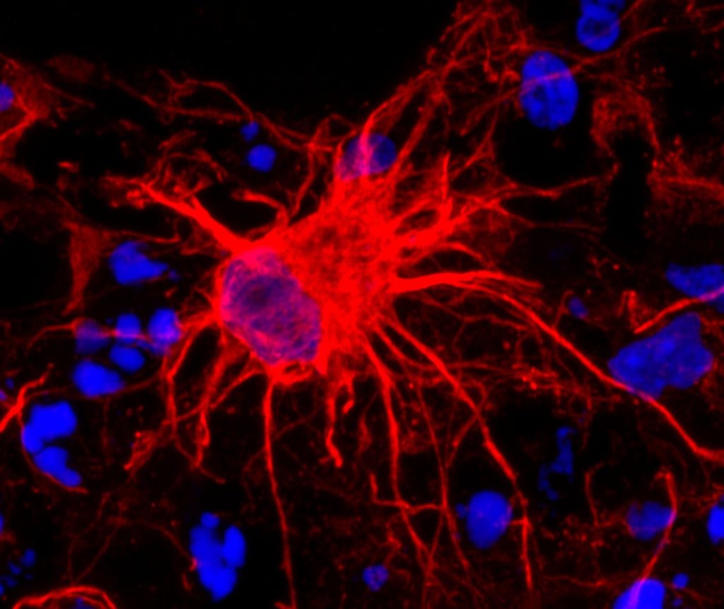
Neurons in the brain of an Alzheimer’s patient, with plaques caused by tau proteins. Credit: NIH

A new study by investigators from Mass General Brigham suggests that whether a person inherits risk of Alzheimer’s disease from their mother or father influences risk of biological changes in the brain that lead to disease. By evaluating 4400 cognitively unimpaired adults ages 65–85, the team found those with a history of Alzheimer’s disease (AD) on either their mother’s side or both parents’ sides had increased amyloid in their brains. Their results are published in JAMA Neurology.

“Our study found if participants had a family history on their mother’s side, a higher amyloid level was observed,” said senior corresponding author Hyun-Sik Yang, MD, a neurologist at Mass General Brigham.

Yang said that previous smaller studies have investigated the role family history plays in Alzheimer’s disease. Some of those studies suggested maternal history represented a higher risk of developing Alzheimer’s, but the group wanted to revisit the question with cognitively normal participants and access to a larger clinical trial data set.

The team examined the family history of older adults from the Anti-Amyloid Treatment in Asymptomatic Alzheimer’s (A4) study, a randomized clinical trial aimed at AD prevention. Participants were asked about memory loss symptom onset of their parents. Researchers also asked if their parents were ever formally diagnosed or if there was autopsy confirmation of Alzheimer’s disease.

“Some people decide not to pursue a formal diagnosis and attribute memory loss to age, so we focused on a memory loss and dementia phenotype,” Yang said.

Researchers then compared those answers and measured amyloid in participants. They found maternal history of memory impairment at all ages and paternal history of early-onset memory impairment was associated with higher amyloid levels in the asymptomatic study participants. Researchers observed that having only a paternal history of late-onset memory impairment was not associated with higher amyloid levels.

“If your father had early onset symptoms, that is associated with elevated levels in the offspring,” said Mabel Seto, PhD, first author and a postdoctoral research fellow in the Department of Neurology at the Brigham. “However, it doesn’t matter when your mother started developing symptoms – if she did at all, it’s associated with elevated amyloid.”

Seto works on other projects related to sex differences in neurology. She said the results of the study are fascinating because Alzheimer’s tends to be more prevalent in women. “It’s really interesting from a genetic perspective to see one sex contributing something the other sex isn’t,” Seto said. She also noted the findings were not affected by whether study participants were biologically male or female.

Yang noted one limitation of the study is some participants’ parents died young, before they could potentially develop symptoms of cognitive impairment. He said social factors like access to resources and education may have also played a role in when someone acknowledged cognitive impairment and if they were ever formally diagnosed.

“It’s also important to note a majority of these participants are non-Hispanic white,” Seto added. “We might not see the same effect in other races and ethnicities.”

Seto said the next steps are to expand the study to look at other groups and examine how parental history affects cognitive decline and amyloid accumulation over time and why DNA from the mother plays a role.

Reisa Sperling, MD, a co-author on the paper, principal investigator of the A4 Study and a neurologist at Mass General Brigham, said the findings could be used soon in clinical translation.

“This work indicates that maternal inheritance of Alzheimer’s disease may be an important factor in identifying asymptomatic individuals for ongoing and future prevention trials,” Sperling said.

Source: Mass General Brigham

Genetic Variants may Predict X Chromosome Loss in Older Women

Photo by Loren Joseph on Unsplash

Researchers have identified inherited genetic variants that may predict the loss of one copy of a woman’s two X chromosomes as she ages, a phenomenon known as mosaic loss of chromosome X, or mLOX. These genetic variants may play a role in promoting abnormal blood cells (that have only a single copy of chromosome X) to multiply, which may lead to several health conditions, including cancer. The study, co-led by researchers at the National Cancer Institute, part of the National Institutes of Health, was published in Nature.

To better understand the causes and effects of mLOX, researchers analysed circulating white blood cells from nearly 900 000 women across eight biobanks, of whom 12% had the condition. The researchers identified 56 common genetic variants – located near genes associated with autoimmune diseases and cancer susceptibility – that influenced whether mLOX developed. In addition, rare variants in a gene known as FBXO10 were associated with a doubling in the risk of mLOX.

In women with mLOX, the investigators also identified a set of inherited genetic variants on the X chromosome that were more frequently observed on the retained X chromosome than on the one that was lost. These variants could one day be used to predict which copy of the X chromosome is retained when mLOX occurs. This is important because the copy of the X chromosome with these variants may have a growth advantage that could elevate the woman’s risk for blood cancer.

The researchers also looked for associations of mLOX with more than 1,200 diseases and confirmed previous findings of an association with increased risk of leukemia and susceptibility to infections that cause pneumonia.

The scientists suggest that future research should focus on how mLOX interacts with other types of genetic variation and age-related changes to potentially alter disease risk.

Source: NIH/National Cancer Institute

Fruit Fly Study Shows Role of Age and Sex-related Head Injury Outcomes in Females

Photo by Fakurian Design on Unsplash

A new study has discovered that even very mild, non-lethal head injuries early in life can lead to neurodegenerative conditions later in life upon ageing. Using fruit flies as a model, the researchers found that chronic immune suppression after mating might make female fruit flies susceptible to delayed brain deterioration following early-life head injuries, which may lead to insights for humans.

The study, published as a Reviewed Preprint in eLife, is described by the editors as fundamental work that advances our understanding of how sex-dependent responses to traumatic brain injury occurs. The work, by a team at Emory University provides what they call compelling results showing the immune and reproductive pathways that may contribute to these differences.

Environmental insults, including mild head trauma, significantly increase the risk of neurodegeneration later in life. However, identifying a causative connection between early-life exposure to mild head trauma and late-life emergence of neurodegeneration is challenging, and it remains unclear as to how sex and age compound the outcomes.

“With their short lives, fruit flies allow scientists to track brain-injury-related changes across their entire lifespan,” says lead author Changtian Ye, a graduate student in the Emory Neuroscience Program, and a member of senior author James Zheng’s lab, at the Emory University School of Medicine. “We recently developed a fruit fly model of mild traumatic brain injury that allows us to deliver mild headfirst impacts and then track what happens in male and female flies from the moment of injury to the occurrence of brain impairments later in life.”

Using their model, Ye and colleagues monitored the impact of mild traumatic brain injury on the flies’ behaviour. Whilst injury initially caused minimal acute deficits in the flies, it led to more profound brain-associated behavioural deficits and degeneration later in life, and these conditions worsened with age. Additionally, they were disproportionately elevated in females, affecting their climbing speed and ability, and leading them to have more damaged brain tissue than their male counterparts.

The researchers also found that female flies that had mated had worse outcomes than unmated (virgin) flies. They identified a protein called ‘sex peptide’ – which is transferred to the female reproductive tract through semen during mating – as a key player in making these flies more susceptible to the harmful effects of brain injury.

“Our analysis of the flies’ RNA data suggested that the chronic suppression of innate immune defence networks in mated females exposed to sex peptide makes them disproportionately vulnerable to neurodegeneration after mild head trauma,” Ye explains.

Together, the findings support the idea that a head injury can pose a major threat for brain health, even if it is mild, and that females can be disproportionately affected. The authors say that additional studies are now needed to determine if similar processes occur in other species.

“Our work establishes a causal relationship between early head trauma and late-life neurodegeneration, emphasising sex differences in injury response and the impact of age during and after injury,” concludes senior author James Zheng, Principle Investigator at the Zheng Lab, Emory University School of Medicine. “It will be interesting to understand if this relationship occurs in other organisms, and to dissect the genetic components and molecular players involved in the sex-different development of neurodegenerative conditions following mild head trauma.”

Source: eLife

Difference in Brain Structures may Explain Concussion Outcomes for Males and Females

Coup and contrecoup brain injury. Credit: Scientific Animations CC4.0

Important brain structures that are key for signalling in the brain are narrower and less dense in females, and more likely to be damaged by brain injuries, such as concussion. Long-term cognitive deficits occur when the signals between brain structures weaken due to the injury. These structural differences in male and female brains might explain why females are more prone to concussions and experience longer recovery from the injury than their male counterparts, according to a University of Pennsylvania-led preclinical study published in Acta Neuropathologica.

Each year, approximately 50 million individuals worldwide suffer a concussion, also referred to as mild traumatic brain injury (TBI). For more than 15% of individuals who suffer persisting cognitive dysfunction, which includes difficulty concentrating, learning and remembering new information, and making decisions.

Although males make up the majority of emergency department visits for concussion, this has been primarily attributed to their greater exposure to activities with a risk of head impacts compared to females. In contrast, it has recently been observed that female athletes have a higher rate of concussion and appear to have worse outcomes than their male counterparts participating in the same sport.

“Clinicians have observed for a long time that females suffer from concussion at higher rates than males in the same sports, and that they take longer to recover cognitive function, but couldn’t explain the underlying mechanisms of this phenomenon,” said senior author Douglas Smith, MD, a professor of Neurosurgery and director of Penn’s Center for Brain Injury and Repair. “The variances in brain structures of females and males not only illuminate why this disparity exists, but also exposes biomarkers, such as axon protein fragments, that can be measured in the blood to determine injury severity, monitor recovery, and eventually help identify and develop treatments that help patients repair these damaged structures and restore cognitive function.”

Axons connect neurons, allowing communication across the brain. These axons form bundles that make up white matter in the brain and play a large role in learning and communication between different brain regions. Axons are delicate structures and are vulnerable to damage from concussion.

Communication between axons in the brain is powered by sodium channels that serve as the brain’s electric grid. When axons are damaged, these sodium channels are also impaired, which causes loss of signaling in the brain. The loss of signaling causes the cognitive impairment experienced by individuals after concussion.

In this study, researchers used large animal models of concussion to identify differences in brains of males and females after a concussion. They found that females had a higher population of smaller axons, which researchers demonstrated are more vulnerable to injury. They also reported that in these models, females had greater loss of sodium channels after concussion.

“The differences in brain structure not only tell us a lot about how brain injury affects males and females differently but could offer insights in other brain conditions that impact axons, like Alzheimer’s and Parkinson’s disease,” said Smith. “If female brains are more vulnerable to damage from concussion, they might also be more vulnerable to neurodegeneration, and it’s worth further research to understand how sex influences the structure and functions of the brain.”

Source: University of Pennsylvania School of Medicine