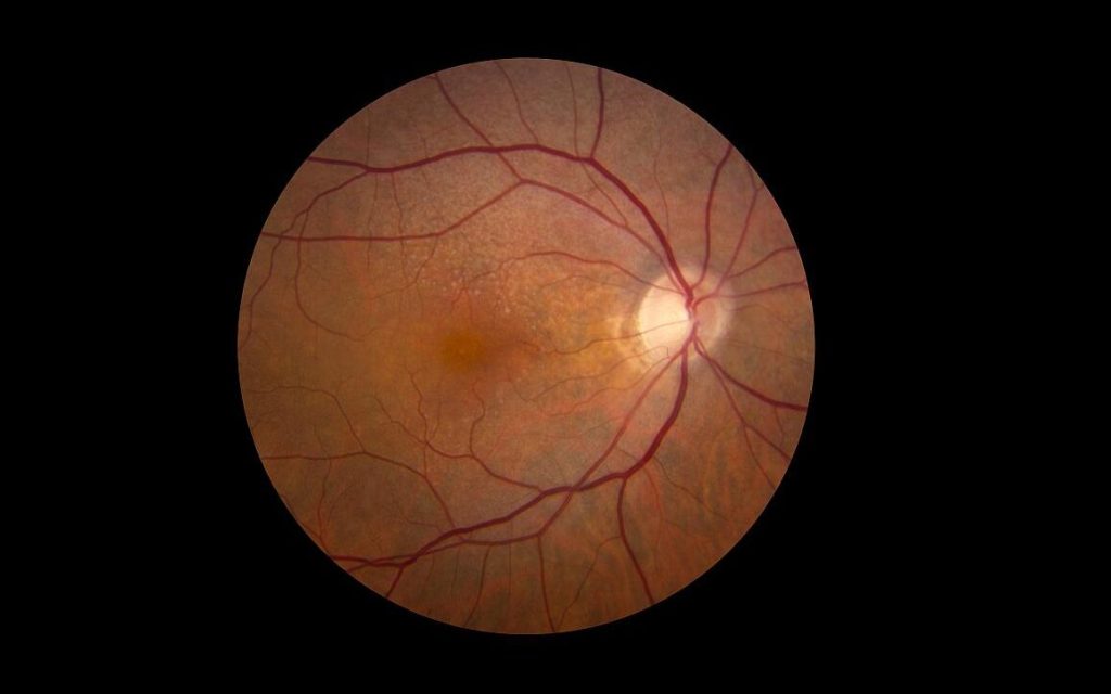
Tag: retina

SARS-CoV-2 can Cross the Blood–Retinal Barrier, Infecting the Eyes

Photoreceptor cells in the retina. Credit: Scientific Animations

The blood-retinal barrier is designed to protect vision from infections by preventing microbial pathogens from reaching the retina where they could trigger an inflammatory response with potential vision loss. But researchers at the University of Missouri School of Medicine have discovered that SARS-CoV-2 can breach this protective retinal barrier with potential long-term consequences in the eye. Their findings are reported in PLOS Pathogens.

Pawan Kumar Singh, PhD, an assistant professor of ophthalmology, leads a team researching new ways to prevent and treat ocular infectious diseases. Using a humanised ACE2 mice model, the team found that SARS-CoV-2, can infect the inside of the eyes even when the virus doesn’t enter the body through the surface of the eyes. Instead, they found that when viruses enter the body through inhalation, it not only infects organs like lungs, but also reaches highly protected organs like eyes through the blood-retinal barrier by infecting the cells lining this barrier.

This finding is important as we increase our understanding of the long-term effects of SARS-CoV-2 infection,” said Singh. “Earlier, researchers were primarily focused on the ocular surface exposure of the virus. However, our findings reveal that SARS-CoV-2 not only reaches the eye during systemic infection but induces a hyperinflammatory response in the retina and causes cell death in the blood-retinal barrier. The longer viral remnants remain in the eye, the risk of damage to the retina and visual function increases.”

Singh also discovered that extended presence of SARS-CoV-2 spike antigen can cause retinal microaneurysm, retinal artery and vein occlusion, and vascular leakage.

“For those who have been diagnosed with COVID-19, we recommend you ask your ophthalmologist to check for signs of pathological changes to the retina,” Singh said. “Even those who were asymptomatic could suffer from damage in the eyes over time because of COVID-19 associated complications.”

While viruses and bacteria have been found to breach the blood-retinal-barrier in immunocompromised people, this research is the first to suggest that the virus that causes COVID-19 could breach the barrier even in otherwise healthy individuals, leading to an infection that manifests inside the eye itself. Immunocompromised patients or those with hypertension or diabetes may experience worse outcomes if they remain undiagnosed for COVID-19 associated ocular symptoms.

“Now that we know the risk of COVID-19 to the retina, our goal is to better understand the cellular and molecular mechanisms of how this virus breaches the blood-retinal barrier and associated pathological consequences in hopes of informing development of therapies to prevent and treat COVID-19 induced eye complications before a patient’s vision is compromised,” Singh said.

Source: University of Missouri-Columbia

Newly Found Retinal Cells may Paint a Complete Picture of Colour Vision

Photo by Jeffrey Riley on Unsplash

Scientists have long studied just how the eye’s three cone photoreceptor types work together to allow humans to perceive colour. In a new study in the Journal of Neuroscience, researchers at the University of Rochester used adaptive optics to identify rare retinal ganglion cells (RGCs) that could help fill in the gaps in existing theories of colour perception.

The retina has three types of cones to detect colour that are sensitive to either short, medium, or long wavelengths of light. Retinal ganglion cells transmit input from these cones to the central nervous system.

In the 1980s, David Williams, the William G. Allyn Professor of Medical Optics, helped map the “cardinal directions” that explain colour detection. However, there are differences in the way the eye detects colour and how colour appears to humans. Scientists suspected that while most RGCs follow the cardinal directions, they may work in tandem with small numbers of non-cardinal RGCs to create more complex perceptions.

Recently, a team of researchers from Rochester’s Center for Visual Science, the Institute of Optics, and the Flaum Eye Institute identified some of these elusive non-cardinal RGCs in the fovea that could explain how humans see red, green, blue, and yellow.

“We don’t really know anything for certain yet about these cells other than that they exist,” says Sara Patterson, a postdoctoral researcher at the Center for Visual Science who led the study. “There’s so much more that we have to learn about how their response properties operate, but they’re a compelling option as a missing link in how our retina processes colour.”

Adaptive optics peer past the eye’s natural distorations

The team took advantage of adaptive optics, which uses a deformable mirror to overcome light distortion and was first developed by astronomers to reduce atmospheric blurring in ground-based telescopes. In the 1990s, Williams and his colleagues began applying adaptive optics to study the human eye. They created a camera that compensated for distortions caused by the eye’s natural aberrations, producing a clear image of individual photoreceptor cells.

“The optics of the eye’s lens are imperfect and really reduce the amount of resolution you can get with an ophthalmoscope,” says Patterson. “Adaptive optics detects and corrects for these aberrations and gives us a crystal-clear look into the eye. This gives us unprecedented access to the retinal ganglion cells, which are the sole source of visual information to the brain.”

Patterson says improving our understanding of the retina’s complex processes could ultimately help lead to better methods for restoring vision for people who have lost it.

“Humans have more than 20 ganglion cells and our models of human vision are only based on three,” says Patterson. “There’s so much going on in the retina that we don’t know about. This is one of the rare areas where engineering has totally outpaced visual basic science. People are out there with retinal prosthetics in their eyes right now, but if we knew what all those cells do, we could actually have retinal prosthetics drive ganglion cells in accordance with their actual functional roles.”

Source: University of Rochester

In Vitro Experiment Explains Why Humans Have Full Colour Vision and Dogs Don’t

Photo by Victor Freitas on Pexels

With human retinas grown in a petri dish, researchers discovered how retinoic acid, a metabolite of vitamin A, generates the specialised cells that enable people to see millions of colours, an ability that dogs, cats, and most other mammals do not have.

“These retinal organoids allowed us for the first time to study this very human-specific trait,” said author Robert Johnston, an associate professor of biology. “It’s a huge question about what makes us human, what makes us different.”

The findings, published in PLOS Biology, increase understanding of colour blindness, age-related vision loss, and other diseases linked to photoreceptor cells. They also demonstrate how genes instruct the human retina to make specific colour-sensing cells, a process scientists thought was controlled by thyroid hormones.

By tweaking the cellular properties of the organoids, the research team found that a vitamin A1 metabolite, retinoic acid, determines whether a cone will specialise in sensing red or green light.

Only humans with normal vision and closely related primates develop the red sensor.

For decades, it was that thought red cones formed through a coin toss mechanism where the cells haphazardly commit to sensing green or red wavelengths – and research from Johnston’s team recently hinted that the process could be controlled by thyroid hormone levels.

Instead, the new research suggests red cones materialise through a specific sequence of events orchestrated by retinoic acid within the eye.

The team found that high levels of retinoic acid in early development of the organoids correlated with higher ratios of green cones. Similarly, low levels of the acid changed the retina’s genetic instructions and generated red cones later in development.

“There still might be some randomness to it, but our big finding is that you make retinoic acid early in development,” Johnston said.

“This timing really matters for learning and understanding how these cone cells are made.”

Green and red cone cells are remarkably similar except for a protein called opsin, which detects light and tells the brain what colors people see.

Different opsins determine whether a cone will become a green or a red sensor, though the genes of each sensor remain 96% identical.

With a breakthrough technique that spotted those subtle genetic differences in the organoids, the team tracked cone ratio changes over 200 days.

“Because we can control in organoids the population of green and red cells, we can kind of push the pool to be more green or more red,” said author Sarah Hadyniak, who conducted the research as a doctoral student in Johnston’s lab and is now at Duke University.

“That has implications for figuring out exactly how retinoic acid is acting on genes.”

The researchers also mapped the widely varying ratios of these cells in the retinas of 700 adults.

Seeing how the green and red cone proportions changed in humans was one of the most surprising findings of the new research, Hadyniak said. Scientists still don’t fully understand how the ratio of green and red cones can vary so greatly without affecting someone’s vision.

If these types of cells determined the length of a human arm, the different ratios would produce “amazingly different” arm lengths, Johnston said.

To build understanding of diseases like macular degeneration, which causes loss of light-sensing cells near the center of the retina, the researchers are working with other Johns Hopkins labs.

The goal is to deepen their understanding of how cones and other cells link to the nervous system.

“The future hope is to help people with these vision problems,” Johnston said.

“It’s going to be a little while before that happens, but just knowing that we can make these different cell types is very, very promising.”

Source: Johns Hopkins University

Rebuilding Retinas with Nanotechnology ‘Scaffolds’

Anglia Ruskin University (ARU) researchers have found a to create a 3D ‘scaffold’ to grow cells from the retina -paving the way for potential new ways of treating a common cause of blindness. Their nanotechnology-based approached is detailed in the journal Materials & Design.

The researchers have been working on a way to successfully grow retinal pigment epithelial (RPE) cells that stay healthy and viable for up to 150 days. RPE cells sit just outside the neural part of the retina and, when damaged, can cause vision to deteriorate.

It is the first time this technology, called ‘electrospinning’, has been used to create a scaffold on which the RPE cells could grow, and could revolutionise treatment for one of age-related macular degeneration, one of the world’s most common vision complaints.

When the scaffold is treated with a steroid called fluocinolone acetonide, which protects against inflammation, the resilience of the cells appears to increase, promoting growth of eye cells. These findings are important in the future development of ocular tissue for transplantation into the patient’s eye.

Age-related macular degeneration (AMD) is a leading cause of blindness in the developed world and is expected to increase in the coming years due to an ageing population. Recent research predicted that 77 million people in Europe alone will have some form of AMD by 2050.

AMD can be caused by changes in the Bruch’s membrane, which supports the RPE cells, and breakdown of the choriocapillaris, the rich vascular bed that is adjacent to the other side of the Bruch’s membrane.

In Western populations, the most common way sight deteriorates is due to an accumulation of lipid deposits called drusen, and the subsequent degeneration of parts of the RPE, the choriocapillaris and outer retina. In the developing world, AMD tends to be caused by abnormal blood vessel growth in the choroid and their subsequent movement into the RPE cells, leading to haemorrhaging, RPE or retinal detachment and scar formation.

The replacement of the RPE cells is among several promising therapeutic options for effective treatment of sight conditions like AMD, and researchers have been working on efficient ways to transplant these cells into the eye.

Lead author Professor Barbara Pierscionek, Deputy Dean (Research and Innovation) at Anglia Ruskin University (ARU) said: “This research has demonstrated, for the first time, that nanofibre scaffolds treated with the anti-inflammatory substance such as fluocinolone acetonide can enhance the growth, differentiation, and functionality of RPE cells.

“In the past, scientists would grow cells on a flat surface, which is not biologically relevant. Using these new techniques. the cell line has been shown to thrive in the 3D environment provided by the scaffolds.

“This system shows great potential for development as a substitute Bruch’s membrane, providing a synthetic, non-toxic, biostable support for transplantation of the retinal pigment epithelial cells. Pathological changes in this membrane have been identified as a cause of eye diseases such as AMD, making this an exciting breakthrough that could potentially help millions of people worldwide.”

Source: Angela Ruskin University

Thinning of the Retina is an Early Marker of MS

Retina and nerve cells. Credit: NIH

For the first time, a study has shown that diagnosis of multiple sclerosis (MS) can be significantly improved by additionally measuring the thickness of retinal layers in the eye in a currently existing procedure. Use of the procedure helps to detect the condition at an earlier stage and predict its progression more accurately, which can help to improve patient outcomes. The findings have been published in the journal Neurology.

As part of their investigation, the research team headed by Gabriel Bsteh and Thomas Berger collaborated with ophthalmology colleagues examine 267 MS patients over five years. Their research built on study results published in 2022, which showed that MS relapse-related damage to the retina reflects the degree of damage caused to the patient’s brain. The previous study also demonstrated that a 5µm reduction in the thickness of the retinal layer following optic neuritis indicated a doubling of the risk of permanent disability after the next relapse. Thanks to the latest research with the large cohort of MS patients, the research team has confirmed that the thickness of the retinal layer can be used as a precise biomarker to assist early diagnosis.

Diagnostic procedure already available

The researchers used a procedure known as optical coherence tomography (OCT) to measure the thickness of the retinal layer. An imaging method that uses infrared light, OCT allows for the generation of high-resolution, three-dimensional images of extremely thin layers of tissue measuring just a few µm. OCT is also a tool for diagnosing and evaluating the progression of eye diseases such as glaucoma. “So we already have this procedure at our disposal,” commented Gabriel Bsteh, first author of the study. He added: “If we use optical coherence tomography alongside the current criteria to diagnose MS, we obtain significantly more accurate results at a much earlier stage. This means we can initiate treatment measures sooner, which considerably improves the long-term prognosis for patients.”

The retina: a window to the brain

Multiple sclerosis is an autoimmune, chronic inflammatory disease that causes inflammation and loss of nerve cells throughout the nervous system. For the most part, patients are unable to feel the consequences of this damage to begin with, so the condition often goes undiagnosed until a late stage, meaning that valuable time is lost during which effective treatment could have been administered. Given that early detection and prognosis of the disease’s progression play a decisive role in MS cases, medical researchers have been trying to find improved detection methods for some time now to help avert serious consequences such as impaired mobility and blindness as far as possible. “We have identified a new biomarker for MS diagnosis, namely the retinal layer thickness, which can be likened to a window to the brain,” said Gabriel Bsteh, summing up the study’s key finding. In the next phases of research, the focus will turn to the importance of retinal layer thickness in measuring responses to MS treatment.

Source: Medical University of Vienna

Optical Illusions Originate in the Retina, not the Brain

The bar in the middle is all one grey level, but it appears lighter on the left and darker on the right due to the background. Credit Jolyon Troscianko

Numerous visual illusions are caused by limits in the way our eyes and visual neurones work – rather than more complex psychological processes, as demonstrated by new research published in PLOS Computational Biology.

Researchers examined illusions in which an object’s surroundings affect the way we see its colour or pattern. Scientists and philosophers have long debated whether these illusions are caused by neural processing in the eye and low-level visual centres in the brain, or involve higher-level mental processes such as context and prior knowledge.

In the new study Dr Jolyon Troscianko, from the University of Exeter, co-developed a model that suggests simple limits to neural responses – not deeper psychological processes – explain these illusions.

“Our eyes send messages to the brain by making neurones fire faster or slower,” said Dr Troscianko. “However, there’s a limit to how quickly they can fire, and previous research hasn’t considered how the limit might affect the ways we see colour.”

The model combines this “limited bandwidth” with information on how humans perceive patterns at different scales, together with an assumption that our vision performs best when we are looking at natural scenes.

The model was developed by researchers from the Universities of Exeter and Sussex to predict how animals see colour, but it was also found to correctly predict many visual illusions seen by humans.

“This throws into the air a lot of long-held assumptions about how visual illusions work,” Dr Troscianko said.

He said the findings also shed light on the popularity of high-definition televisions.

“Modern high dynamic range televisions create bright white regions that are over 10 000 times brighter than their darkest black, approaching the contrast levels of natural scenes,” Dr Troscianko added.

“How our eyes and brains can handle this contrast is a puzzle because tests show that the highest contrasts we humans can see at a single spatial scale is around 200:1.

“Even more confusingly, the neurones connecting our eyes to our brains can only handle contrasts of about 10:1.

“Our model shows how neurones with such limited contrast bandwidth can combine their signals to allow us to see these enormous contrasts, but the information is ‘compressed’ – resulting in visual illusions.

“The model shows how our neurones are precisely evolved to use of every bit of capacity.

“For example, some neurones are sensitive to very tiny differences in grey levels at medium-sized scales, but are easily overwhelmed by high contrasts.

“Meanwhile, neurones coding for contrasts at larger or smaller scales are much less sensitive, but can work over a much wider range of contrasts, giving deep black-and-white differences.

“Ultimately this shows how a system with a severely limited neural bandwidth and sensitivity can perceive contrasts larger than 10 000:1.”

Source: University of Exeter

Adult Brains can Rewire to Recover from Inherited Blindness

Eye
Source: Daniil Kuzelev on Unsplash

A recent discovery has revealed that the adult brain has far greater potential to recover from inherited blindness than previously believed, with important implications for visually impaired people. The paper appears in Current Biology.

The research team was examining treatment for Leber congenital amaurosis (LCA), a group of inherited retinal diseases distinguished by severe visual impairment at birth. The condition, caused by mutations in any of over two dozen genes, results in degeneration or dysfunction in the retina’s photoreceptors.

Administering chemical compounds that target the retina, called synthetic retinoids, can restore a notable amount of vision in children with LCA. The UCI team wanted to find out if the treatment could make a difference for adults who have the condition.

“Frankly, we were blown away by how much the treatment rescued brain circuits involved in vision,” said corresponding author Sunil Gandhi, professor of neurobiology and behaviour. “Seeing involves more than intact and functioning retinae. It starts in the eye, which sends signals throughout the brain. It’s in the central circuits of the brain where visual perception actually arises.” Until now, scientists believed that the brain must receive those signals in childhood so that central circuits could wire themselves correctly.

The researchers were surprised by what they found in rodent models of LCA. “The central visual pathway signalling was significantly restored in adults, especially the circuits that deal with information coming from both eyes,” Prof Gandhi said. “Immediately after the treatment, the signals coming from the opposite-side eye, which is the dominant pathway in the mouse, activated two times more neurons in the brain. What was even more mind-blowing was that the signals coming from the same-side eye pathway activated five-fold more neurons in the brain after the treatment and this impressive effect was long-lasting. The restoration of visual function at the level of the brain was much greater than expected from the improvements we saw at the level of the retinae. The fact that this treatment works so well in the central visual pathway in adulthood supports a new concept, which is that there is latent potential for vision that is just waiting to be triggered.”

The finding opens exciting research possibilities. “Whenever you have a discovery that breaks with your expectations about the possibility for the brain to adapt and rewire, it teaches you a broader concept,” Prof Gandhi said. “This new paradigm could aid in the development of retinoid therapies to more completely rescue the central visual pathway of adults with this condition.”

Source: University of California – Irvine

Retinal Cells a Haven for Ebola and Other Viruses

Credit: National Eye Institute

A specific cell within the retina, the retinal pigment epithelial cell, appears to be particularly good at housing Ebola and other viruses, according to new research published in the journal Frontiers in Virology.

“Inflammation of the eye, known as uveitis, is very common following infection with Ebola and we know the cells within the iris, at the front of the eye, as well as the retina have the capacity to play a major role in uveitis and act as hosts for microorganisms,” explained study senior author Professor Justine Smith at Flinders University.

“However, what we didn’t know was which out of the two was most responsible in the case of Ebola.”

“Patients with Ebola eye disease have characteristic retinal scars”

Professor Justine Smith

The study used cells from human eyes donated from the South Australia Eye Bank to investigate the ability of iris and retinal pigment epithelial cells to be infected by Ebola.

Cells were infected with Ebola virus, Reston virus (a type of ebolavirus that does not cause disease in humans) or Zika virus (another type of virus, but one that also can cause uveitis), while some were left uninfected for the duration of the trial.

While both types of cells allow replication of the Ebola virus, it was the retinal cells that showed much higher levels of infection.

“We also found similar results when looking at the cells infected with Reston virus and Zika virus,” said Professor Smith.

“Patients with Ebola eye disease have characteristic retinal scars, suggesting the retinal pigment epithelium is involved in the disease, so this finding is consistent with what eye doctors are seeing in the clinic.

“These retinal cells are good at eating things – called phagocytosis – and they play an essential part in the visual cycle by recycling our photoreceptors, so it makes sense that these cells would be a receptive haven for Ebola, as well as other viruses.”

The researchers say the study demonstrates an important target cell for Ebola infection in the eye and suggests the potential for these cells to be monitored during acute viral infection to identify patients at highest risk of uveitis.

“Amongst other issues, including pain and blurred vision, uveitis can ultimately lead to vision loss, so it’s important we find ways to diagnose it as early as possible to enable swift treatment,” said Professor Smith.

Source: Flinders University

Retinal Scans May be Able to Detect ASD and ADHD

Eye
Source: Daniil Kuzelev on Unsplash

By measuring the electrical activity of the retina in responses to a light stimulus, researchers found that they may be able to neurodevelopmental disorders such as ASD and ADHD, as reported in new research published in Frontiers in Neuroscience.

In this groundbreaking study, researchers found that recordings from the retina could identify distinct signals for both Attention Deficit Hyperactivity Disorder (ADHD) and Autism Spectrum Disorder (ASD) providing a potential biomarker for each condition.

Using the ‘electroretinogram’ (ERG) – a diagnostic test that measures the electrical activity of the retina in response to a light stimulus – researchers found that children with ADHD showed higher overall ERG energy, whereas children with ASD showed less ERG energy.

Research optometrist at Flinders University, Dr Paul Constable, said the preliminary findings indicate promising results for improved diagnoses and treatments in the future.

“ASD and ADHD are the most common neurodevelopmental disorders diagnosed in childhood. But as they often share similar traits, making diagnoses for both conditions can be lengthy and complicated,” Dr Constable says.

“Our research aims to improve this. By exploring how signals in the retina react to light stimuli, we hope to develop more accurate and earlier diagnoses for different neurodevelopmental conditions.

“Retinal signals have specific nerves that generate them, so if we can identify these differences and localise them to specific pathways that use different chemical signals that are also used in the brain, then we can show distinct differences for children with ADHD and ASD and potentially other neurodevelopmental conditions.”

“This study delivers preliminary evidence for neurophysiological changes that not only differentiate both ADHD and ASD from typically developing children, but also evidence that they can be distinguished from each other based on ERG characteristics.”

According to the World Health Organization, one in 100 children has ASD, with 5–8% of children diagnosed with ADHD.

Attention Deficit Hyperactivity Disorder (ADHD) is a neurodevelopmental condition characterised by being overly active, struggling to pay attention, and difficulty controlling impulsive behaviours. Autism spectrum disorder (ASD) is also a neurodevelopmental condition where children behave, communicate, interact, and learn in ways that are different from most other people.

Co-researcher and expert in human and artificial cognition at the University of South Australia, Dr Fernando Marmolejo-Ramos, says the research has potential to extend across other neurological conditions.

“Ultimately, we’re looking at how the eyes can help us understand the brain,” Dr Marmolejo-Ramos says.

“While further research is needed to establish abnormalities in retinal signals that are specific to these and other neurodevelopmental disorders, what we’ve observed so far shows that we are on the precipice of something amazing.

“It is truly a case of watching this space; as it happens, the eyes could reveal all.”

Source: Flinders University

Cell Fusion Jump-starts Retinal Regeneration

Genetics
Image source: Pixabay

Researchers have reported that they have successfully fused human retinal cells with adult stem cells, in a novel potential regenerative therapy to treat retinal damage and visual impairment.

The resulting hybrid cells stimulate the regenerative potential of human retinal tissue, something previously only thought to be found in cold-blood vertebrates.

Cell fusion events, where two different cells combine into one single entity, are known to be a possible mechanism contributing to tissue regeneration. These cell fusions result in four sets of chromosomes instead of the usual two. Though a rare phenomenon in humans, it has been reliably detected in the liver, brain, and gastrointestinal tract. Now, cell fusion events have been found also take place in the human retina, as reported in eBioMedicine.

Seeking to see if cell fusion events could differentiate into neurons, the researchers fused Müller glia, cells that play a secondary but important role in maintaining the structure and function of the retina, with adult stem cells.

“We were able to carry out cell fusion in vitro, creating hybrid cells. Importantly, the process was more efficient in the presence of a chemical signal transmitted from the retina in response to damage, resulting in rates of hybridisation increasing twofold. This gave us an important clue for the role of cell fusion in the retina,” said first author Sergi Bonilla.

The hybrid cells were injected into a growing retinal organoid, a model that closely resembles the function of the human retina. The researchers found that the hybrid cells successfully engrafted into the tissue and differentiated into cells that closely resemble ganglion cells, a type of neuron essential for vision.

“Our findings are important because they show that the Müller Glia in the human retina have the potential to regenerate neurons,” said lead researcher Professor Pia Cosma. “Salamanders and fish can repair damage caused to the retina thanks to their Müller glia, which differentiate into neurons that rescue or replace damaged neurons. Mammalian Müller glia have lost this regenerative capacity, which means retinal damage or degradation can lead to visual impairment for life. Our findings bring us one step closer to recovering this ability.”

Further work will be to understand why these hybrid cells, which have four complete sets of chromosomes, don’t result in chromosomal instability and cancer development. The authors of the study believe the retina may have a mechanism regulating chromosome segregation similar to the liver, which contains tetraploid cells that act as a genetic reservoir, undergoing mitosis in response to stress and injury.

Source: Center for Genomic Regulation