Tag: regulatory T cells

Peacekeeper Cells Protect the Body from Autoimmunity During Infection
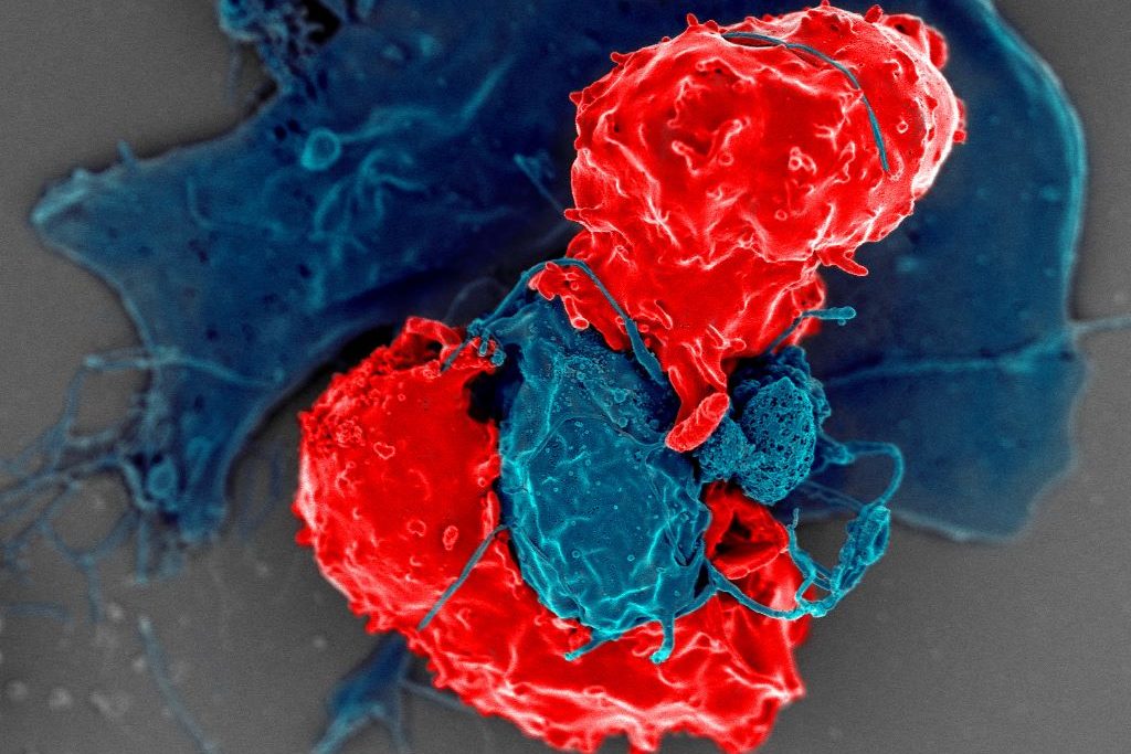
Scanning electron microscope image of T regulatory cells (red) interacting with antigen-presenting cells (blue). T regulatory cells can suppress responses by T cells to maintain homeostasis in the immune system. Credit: National Institute of Allergy and Infectious Diseases/NIH

In the flurry of immune activity in an infection, immune cells need to be prevented from mistakenly attacking each other. New research from the University of Chicago shows how a specially trained population of immune cells keeps the peace by preventing other immune cells from attacking their own. The study, published in Science, provides a better understanding of immune regulation during infection and could provide a foundation for interventions to prevent or reverse autoimmune diseases.

Several groups of white blood cells help coordinate immune responses. Dendritic cells take up proteins from foreign pathogens, chop them up into peptides called antigens, and display them on their surface. CD4+ conventional T (Tconv) cells, or helper T cells, inspect the peptides presented by dendritic cells. If the peptides are foreign antigens, the T cells expand in numbers and transform into an activated state, specialized to eradicate the pathogen. If the dendritic cell is carrying a “self-peptide,” or peptides from the body’s own tissue, the T cells are supposed to lay off.

During an autoimmune response, the helper T cells don’t distinguish between foreign peptide antigens and self-peptides properly and go on the attack no matter what. To prevent this from happening, another group of T cells called CD4+ regulatory T (Treg) cells, are supposed to intervene and prevent friendly fire from the Tconv cells.

“You can think of them [Treg cells] as peacekeeper cells,” said Pete Savage, PhD, Professor of Pathology at UChicago and senior author of the new study. Tregs obviously do their job well most of the time, but Savage said that it has never been clear how they know when to intervene and prevent helper T cells from starting an autoimmune response, and when to hold back and let them fight an infection.

So, Savage and his team, led by David Klawon, PhD, a former graduate student in his lab who is now a postdoctoral fellow at the Massachusetts Institute of Technology (MIT), wanted to explore this property of the immune system, known in the field as self-nonself discrimination. T cells are produced in the thymus, a specialised organ of the immune system. During development, Treg cells are trained to recognise specific peptides, including self-peptides from the body. When dendritic cells present a self-peptide, the Treg cells trained to spot them intervene to stop helper T cells from getting triggered.

For the study, Savage and Klawon worked in close collaboration with co-first author Nicole Pagane, a graduate student at MIT, as well as co-corresponding authors Harikesh Wong at the Ragon Institute of the Massachusetts General Hospital, MIT and Harvard University, and Ron Germain at the National Institutes of Health.

T cell specificity is what the team found makes a crucial difference in self-nonself discrimination. The researchers experimentally depleted Treg cells in mice that were specific to a single self-peptide from the prostate. In healthy mice in the absence of infection, this change did not trigger autoimmunity to the prostate. When the researchers infected mice with a bacterium that expressed the prostate self-peptide, however, the absence of matched, prostate-specific Treg cells triggered prostate-reactive T helper cells and introduced autoimmunity to the prostate.

Interestingly though, this alteration did not impair the ability of helper T cells to control the bacterial infection by responding to foreign peptides.

“It’s like a doppelganger population of T cells. The CD4 helper cells that could induce disease by attacking the self share an equivalent, matched population of these peacekeeper Treg cells,” Savage said. “When we removed Treg cells reactive to a single self-peptide, the T helper cells reactive to that self-peptide were no longer controlled, and they induced autoimmunity.”

The root causes of autoimmune disease are a complex interaction of genetics, the environment, lifestyle, and the immune system. Classic, conventional thinking in the immunology field promoted the idea that the immune system establishes self-nonself discrimination by purging the body of helper T cells that are reactive to self-peptides, thereby preventing autoimmunity. Savage said this study shows that purging is inefficient though, and that specificity matching by Treg cells may be equally as important.

“The idea is that specificity matters, and for a fully healthy immune system, you need to have a good collection of these doppelganger Treg cells,” he said. As long as the immune system generates enough matched Treg cells, they can prevent autoimmune responses without impacting responses to infections.

“It’s like flipping the idea of self-nonself discrimination upside down. Instead of having to delete all helper T cells reactive to self-antigens, you simply generate enough of these Treg peacekeeper cells instead,” Savage said.

Source: University of Chicago

Salt Cuts off Regulatory T Cells’ Energy Supply

Spilled salt shaker
Source: Pixabay CC0

Regulatory T cells ensure that immune responses happen in a controlled way. But eating too much salt weakens these cells’ energy supply, thus rendering them temporarily dysfunctional. This salt-induced ‘load shedding’ may have implications for autoimmunity, researchers report in Cell Metabolism.

Excessive salt consumption not only causes cardiovascular problems, it could also adversely impact the immune system. The study found that salt can disrupt regulatory T cells by impairing their energy metabolism. The findings may provide new avenues for exploring the development of autoimmune and cardiovascular diseases.

A few years ago, research by teams led by Professor Dominik Müller and Professor Markus Kleinewietfeld revealed that excess salt in the diet can negatively affect the metabolism and energy balance in certain types of innate immune cells called monocytes and macrophages and stop them from working properly. They further showed that salt triggers malfunctions in the mitochondria. Inspired by these findings, the research groups wondered whether excessive salt intake might also create a similar problem in adaptive immune cells like regulatory T cells.

Important immune regulators

Regulatory T cells, also known as Tregs, are an essential part of the adaptive immune system. They are responsible for maintaining the balance between normal function and unwanted excessive inflammation.

Scientists believe that the deregulation of Tregs is linked to the development of autoimmune diseases like multiple sclerosis. Recent research has identified problems in mitochondrial function of Tregs from patients with autoimmunity, yet the contributing factors remain elusive.

“Considering our previous findings of salt affecting mitochondrial function of monocytes and macrophages as well as the new observations on mitochondria in Tregs from autoimmune patients, we were wondering if sodium might elicit similar issues in Tregs of healthy volunteers,” says Müller, who co-heads the Hypertension-Mediated End-Organ Damage Lab at the Max Delbrück Center and the ECRC.

Previous research has also shown that excess salt could impact Treg function by inducing an autoimmune-like phenotype. In other words, too much salt makes the Treg cells look like those involved in autoimmune conditions. However, exactly how sodium impairs Treg function had not yet been uncovered.

Salt interferes with mitochondrial function of Tregs

The new international study led by Kleinewietfeld and Müller has now discovered that sodium disrupts Treg function by altering cellular metabolism through interference with mitochondrial energy generation. This mitochondrial problem seems to be the initial step in how salt modifies Treg function, leading to changes in gene expression that showed similarities to those of dysfunctional Tregs in autoimmune conditions.

Even a short-term disruption of mitochondrial function had long-lasting consequences for the fitness and immune-regulating capacity of Tregs in various experimental models. The new findings suggest that sodium may be a factor that could contribute to Treg dysfunction, potentially playing a role in different diseases, although this needs to be confirmed in further studies.

“The better understanding of factors and underlying molecular mechanisms contributing to Treg dysfunction in autoimmunity is an important question in the field. Since Tregs also play a role in diseases such as cancer or cardiovascular disease, the further exploration of such sodium-elicited effects may offer novel strategies for altering Treg function in different types of diseases,” says Kleinewietfeld, who heads the VIB Laboratory for Translational Immunomodulation. “However, future studies are needed to understand the molecular mechanisms in more detail and to clarify their potential relationship to disease.”

Source: Max Delbrück Center for Molecular Medicine in the Helmholtz Association

Regulatory T Cells Play a Surprising Role in Hair Growth

Photo by Engin Akyurt on Unsplash

In an unexpected finding in studying alopecia, scientists have uncovered an unexpected link between T cells and hair growth, which could potentially be used to treat the condition. The findings, published in Nature Immunology, describe how regulatory T cells interact with skin cells using a hormone as a messenger to generate new hair follicles and hair growth.

Alopecia is an autoimmune condition where the immune system attacks the hair follicles, resulting in hair loss.

“For the longest time, regulatory T cells have been studied for how they decrease excessive immune reactions in autoimmune diseases,” explained Ye Zheng, associate professor at the Salk Institute and the paper’s corresponding author. “Now we’ve identified the upstream hormonal signal and downstream growth factor that actually promote hair growth and regeneration completely separate from suppressing immune response.”

Initially, the researchers were investigating the roles of regulatory T (Treg) cells and glucocorticoid hormones in autoimmune diseases. (Glucocorticoid hormones are cholesterol-derived steroid hormones produced by the adrenal gland and other tissues.) They first investigated how these immune components functioned in multiple sclerosis, Crohn’s disease and asthma.

They found that glucocorticoids and Treg cells did not function together to play a significant role in any of these conditions. So, they thought they’d have more luck looking at environments where Treg cells expressed particularly high levels of glucocorticoid receptors (which respond to glucocorticoid hormones), such as in skin tissue. The scientists induced hair loss in normal mice and mice lacking glucocorticoid receptors in their Treg cells.

“After two weeks, we saw a noticeable difference between the mice — the normal mice grew back their hair, but the mice without glucocorticoid receptors barely could,” says first author Zhi Liu, a postdoctoral fellow in Associate Prof Zheng’s lab. “It was very striking, and it showed us the right direction for moving forward.”

The findings suggested that some sort of communication must be occurring between Treg cells and hair follicle stem cells to allow for hair regeneration.

The scientists then investigated how the regulatory T cells and glucocorticoid receptors behaved in skin tissue samples, and found that glucocorticoids instruct the Treg cells to activate hair follicle stem cells, leading to hair growth. This crosstalk between the T cells and the stem cells depends on a mechanism whereby glucocorticoid receptors induce production of the protein TGF-beta3, all within the regulatory T cells. TGF-beta3 then activates the hair follicle stem cells to differentiate into new hair follicles, promoting hair growth. Additional analysis confirmed that this pathway was completely independent of regulatory T cells’ ability to maintain immune balance.

However, Treg cells don’t normally produce TGF-beta3, as they did here. A database search revaled that this phenomenon occurs in injured muscle and heart tissue, similar to how hair removal simulated a skin tissue injury in this study.

“In acute cases of alopecia, immune cells attack the skin tissue, causing hair loss. The usual remedy is to use glucocorticoids to inhibit the immune reaction in the skin, so they don’t keep attacking the hair follicles,” said Associate Prof Zheng. “Applying glucocorticoids has the double benefit of triggering the regulatory T cells in the skin to produce TGF-beta3, stimulating the activation of the hair follicle stem cells.”

This study revealed that Treg cells and glucocorticoid hormones are not just immunosuppressants but also have a regenerative function. Next, the scientists will look at other injury models and isolate Treg cells from injured tissues to monitor increased levels of TGF-beta3 and other growth factors.

Source: Salk Institute