Tag: regeneration

Cutting Down on Salt Levels Stimulates Kidney Regeneration
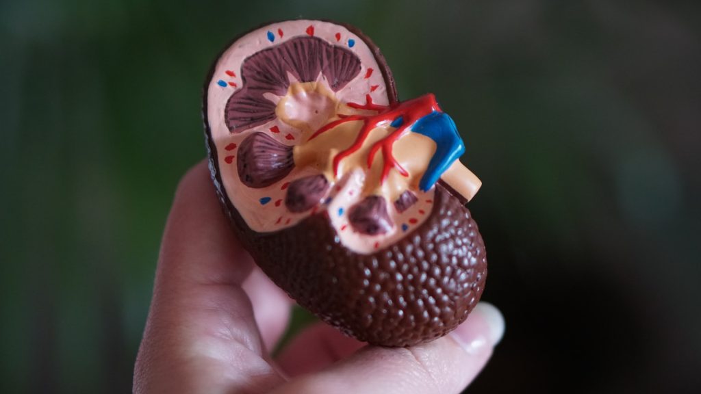
Photo by Robina Weermeijer on Unsplash

A loss of salt and body fluid can stimulate kidney regeneration and repair in mice, according to a study published in The Journal of Clinical Investigation. This innate regenerative response relies on a small population of kidney cells in a region known as the macula densa (MD), which senses salt and exerts control over filtration, hormone secretion, and other key functions of this vital organ.

“Our personal and professional mission is to find a cure for kidney disease, a growing global epidemic affecting one out of seven adults, which translates to 850 million people worldwide…” said study leader Janos Peti-Peterdi, a professor of physiology, neuroscience and medicine at the Keck School of Medicine of USC. “Currently, there is no cure for this silent disease. By the time kidney disease is diagnosed, the kidneys are irreversibly damaged and ultimately need replacement therapies, such as dialysis or transplantation.”

To address this growing epidemic, Peti-Peterdi, first author Georgina Gyarmati, and their colleagues took a highly non-traditional approach. As opposed to studying how diseased kidneys fail to regenerate, the scientists focused on how healthy kidneys originally evolved.

“From an evolutionary biology perspective, the primitive kidney structure of the fish turned into more complicated and more efficiently working kidneys to absorb more salt and water,” said Peti-Peterdi. “This was necessary for adaptation to the dry land environment when the animal species moved from the salt-rich seawater. And that’s why birds and mammals have developed MD cells and this beautiful, bigger, and more efficient kidney structure to maintain themselves and functionally adapt to survive. These are the mechanisms that we are targeting and trying to mimic in our research approach.”

With this evolutionary history in mind, the research team fed lab mice a very low salt diet, along with a commonly prescribed drug called an ACE inhibitor that furthered lowered salt and fluid levels. The mice followed this regimen for up to two weeks, since extremely low salt diets can trigger serious health problems if continued long term.

In the region of the MD, the scientists observed regenerative activity, which they could block by administering drugs that interfered with signals sent by the MD. This underscored the MD’s key role in orchestrating regeneration.

When the scientists furthered analysed mouse MD cells, they identified both genetic and structural characteristics that were surprisingly similar to nerve cells. This is an interesting finding, because nerve cells play a key role in regulating the regeneration of other organs such as the skin.

In the mouse MD cells, the scientists also identified specific signals from certain genes, including Wnt, NGFR, and CCN1, which could be enhanced by a low-salt diet to regenerate kidney structure and function. In keeping with these findings in mice, the activity of CCN1 was found to be greatly reduced in patients with chronic kidney disease (CKD).

To test the therapeutic potential of these discoveries, the scientists administered CCN1 to mice with a type of CKD known as focal segmental glomerulosclerosis. They also treated these mice with MD cells grown in low-salt conditions. Both approaches were successful, with the MD cell treatment producing the biggest improvements in kidney structure and function. This might be due to the MD cells secreting not only CCN1, but also additional unknown factors that promote kidney regeneration.

“We feel very strongly about the importance of this new way of thinking about kidney repair and regeneration,” said Peti-Peterdi. “And we are fully convinced that this will hopefully end up soon in a very powerful and new therapeutic approach.”

Source: Keck School of Medicine of USC

Probing the Gut’s Ability to Change Size According to Nutrient Intake

Source: CC0

The gut has considerable plasticity among animals, shrinking as much 50% in cases of fasting such as hibernating and able to rapidly return to normal size on refeeding. Now, scientists from the University of Copenhagen used fruit flies to investigate the signalling mechanisms and cellular changes that regulate this rapidly renewable tissue, which could reveal insights into diseases such as colorectal cancer. Their results are published in Nature Communications.

“Taking advantage of the broad genetic toolbox available in the fruit fly, we have investigated the mechanisms underpinning nutrient-dependent gut resizing,” says Dr Ditte S. Andersen.

The results show that nutrient deprivation results in an accumulation of progenitor cells that fail to differentiate into the mature cells causing the gut to shrink.

Upon refeeding these stalled progenitor cells readily differentiate into mature cells to promote regrowth of the gut.

Ditte S. Andersen continues: “We have identified activins as critical regulators of this process. In nutrient restrictive conditions, activin signalling is strongly repressed, while it is reactivated and required for progenitor maturation and gut resizing in response to refeeding. Activin-dependent resizing of the gut is physiologically important as inhibition of activin signalling reduces survival of flies to intermittent fasting.”

Regulators of organ plasticity are essential for host adaptation to an ever-changing environment, however, the same signals are often deregulated in cancers. Indeed, mutations affecting activin signalling are frequent in cancer cells in a variety of tissues. This study provides a starting point for investigating the link between aberrant activin signalling and the development of colorectal cancers and sets the stage for exploring the efficiency of anti-activin therapeutic strategies in treating colorectal cancers.

Source: University of Copenhangen

Neuroscientists Regenerate Neurons in Mice with Spinal Cord Injury

Source: CC0

In a new study using mice, neuroscientists have uncovered a crucial component for restoring functional activity after spinal cord injury. In the study, published in Science, the researchers showed that re-growing specific neurons back to their natural target regions led to recovery, while random regrowth was not effective.

In a 2018 study in Naturethe team identified a treatment approach that triggers axons to regrow after spinal cord injury in rodents. But even as that approach successfully led to the regeneration of axons across severe spinal cord lesions, achieving functional recovery remained a significant challenge.

For the new study, the team of researchers from UCLA, the Swiss Federal Institute of Technology, and Harvard University aimed to determine whether directing the regeneration of axons from specific neuronal subpopulations to their natural target regions could lead to meaningful functional restoration after spinal cord injury in mice. They first used advanced genetic analysis to identify nerve cell groups that enable walking improvement after a partial spinal cord injury.

The researchers then found that merely regenerating axons from these nerve cells across the spinal cord lesion without specific guidance had no impact on functional recovery. However, when the strategy was refined to include using chemical signals to attract and guide the regeneration of these axons to their natural target region in the lumbar spinal cord, significant improvements in walking ability were observed in a mouse model of complete spinal cord injury.

“Our study provides crucial insights into the intricacies of axon regeneration and requirements for functional recovery after spinal cord injuries,” said Michael Sofroniew, MD, PhD, professor of neurobiology at the David Geffen School of Medicine at UCLA and a senior author of the new study. “It highlights the necessity of not only regenerating axons across lesions but also of actively guiding them to reach their natural target regions to achieve meaningful neurological restoration.”

The authors say understanding that re-establishing the projections of specific neuronal subpopulations to their natural target regions holds significant promise for the development of therapies aimed at restoring neurological functions in larger animals and humans. However, the researchers also acknowledge the complexity of promoting regeneration over longer distances in non-rodents, necessitating strategies with intricate spatial and temporal features. Still, they conclude that applying the principles laid out in their work “will unlock the framework to achieve meaningful repair of the injured spinal cord and may expedite repair after other forms of central nervous system injury and disease.”

Source: University of California – Los Angeles Health Sciences

Signalling Control Explains Why Adult Hearts Cells Don’t Regenerate

Source: Pixabay CC0

New research published in Developmental Cell has uncovered a possible explanation for why explain why adult heart cells lack regeneration capacity. As heart cells mature in mice, the number of communication pathways called nuclear pores dramatically decreases. While this might protect the organ from damaging signals, it could also prevent adult heart cells from regenerating, the researchers found.

The study, from University of Pittsburgh and UPMC scientists, suggests that quieting communication between heart cells and their environment protects this organ from harmful signals related to stresses such as high blood pressure, but at the cost of preventing heart cells from receiving signals that promote regeneration.

“This paper provides an explanation for why adult hearts do not regenerate themselves, but newborn mice and human hearts do,” said senior author Bernhard Kühn, MD, professor of paediatrics. “These findings are an important advance in fundamental understanding of how the heart develops with age and how it has evolved to cope with stress.”

While skin and many other tissues of the human body retain the ability to repair themselves after injury, the same isn’t true of the heart. During human embryonic and foetal development, heart cells undergo cell division to form the heart muscle. But as heart cells mature in adulthood, they enter a terminal state in which they can no longer divide.

To understand more about how and why heart cells change with age, Prof Kühn teamed up with fellow Pitt researchers to look at nuclear pores. These perforations in the lipid membrane that surround a cell’s DNA regulate the passage of molecules to and from the nucleus.

“The nuclear envelope is an impermeable layer that protects the nucleus like asphalt on a highway,” said Prof Kühn. “Like manholes in this asphalt, nuclear pores are pathways that allow information to get through the barrier and into the nucleus.”

Using super-resolution microscopy, the researchers visualised and counted the number of nuclear pores in mouse heart cells, or cardiomyocytes. The number of pores decreased by 63% across development, from an average of 1,856 in foetal cells to 1040 in infant cells to just 678 in adult cells. These findings were validated with electron microscopy to show that nuclear pore density decreased across heart cell development.

In previous research, Prof Kühn and his team showed that a gene called Lamin b2, which is highly expressed in newborn mice but declines with age, is important for cardiomyocyte regeneration.

In the new study, they show that blocking expression of Lamin b2 in mice led to a decrease in nuclear pore numbers. Mice with fewer nuclear pores had diminished transport of signalling proteins to the nucleus and decreased gene expression, suggesting that reduced communication with age may drive a decrease in cardiomyocyte regenerative capacity.

“These findings demonstrate that the number of nuclear pores controls information flux into the nucleus,” explained Prof Kühn. “As heart cells mature and the nuclear pores decrease, less information is getting to the nucleus.”

In response to stress such as high blood pressure, a cardiomyocyte’s nucleus receives signals that modify gene pathways, leading to structural remodelling of the heart. This remodelling is a major cause of heart failure.

The researchers used a mouse model of high blood pressure to understand how nuclear pores contribute to this remodelling process. Mice that were engineered to express fewer nuclear pores showed less modulation of gene pathways involved in harmful cardiac remodelling. These mice also had better heart function and survival than their peers with more nuclear pores.

“We were surprised at the magnitude of the protective effect of having fewer nuclear pores in mice with high blood pressure,” said Prof Kühn. “However, having fewer communication pathways also limits beneficial signals such as those that promote regeneration.”

Source: University of Pittsburgh

Macrophage Role in Liver Regeneration Identified

A macrophage engulfing a yeast cell. Source: CDC

Researchers have found out what role macrophages play in liver regeneration after resection. The results are published in the journal Biomedicine & Pharmacotherapy.

In mammals, the liver is the most regenerative internal organ. It can restore its original size with as little as 25% of the original tissue remaining. Macrophages play an important role in this process. It is known, for example, that if the liver is affected by foreign substances, including drugs, macrophages migrate to the liver, absorb harmful microorganisms and dead cells, cause inflammation and thus contribute to the restoration of the organ. However, it is still unknown unambiguously how macrophages affect the growth of the liver after its resection, ie when a large part of the organ is removed. RUDN University doctors investigated this issue in an experiment with laboratory mice.

“The role of macrophages in the liver growth after massive resections is uncertain. Some studies reveal the lack of immigration of macrophages to the liver during its recovery from partial resection, whereas other studies demonstrate such possibility. So, we focused our study on the macrophage population dynamics after 70% liver resection in mouse mode”, Andrey Elchaninov, MD, PhD, researcher at the Department of Histology, Cytology and Embryology of RUDN University.

The researchers removed 70% of the liver of a number of lab mice. Immediately after that, then a day later, three days later, and a week later, the scientists took liver samples for analysis. The resulting cells were studied using an immunohistochemical method. The sections were labelled with specific antibodies to the glycoproteins CD68, CD206 and other compounds that are found on the surface of macrophages. To count them, the antibodies are labelled with fluorescent dyes and glow when attached to macrophages. The researchers also measured the rate of reproduction and cell death of macrophages.

The researchers found that after resection, macrophages migrate to the liver in large numbers. A day after surgery, the number of macrophages with CD68 in the liver doubles, which persists after a week. It also turned out that the resection led to significant changes in the ratio of different types of macrophages. For example, the proportion of Ly6C cells in the week after surgery increased 4-fold — from 5% to 22%, and the proportion of CD86 droppedfrom 50% to 15%. The role of macrophages is ambiguous. On the one hand, they release chemicals (chemoattractors) that attract white blood cells responsible for the body’s inflammatory response, but on the other hand, they regulate the reproduction of liver cells and the metabolism in the organ.

“Corresponding profiles of macrophages in the regeneration of the liver cannot be unambiguously defined as pro- or anti-inflammatory,” the researchers said. “Their typical features include elevated expression of leukocyte chemoattractant factors, and many of the differentially expressed sequences are related to the control of cell growth and metabolic processes in the liver. Our findings revealed essential roles of macrophages and macrophages proliferation in the mouse liver during its recovery from a massive resection.”

Source: EurekAlert!

Journal reference: Elchaninov, A., et al. (2021) Macro- and microtranscriptomic evidence of the monocyte recruitment to regenerating liver after partial hepatectomy in mouse model. Biomedicine & Pharmacotherapy. doi.org/10.1016/j.biopha.2021.111516.

Antibodies May Hold The Key to Tooth Regeneration

It may be possible to regenerate missing teeth using monoclonal antibodies, according to a new study by scientists at Kyoto University and the University of Fukui. 

The team reported that an antibody for one gene—uterine sensitisation associated gene-1 or USAG-1—can stimulate tooth growth in mice suffering from tooth agenesis, a congenital condition. The paper was published in Science Advances. Monoclonal antibodies are often used to treat cancers, arthritis, and vaccine development.

Although the normal adult mouth has 32 teeth, about 1% of the population has more or fewer due to congenital conditions; adults with too many teeth are of interest because they could hold genetic clues to tooth regeneration.

Katsu Takahashi, one of the lead authors of the study and a senior lecturer at the Kyoto University Graduate School of Medicine, said that the fundamental molecules responsible for tooth development have already been identified.

“The morphogenesis of individual teeth depends on the interactions of several molecules including BMP, or bone morphogenetic protein, and Wnt signaling,” said Takahashi.

BMP and Wnt are also involved in the development of organs when humans are mere embryos. This means that drugs directly affecting their activity are usually avoided, as side effects could impact the entire body. The team considered the gene USAG-1, as they guessed that it could be safer to target the factors that antagonise BMP and Wnt specifically in tooth development .

“We knew that suppressing USAG-1 benefits tooth growth. What we did not know was whether it would be enough,” added Takahashi.

The scientists therefore investigated the effects of several monoclonal antibodies for USAG-1. Since USAG-1 interacts with both BMP and Wnt, many of the antibodies resulted in poor birth and survival rates of mice, showing that BMP and Wnt are important for whole body growth. However one antibody managed to disrupt the interaction of USAG-1 with BMP only.

Experimentation showed that BMP signalling is necessary for the number of teeth in mice, and a single administration was enough to generate an entire tooth. The same effects were seen in ferrets.

“Ferrets are diphyodont animals with similar dental patterns to humans. Our next plan is to test the antibodies on other animals such as pigs and dogs,” explains Takahashi.

This is the first study to show the benefits of monoclonal antibodies on tooth regeneration, and offers new alternatives to implants.

“Conventional tissue engineering is not suitable for tooth regeneration. Our study shows that cell-free molecular therapy is effective for a wide range of congenital tooth agenesis,” concluded Manabu Sugai of the University of Fukui, another author of the study.

Source: Medical Xpress

Journal information: A. Murashima-Suginami et al, Anti–USAG-1 therapy for tooth regeneration through enhanced BMP signaling, Science Advances (2021). DOI: 10.1126/sciadv.abf1798