Tag: red blood cells

Not Just for Respiration: Lungs Also Produce Blood Cells
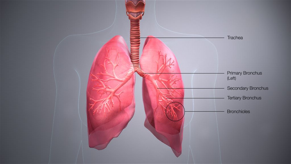
Credit: Scientific Animations CC4.0

For many years, scientists assumed that blood production took place in the bone marrow, providing the 200 billion blood cells needed per day. But now, researchers at UCSF are showing it’s also happening in the lungs. 

They found haematopoietic stem cells (HSCs) in human lung tissue that make red blood cells, as well as megakaryocytes, which produce the platelets that form blood clots. The findings appear in the journal Blood.

The work, which was supported by the National Heart, Lung, and Blood Institute (NHLBI) of the National Institutes of Health (NIH), suggests the lungs could be a potent source for life-saving stem cell transplants.

“For decades, bone marrow transplants have been a lynchpin in the treatment of cancers like leukemia,” said Mark Looney, MD, professor of medicine and laboratory medicine at UCSF and senior author of the paper. “The lung HSCs could prove to be a second and significant reservoir of these precious stem cells.”

From mouse to human

In 2017, the UCSF team found cells in the mouse lung making 50% of the mouse’s platelets

They also discovered lung stem cells in mice that made all the constituents of blood, including red blood cells, megakaryocytes and several types of immune cells.

Looney’s group wanted to prove this was also happening in people. So, they obtained donated samples of lung, bone marrow and blood, and compared what they found in each tissue.

Screening a golf-ball-sized volume of lung tissue, the scientists found stem cells in the lung that strongly resembled the well-known HSCs of bone marrow. Surprisingly, the HSCs were found at similar rates in both lung and bone marrow. 

“The lung HSCs weren’t one-offs – they were a reliable presence in the lungs,” said Catharina Conrad, MD, PhD, postdoctoral scholar in Looney’s lab and first author of the paper. “But we still needed to know that they were actually capable of making blood.”

So, the scientists coaxed lung and bone marrow HSCs to mature in petri dishes and found the lung HSCs were productive just like the bone marrow HSCs.

“Both types of HSCs thrived in our gold-standard stem cell experiment, but the lung HSC colonies made more red blood cells and megakaryocytes, while the bone marrow colonies tended to make more immune cells,” Looney said.

The human lung HSCs also could restore bone marrow in HSC-deficient mice. The discovery confirmed Looney’s earlier discovery that the mouse lung and bone marrow complemented one another in producing blood, even sending stem cells to restore one another.

“We think these HSCs could be a reservoir of haematopoiesis in a particular organ, in this case the lung, that gets activated whenever the body needs more of any part of the blood, whether it’s platelets, red blood cells or immune cells,” Looney said.

Getting to know the new HSC in town

To show that the lung HSCs truly resided in the lung, and weren’t just escapees from the bone marrow, Conrad and Looney looked for the HSCs in human lung tissue samples.

They found them between blood vessels in an arrangement that was reminiscent of what’s seen in bone marrow.

“They really seem to live there and aren’t just passing through,” Conrad said. 

Lastly, the team analysed the output of routine bone marrow transplants, which today begin with a blood draw from a donor followed by a screen for stem cells. 

Remarkably, nearly a fifth of the stem cells isolated for bone marrow transplant carried the signature of lung HSCs – suggesting that cells in “bone marrow transplants” aren’t only from bone marrow.

There’s a lot more to learn about the lung HSCs. Could the different pools of HSCs serve different therapeutic roles in medicine? Why do the lungs themselves need to make blood?

“The lungs are critical to blood circulation, so it’s tantalising to see the lung HSCs as an emergency reservoir for red blood cell and platelet production,” Looney said. “Now that we know they exist, it opens up a lot of new opportunities for a therapy, hematopoietic stem cell transplantation, that is very commonly used for patients with the need.”

Source: EurekAlert!

Vascular Damage in Diabetes Arises from Red Blood Cell Changes

Photo by National Cancer Institute on Unsplash

Altered function of the red blood cells leads to vascular damage in type 2 diabetes, and new research shows that this effect is caused by low levels of an important red blood cell molecule. 

Patients with type 2 diabetes have an increased risk of cardiovascular disease, and type 2 diabetes may over time damage blood vessels, raising the risk for heart attack and stroke. However, the disease mechanisms underlying cardiovascular injury in type 2 diabetes are largely unknown and treatments to prevent such injuries are lacking.

Research has shown that red blood cells become dysfunctional in type 2 diabetes and can act as mediators of vascular complications. In this study, published in Diabetes, researchers examined cells from patients with type 2 diabetes and mice to see if molecular changes in the red blood cells could explain these harmful effects in type 2 diabetes.

The researchers found that levels of the small molecule microRNA-210 were markedly reduced in red blood cells from 36 patients with type 2 diabetes compared to healthy controls. Micro-RNAs belong to a group of molecules that serve as regulators of vascular function in diabetes and other conditions. The reduction in microRNA-210 caused alterations in specific vascular protein levels, and impaired blood vessel endothelial cell function. In laboratory experiments, restoration of microRNA-210 levels in red blood cells prevented the development of vascular injury via specific molecular changes.

“The findings demonstrate a previously unrecognised cause of vascular injury in type 2 diabetes,” said Zhichao Zhou, researcher at the Department of Medicine, Solna, Karolinska Institutet. “We hope that the results will pave the way for new therapies that increase red blood cell microRNA-210 levels and thereby prevent vascular injury in patients with type 2 diabetes.”

Source: Karolinska Institutet

A Key Immune Function in Red Blood Cells Has Been Discovered

Source: Wikimedia CC0

Red blood cells have been discovered to have a critical function as immune sensors by binding cell-free DNA (nucleic acid) present in the body’s circulation during sepsis and COVID. 

This DNA-binding capability triggers their removal from circulation, driving inflammation and anaemia during severe illness and playing a much larger role in the immune system than previously thought. Scientists have long known that red blood cells also interacted with the immune system, but not whether they directly altered inflammation, until now. The study appears in Science Translational Medicine.

“Anaemia is common, affecting about a quarter of the world’s population. Acute inflammatory anaemia is often seen early after an infection such as parasitic infections that cause malaria,” said senior author Nilam Mangalmurti, MD, an assistant professor of Medicine at Penn. “For a long time we haven’t known why people, when they are critically ill from sepsis, trauma, COVID, a bacterial infection, or parasite infection, develop an acute anaemia. These findings explain one of the mechanisms for the development of acute inflammatory anaemia for the first time.”

Toll-like receptors (TLRs) play a key role in the immune system by activating immune responses like cytokine production. Analysing the red blood cells of about 50 sepsis patients and 100 COVID patients the study found that, during these illnesses, red blood cells express more TLR9 on their surface.

When the red blood cells bind too much inflammation-causing nucleic acid, they lose their normal structure, causing the body to no longer recognise them, prompting macrophages to engulf them. When this happens, it causes the immune system to become activated in otherwise unaffected organs, creating inflammation. The discovery of this mechanism will allow research on blocking this specific receptor and creating targeted therapies for autoimmune diseases, infectious diseases, and various inflammatory illnesses associated with acute anaemia.

“Right now when patients in the ICU become anaemic, which is almost all of our critically ill patients, the standard is to give them blood transfusions, which has long been known to be accompanied by a host of issues including acute lung injury and increased risk of death,” Prof Mangalmurti said. “Now that we know more about the mechanism of anaemia, it allows us to look at new therapies for treating acute inflammatory anaemia without transfusions, such as blocking TLR9 on the red blood cells. Targeting this TLR9 may also be a way to dampen some of the innate immune activation without blocking this receptor in immune cells, which are very important for the host when fighting a pathogen or injury.”

This DNA-binding discovery could also have implications for research into using red blood cells in diagnostics, Prof Mangalmurti said. For example, a physician might be able to take red blood cells from a patient with pneumonia, sequence the nucleic acid absorbed from the infection, and identify the specific kind of pathogen to better determine what kind of antibiotic to prescribe.

Prof Mangalmurti and colleagues are looking at whether this is a valid option in diagnosing infection in critically ill patients and if this DNA-binding mechanism by red blood cells is a universal mechanism of anaemia in parasitic infections.

Source: Perelman School of Medicine at the University of Pennsylvania