Tag: radiotherapy

Greater Precision with MRI-guided Prostate Cancer Radiotherapy

Credit: Darryl Leja / National-Human-Genome Research Institute / National Institutes of Health

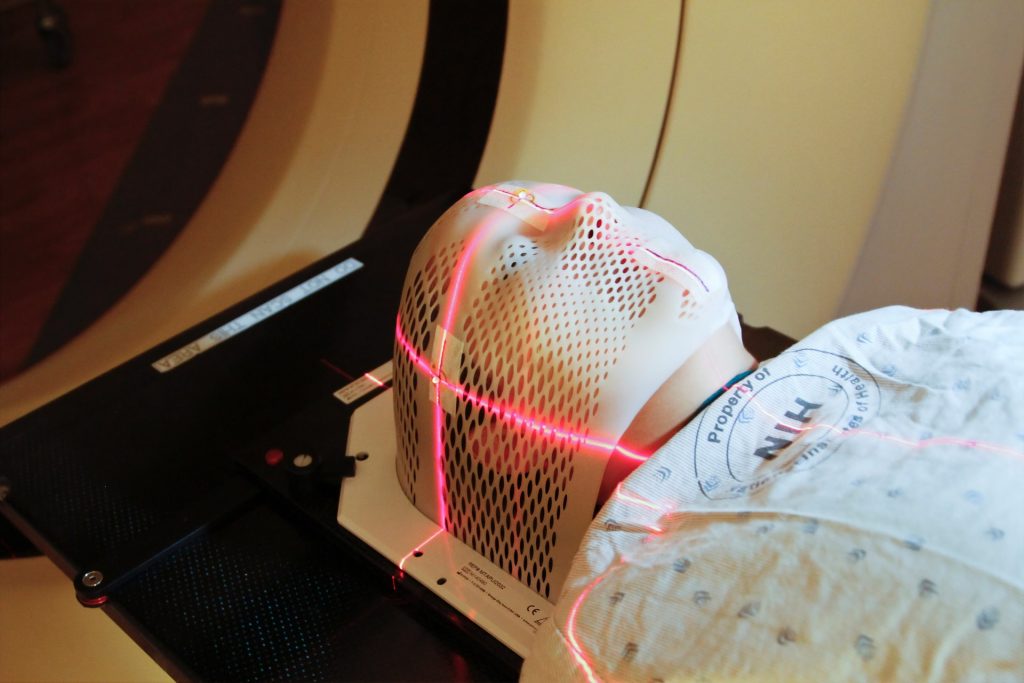
Men undergoing MRI-guided radiotherapy for localised prostate cancer, had fewer toxicities and better quality of life, according to new research published in JAMA Oncology. This was the first randomised phase III clinical trial to directly compare MRI-guided stereotactic body radiotherapy (SBRT) with the same therapy guided by CT.

“MRI guidance offers several advantages over standard CT guidance, most notably the ability to dramatically reduce planning margins, providing more focused treatment with less injury to nearby normal tissues and organs,” said lead author Amar Kishan, MD, a radiation oncologist. “MRI technology is more costly than CT, both in terms of upfront equipment expenses and longer treatment times, which is one reason our study set out to determine if MRI-guided technology offers tangible benefits for patients.”

SBRT for prostate cancer usually delivers radiation in five or fewer precisely targeted doses. It is an established and generally well tolerated form of treatment, but it can cause toxicities resulting in urinary, bowel and sexual dysfunction. This UCLA-led clinical trial included 154 analysable patients with prostate cancer who were randomised to either a CT-guidance arm (76 patients) or an MRI-guidance arm (78 patients).

This study included assessment by both physicians and patients. From both perspectives, MRI-guided therapy was associated with fewer side effects and better quality of life over at least three months of follow-up.

“In this trial, we demonstrated that the reduction in treatment volumes facilitated by MRI guidance leads to a significant reduction in moderate physician-scored toxicity and to a reduction in the proportion of patients noting significant decrements in patient-reported outcome metrics in the near term,” said Dr Kishan. “Although additional studies will need to confirm these benefits over time, we’re hopeful that these results will lead to better outcomes for men with prostate cancer.”

The 2mm margin used with MRI-guidance in the trial is narrower than has been used in any previous large study. Unlike CT, MRI technology can monitor prostate motion directly, and it offers improved soft tissue contrast, improving the accuracy of alignment prior to radiation.

Source: University of California – Los Angeles Health Sciences

Exercise Improves Quality of Life in Breast Cancer Radiotherapy

Tired woman after exercise
Photo by Ketut Subiyanto on Pexels

Radiotherapy is an important part of breast cancer treatment but can lead to cancer-related fatigue and negatively impact patients’ health-related quality of life. Fortunately, latest research by has revealed exercise may make radiotherapy more tolerable for patients, offering benefits for their emotional, physical and social wellbeing.

Researchers at Edith Cowan University (ECU) included 89 women in the study, with 43 completing a home-based 12-week program, consisting of a weekly exercise regime of one to two resistance training sessions and an accumulated 30–40 minutes of aerobic exercise. The 46 controls did not participate in the exercise regime.

The results, published in Breast Cancer, showed that patients who exercised recovered from cancer-related fatigue quicker during and after radiotherapy compared to the control group and saw a significant increase in health-related quality of life post radiotherapy with no reported adverse effects.

Study supervisor Professor Rob Newton said this showed home-based resistance and aerobic exercise during radiotherapy is safe, feasible and effective in accelerating recovery from cancer-related fatigue and improving health-related quality of life.

“A home-based protocol might be preferable for patients, as it is low-cost, does not require travel or in-person supervision and can be performed at a time and location of the patient’s choosing,” he said.

“These benefits may provide substantial comfort to patients.”

Important changes observed

Australia’s current national guidelines for cancer patients recommend moderately intense aerobic exercise for 30 minutes per day, five days a week, or vigorously intense aerobic exercise for 20 minutes a day for three days a week.

They also call for 8–10 strength-training exercises with 8–12 repetitions per exercise, for two-to-three days per week.

However, study lead Dr Georgios Mavropalias said benefits were still observed with less exercise.

“The amount of exercise was aimed to increase progressively, with the ultimate target of participants meeting the national guideline for recommended exercise levels,” he said.

“However, the exercise programmes were relative to the participants’ fitness capacity, and we found even much smaller dosages of exercise than those recommended in the national guidelines can have significant effects on cancer-related fatigue and health-related quality of living during and after radiotherapy.”

The study also found participants good adherence to exercise programmes once they started. The exercise group reported significant improvements in mild, moderate and vigorous physical activity up to 12 months after the supervised exercise programme finished.

“The exercise programme in this study seems to have induced changes in the participants’ behaviour around physical activity,” Dr Mavropalias said.

“Thus, apart from the direct beneficial effects on reduction in cancer-related fatigue and improving health-related quality of life during radiotherapy, home-based exercise protocols might result in changes in the physical activity of participants that persist well after the end of the program.”

Source: Edith Cowan University

Life Oncology Launches First AI-driven Ethos™ Radiotherapy Cancer Treatment in Sub-Saharan Africa

The Life Oncology unit at Life Vincent Pallotti Hospital today launched the Ethos™  radiotherapy system, a revolutionary new technology, the first in Sub-Saharan Africa, which uses artificial intelligence (AI) and adaptive treatment to adjust cancer treatments in response to patients’ unique, changing needs.

The Ethos™ radiotherapy system allows radiotherapy specialists to treat cancer patients with the most accurate, precision techniques. “Adaptive therapy provides the ability to adapt the treatment plan based on tumour and anatomical changes. The goal is to better target the tumour, reduce radiation dose of healthy tissue, and potentially improve overall outcomes”, says Dr Louis Kathan, Life Healthcare’s Chief Medical Officer in South Africa.

Dr Kathan, who is also a radiation oncologist with close to 15 years of patient treatment experience, explains that the streamlined workflow of Ethos™ radiotherapy is enabled by its AI-driven planning and contouring capabilities. “The system allows us to use AI to adapt to a patient’s anatomy. This allows us to make decisions more efficiently on a daily basis based on each patient’s individual needs. The ability of the machine to deliver on-couch adaptive treatment puts the patient at the centre of care”, he added.

“Clinicians globally have waited for the day when they have the ability to adapt radiotherapy treatments to changes in patient anatomy. Typically changes to a patient’s treatment plan due to changes in anatomy require time-consuming re-scanning and re-planning between treatment sessions, which could take up to two days. The Ethos™ radiotherapy adaptive technology allows the treatment plan to be adjusted daily, in real-time and the treatment to be planned and administered, all within 20 to 30 minutes”, says Kulthum Ismail, unit manager: radiation therapy at the Life Oncology unit based at Life Vincent Pallotti Hospital.

The planning and treatment all happen in one session, making it a faster process compared to existing radiotherapy technology, although Kulthum added that there is still a big role to play for current systems being used.

Kulthum Ismail, unit manager: radiation therapy at the Life Oncology unit based at Life Vincent Pallotti Hospital

The Life Oncology unit at Life Vincent Pallotti Hospital is committed to delivering the very best oncology care with substantial investments made in recent years to technologies to place the hospital at the forefront of advanced treatments in surgical-, medical-, gynaecological- and radiation oncology. These include:

  • The Novalis Tx delivery system
  • Image guided radiosurgery for tumour motion monitoring
  • Respiratory gating (respiration triggered dose delivery)
  • Minimally invasive frameless radiosurgery for patient comfort
  • Radiofrequency ablation
  • Elements Multiple Brain Mets Stereotactic Radiosurgery System which delivers powerful treatment that targets multiple brain metastases (more than one cancer lesion in the brain) simultaneously, faster and more precisely

The Ethos™ radiotherapy system at Life Vincent Pallotti Hospital is already being used to treat patients with cervical, rectum, lung and prostate cancer. In time, other cancers are hoped to be included in the treatment programme. 

“The Ethos™ radiotherapy system offers a faster and more personalised, targeted approach to radiotherapy treatment which means we really are placing the patient at the centre of care,” said Adam Pyle, Life Healthcare’s CEO for South Africa. “It’s our way of navigating our patients into the future of oncology care as we continue to live our purpose of making life better.”

Pete Wharton-Hood, Group Chief Executive for Life Healthcare added that the Group’s aspiration to be a leading cancer care provider has taken a substantial step forward with the expansion at the Life Oncology unit at Life Vincent Pallotti Hospital in Cape Town. “As a people and patient-centric organisation, we continuously seek ways to build on our existing technological offerings and services to consistently improve patient outcomes, enhance our patient experience and drive accessible and affordable healthcare. This means we get to better support our specialists who we partner with, so that together we continue delivering improved patient quality, patient experience, efficiency, and clinical excellence.”

A New Combined Therapy Extends Prostate Cancer Survival

Credit: Darryl Leja / National Human Genome Research Institute / National Institutes of Health

Practice-changing research published in The Lancet shows that a new combined therapy involving androgen deprivation therapy plus pelvic lymph node radiation kept nearly 90% of clinical trial patients’ prostate cancer at bay for five years.

The study also shows that patients with prostate cancer who didn’t receive androgen deprivation therapy — and who did not receive pelvic lymph node radiation — had a five-year survival of 70%.

“We can now confirm that pelvic lymph node treatment used together with androgen deprivation therapy, or even used as a stand-alone treatment option, greatly improves outcomes in patients with postoperative prostate cancer,” said Howard Sandler, MD, of Cedar Sinai Medical Center, senior author of the study. “These findings are an encouraging step forward, both for the medical community and for the patients and their loved ones seeking curative treatment options.”

The trial enrolled 1716 patients between March 31, 2008, and March 30, 2015. Enrollees were separated into three groups.

Group one received salvage prostate bed radiotherapy — a standard radiation targeted to the area in which the prostate used to exist before its surgical removal. Median five-year survival was 71% in these patients.

The second group received the standard radiation treatment, in combination with androgen deprivation therapy. Median five-year survival was 81%.

The third group received salvage prostate bed radiotherapy, androgen deprivation therapy and pelvic lymph node radiation. These patients had a five-year freedom from progression of just over 87%.

“The combined treatment approach proved to be the most beneficial approach,” said Dr Sandler.

Prostate cancer is the most common non-skin cancer in the US, affecting 1 in every 6 to 7 men. Though early warning signs are rare for prostate cancer, screening tests can catch it early. Diagnosis usually accompanies an elevated level of PSA, an acronym for prostate-specific antigen.

After prostatectomy, a man’s PSA level should be near zero. However, some men start to see their PSA levels rise several years after surgery. This is typically an indication that radiation therapy is needed.

Dr Sandler says men with postoperative prostate cancer can have excellent outcomes, especially if radiation is given early — when PSA levels are at their lowest — and in combination with proven therapies, as suggested in this new research.

Source: Cedars-Sinai Medical Center

Tissue-sparing Radiotherapy for Lung Cancer Brain Metastasis is Effective

MRI or CT machine
Photo by Mart Production on Pexels

A new study appearing in The Lancet Oncology suggests that a targeted radiation therapy is as effective as standard care for patients with lung cancer brain metastasis.

The findings suggests that patients could benefit from this targeted approach as it is known to have have fewer negative cognitive consequences.

In non-small-cell (NSLC) lung cancer, about 57% of patients present with metastatic disease, and 20% present with brain metastases. Brain metastasis is currently treated with whole brain radiation therapy, which targets the entire brain. While this approach treats even microscopic tumours, it results in memory problems and decreases cognitive function. The alternative, stereotactic radiosurgery, spares healthy brain tissue by precisely targeting the tumour, has been shown to have less severe cognitive consequences but has not yet been studied in patients with small cell lung cancer that has metastasised to the brain.

“For many years, it made sense to treat these patients with whole brain radiation because their survival was quite poor,” said Karolina Gaebe, a research student in Dr Sunit Das’s lab, who led the study.

“For them, long-term consequences of the treatment were not as crucial as reducing the impact of disease in the short-term. But now, as treatments for their lung cancer have improved, these patients are surviving much longer.”

The researchers set out to learn more after noticing patients with longer survival times were also living with severe cognitive impairments due to the treatments for their brain metastases. They wanted to understand whether a more targeted brain radiation regimen might be as beneficial for these patients, as has been demonstrated for most other cancer types.

As a first step, they undertook this meta-analysis, reviewing current literature to examine survival and brain outcomes following stereotactic radiosurgery for patients with small cell lung cancer that had spread to the brain. The team analysed data from 31 studies and included 18 130 patients, the largest cohort of small cell lung cancer patients with brain metastases to be studied so far.

The next steps are to conduct a large clinical trial to investigate cognitive outcome differences between stereotactic radiosurgery and whole brain radiation therapy for such patients.

“Because this is a meta-analysis, we can’t use this as absolute evidence that all patients should be treated in this way,” Dr Das said. “But essentially, this means that we need to challenge our standing worldwide paradigms for treating patients with this disease and revisit the idea that these patients should receive whole brain radiation therapy.”

Source: EurekAlert!

Radiation after Breast-conserving Surgery Reduces Cancer Recurrence

Source: National Cancer Institute

A long-term follow up analysis of a trial has shown that breast radiation following lumpectomy significantly reduces incidence of ipsilateral breast recurrence (IBR) for “low risk” DCIS.

NRG-RTOG 9804 is a clinical study conducted by the National Cancer Institute National Clinical Trials Network group NRG Oncology. These results were recently published in JCO.

IBR occurs at a rate of 5-10% after breast-conserving surgery. The NRG-RTOG 9804 study enrolled 636 women with a median age of 58 between December 1999 and July 2006. Patients were randomised to breast radiation (RT) or observation (OBS) treatment groups. All patients who participated in the trial underwent annual mammography and specified clinical exam intervals. For this analysis, the median follow up time was 13.9 years.

Analysis focused on the long-term cumulative incidence of IBR, the primary endpoint in the study. The study hypothesised that radiation would significantly reduce IBR from 6% to 3.5% at 5 years, assuming that the reduction in IBR from RT would be less than previous trials that included higher grades and larger sizes of DCIS. With long-term follow-up, cumulative incidence of IBR remained statistically significantly lower with RT, as compared to OBS. At 10 and 15 years, the cumulative incidence of IBR with OBS was 9.2% and 15.1%, respectively, and was 1.5% and 7.1%, respectively, with RT. The 10 and 15 year invasive IBR incidence, respectively, was 0.4% and 5.4%  with RT; 4.3%  and 9.5% with OBS. A total of 52 IBRs were observed; 14 in the RT arm and 38 in the OBS arm.

No statistically significant differences in mastectomy, distant metastasis, overall or disease-free survival were seen between the two treatment arms.

“Since IBR risk continues to increase through at least 15 years, with radiation conferring both a delay and decrease in this risk, the data presented support the decision to treat patients who wish to minimise their IBR and particularly the invasive cancer risk long term. Factors such as age, life expectancy, and willingness (if oestrogen receptor–positive) to take antioestrogen therapy should be taken into consideration in this patient-doctor shared decision,” stated lead author Beryl McCormick, MD, of the Memorial Sloan Kettering Cancer Center.

Source: NRG Oncology

FLASH Radiation Treatment for Tumours a Step Closer

Heavy ion bombardment in FLASH radiation treatment could be the future of radiotherapy, with encouraging findings from a German lab.

The GSI Helmholtzzentrum für Schwerionenforschung and the future accelerator centre FAIR succeeded in performing a carbon ion FLASH experiment for the first time in their Phase 0 experiment. 

The scientists involved were able to achieve the very high dose rates required to irradiate tumours. The success was a collective effort of the GSI Biophysics Department and the accelerator crew on the GSI/FAIR campus in close collaboration with the German Cancer Research Center DKFZ and the Heidelberg Ion Therapy (HIT) center.

FLASH irradiation involves utra-short and ultra-high radiation, delivering the treatment dose in fractions of a second. Traditional radiation therapy, as well as proton or ion therapy, deliver smaller doses of radiation to a patient over an extended time period, whereas FLASH radiotherapy is thought to require only a few short irradiations, all lasting less than 100 milliseconds.

In the field of electron radiation, recent in vivo investigations have shown that the FLASH method’s ultra-high dose rate is less harmful to healthy tissue, but just as efficient as conventional dose-rate radiation to inhibit tumour growth. Such an effect has not yet been demonstrated for proton and for ion beam irradiation, which is the basis of the tumour therapy with carbon ions developed at GSI. There is still a lot of research to be done here. The results of the current experiment at GSI are now being evaluated and will contribute to new knowledge.

There have also been technical barriers to FLASH radiation. Until now, FLASH technique has only been applicable using electron and proton accelerators. While the required dose rates for electrons and protons can be achieved with a cyclotron (circular accelerator), this is more difficult with the synchrotrons required in heavy ion therapy, such as the SIS18 at GSI.

That is why the current FAIR Phase 0 experiment is a very crucial step: Thanks to the improvements at the existing GSI accelerator facility as part of the preparations for FAIR, the necessary dose rate in millisecond range can now also be achieved for carbon ions. However,  much development work remains to be done before this procedure is mature enough to be routinely used on patients in the field of electron radiation.

Professor Marco Durante, Head of the GSI Biophysics Research Department, was very pleased with this important milestone in  the development of FLASH irradiation:

“It is a forward-looking method that could significantly increase the therapeutic window in radiotherapy. I am very happy that the researchers and the accelerator team were able to demonstrate the possibility to create conditions with carbon beams that are necessary for FLASH therapy of tumors. If we can combine the great effect and precision of heavy ion therapy with FLASH irradiation while maintaining efficacy and causing little damage to healthy tissue, this could pave the way of a future radiation therapy several years from now.”

Professor Paolo Giubellino, The Scientific Managing Director of GSI and FAIR, expressed his delight at the results: “The combination of expertise in biophysics and medicine as well as engineering excellence allows the first world-class experiments FLASH irradiation with ion beams to be performed. This could result in important complements to existing radiation therapies. Applications in tumour therapy are one of the research areas that can benefit from the recent increased intensities of GSI accelerators. However, modern radiobiology will substantially benefit from beams with even higher intensities, such as we will have at the FAIR facility currently under construction. FLASH is a first example of these future directions of work”.

Source: GSI Helmholtzzentrum für Schwerionenforschung GmbH

New Radiotherapy Treatment for Metastatic Cancers is Safe

MRI machine. Image by Michal Jarmoluk from Pixabay

A phase I trial showed that stereotactic body radiotherapy (SBRT) proved safe for treating cancer patients with multiple metastases. However, some late adverse effects underscored the need for long-term follow-up.

SBRT is a new radiotherapy treatment method that delivers high doses of ionising radiation to tumours with great precision, minimising damage to other parts of the body while killing tumour cells.

Increasingly used by clinicians, SBRT can improve survival in cancer patients with multiple metastases, explained first author Steve Chmura, MD, PhD, of University of Chicago Medicine, and colleagues, but there is little evidence as to its safety in this application.

“Existing data include mostly treatment of 1 or 2 metastases separated widely from each other and use of differing radiation doses, toxicity reporting, image guidance, and normal tissue constraints,” they wrote. “Given the critical need, NRG Oncology NRG-BR001 trial sought to determine the safety of delivering curative-intent SBRT to patients with 3 to 4 metastases or 2 metastases within close proximity to each other.”

Patients in the study had metastatic breast, prostate, or non-small-cell lung carcinoma (NSCLC). Each metastasis was assigned to a metastatic location based on the potential for toxicity.

Dose-limiting toxicity (DLT; side effects severe enough to discontinue treatment) was the primary study outcome, defined as specific AEs of grades 3 to 5 related to SBRT within 180 days of treatment. Dose levels were considered to be safe if DLTs were seen in no more than one of six patients per location with metastases.

Of the evaluable 35 patients, 12 (34.3%) had breast cancer, 10 NSCLC (28.6%), and 13 (37.1%) prostate cancer, with a median of three metastases per patient.

DLT analysis WAS based on six evaluable patients in all of the metastatic locations save the liver (five evaluable patients). The authors reported there were no protocol-specified DLTs in any of the seven metastatic locations within 180 days of the initiation of treatment.

There were 50 grade 3 or 4 AEs reported in 18 patients, and eight were deemed to be linked to the treatment. Of those eight, six (including bone pain, pulmonary fibrosis, bronchial fistula, bronchial stenosis, spinal fracture, and humeral fracture) were reported in six patients over 180 days from the start of the treatment. No treatment-related deaths occurred, according to the authors.

The authors suggested that, with the number of late AEs reported in this trial, patients should be monitored closely for late toxic effects.

“Given the potential for ablative radiotherapy to improve outcomes of patients with oligometastatic cancer, the finding that SBRT is safe when delivered to 3 to 4 metastases or 2 metastases in close proximity to one another is important, and serves as the foundation for ongoing randomized trials,” wrote the authors. They noted that these include studies such as the phase II/III NCI-sponsored NRG-BR002 trial.

Source: MedPage Today

Journal information: Chmura S, et al “Evaluation of safety of stereotactic body radiotherapy for the treatment of patients with multiple metastases” JAMA Oncol 2021; DOI: 10.1001/jamaoncol.2021.0687.

Tumour Weakened against Radiation by Tweaking a Certain Protein

The vulnerability of specific tissues to ionising radiation has been linked to the time-varying levels of a tumour-suppressing protein, opening new avenues for cancer combination therapy.

The ability for cells to survive radiation damage has been known to be connected to p53, but tissues with vastly different levels of p53 have been shown to have great differences in sensitivity. In the face of this apparent paradox, researchers from Blavatnik Institute at Harvard Medical School, Massachusetts General Hospital, and the Novartis Institutes for BioMedical Research investigated the behaviour of p53 in irradiated tissues.

“Dynamics matter. How things change over time matters,” said co-corresponding author Galit Lahav, the Novartis Professor of Systems Biology at HMS. “Our ability to understand biology is limited when we only look at snapshots. By seeing how things evolve temporally, we gain much richer information that can be critical for dissecting diseases and creating new therapies.”

Ionising radiation randomly damages a cell’s molecular machinery, causing it to initiate cell death if it is too serious. The arbitrator of cellular suicide is p53, which is also involved in tumour suppression. 

The findings opened new avenues for combination cancer therapies, as they discovered that administering a drug that blocks p53 levels from oscillating resulted in tumours in mice being more susceptible to radiation. 
Administering to the mice an experimental anti-cancer drug that inhibits MDM2, a protein which degrades p53, they forced p53 to stay elevated. The large intestine, which is normally radiation resistant, showed increased vulnerability.

Testing out this enhanced vulnerability on human tumours transplanted into mice, a significant tumour shrinkage was seen following radiation and then MDM2 inhibitor administration.

Co-corresponding author Galit Lahav, Novartis Professor of Systems Biology, Harvard Medical School explained: “By irradiating first, we force the cancer cells to activate p53, and by adding MDM2 inhibitor on top of that, we can keep p53 active longer. This combination has a much stronger effect than either alone.”

The findings showed the importance of understanding the role of p53 in cancer, the dynamic nature of which is not being looked at in studies testing MDM2. More research into the biological pathways of p53 is called for. 
“For a lab studying p53, cancer is always a major motivation. Our goal is to acquire knowledge to help develop better and more efficient therapies,” Lahav said. “Understanding how p53 behaves over time in different conditions is a critical piece of the puzzle.”

Source: News-Medical.Net

Journal information: Stewart-Ornstein, J., et al. (2021) p53 dynamics vary between tissues and are linked with radiation sensitivity. Nature Communications.doi.org/10.1038/s41467-021-21145-z