The Pupil as a Window into the Sleeping Brain

For the first time, researchers have been able to observe how the pupils react during sleep over a period of several hours. A look under the eyelids showed them that more happens in the brain during sleep than was previously assumed.
While eyes are typically closed in sleep, there is a flurry of activity taking place beneath the eyelids: a team of researchers, led by principal investigators Caroline Lustenberger, Sarah Meissner and Nicole Wenderoth from the Neural Control of Movement Lab at ETH Zurich, have observed that the size of the pupil fluctuates constantly during sleep. As they report in Nature Communications, sometimes it increases in size, sometimes it decreases; sometimes these changes occur within seconds, other times over the course of several minutes.
“These dynamics reflect the state of arousal, or the level of brain activation in regions that are responsible for sleep-wake regulation,” says Lustenberger. “These observations contradict the previous assumption that, essentially, the level of arousal during sleep is low.”
Instead, these fluctuations in pupil size show that even during sleep, the brain is constantly switching between a higher and lower level of activation. These new findings also confirm for humans what other research groups have recently discovered in studies on rodents, who also exhibit slow fluctuations in the activation level (known in the field as arousal).
New method for an old mystery
The regions of the brain which control the activation level are situated deep within the brainstem, making it previously difficult to directly measure these processes in humans during sleep. Existing methods are technically demanding and have not yet been established in this context. The ETH researchers’ study therefore relies on pupil measurements. Pupils are known to indicate the activation level when a person is awake. They can therefore be used as markers for the activity in regions situated deeper within the brain.
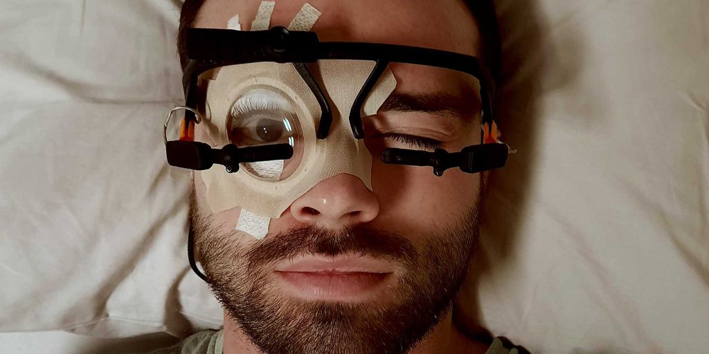
The ETH researchers developed a new method for examining the changes in people’s pupils while asleep: using a special adhesive technique and a transparent plaster, they were able to keep the eyes of the test subjects open for several hours.
“Our main concern was that the test subjects would be unable to sleep with their eyes open. But in a dark room, most people forget that their eyes are still open and they are able to sleep,” explains the study’s lead author, Manuel Carro Domínguez, who developed the technique.
Analysis of the data showed that pupil dynamics is related not just to the different stages of sleep, but also to specific patterns of brain activity, such as sleep spindles and pronounced deep sleep waves – brain waves that are important for memory consolidation and sleep stability. The researchers also discovered that the brain reacts to sounds with varying degrees of intensity, depending on the level of activation, which is reflected in the size of the pupil.
A central regulator of the activation level is a small region in the brainstem, known as the locus coeruleus. In animals, scientists have been able to show that this is important for the regulation of sleep stages and waking. The ETH researchers were unable to prove in this study whether the locus coeruleus is indeed directly responsible for pupil changes. “We are simply observing pupil changes that are related to the level of brain activation and heart activity,” Lustenberger explains.
In a follow-up study, the researchers will attempt to influence the activity of the locus coeruleus using medication, so that they can investigate how this affects pupil dynamics. They hope to discover whether this region of the brain is in fact responsible for controlling the pupils during sleep, and how changes in the level of activation affect sleep and its functions.
Using pupillary dynamics to diagnose illnesses
Understanding pupil dynamics during sleep could also provide important insights for the diagnosis and treatment of sleep disorders and other illnesses. The researchers therefore want to investigate whether pupil changes during sleep can provide indications of dysfunctions of the arousal system. These include disorders such as insomnia, post-traumatic stress disorder and possibly Alzheimer’s. “These are just hypotheses that we want to investigate in the future,” says Lustenberger.
Another goal is to make the technology usable outside of sleep laboratories, such as in hospitals where it could help to monitor waking in coma patients or to diagnose sleep disorders more accurately. The pupil as a window onto the brain could thus pave the way for new opportunities in sleep medicine and neuroscience.
Source: ETH Zurich


