New research published in Developmental Medicine & Child Neurology reveals that children born preterm are more likely to screen positive for autism than full-term children.
For the study, 9725 toddlers were screened at 15-, 18-, or 24-month well child visits using a test called the Modified Checklist for Autism in Toddlers, Revised.
Screening results that were positive for autism were most common among children born extremely preterm (51.35%) and least common among those born full-term (6.95%). Subsequent evaluations after positive screening revealed the following rates of autism diagnoses: 16.05% of extremely preterm, 2.00% of very preterm, 2.89% of moderately preterm, and 1.49% of full-term births.
Utilising the screening test at ages unadjusted for early birth was effective for identifying autism, as only a small number of preterm children (1.90%) who screened positive with the test did not receive a diagnosis of autism or other developmental delay following evaluation.
“With this research, we are hoping to help dissipate doubts that clinicians might have about the utility of screening for autism in toddlers born preterm,” said corresponding author Georgina Perez Liz, MD, of the AJ Drexel Autism Institute. “Low-cost, universal public health strategies such as screening can lead to less disparity in autism detection and help children on the spectrum start specific intervention and supports earlier in life.”
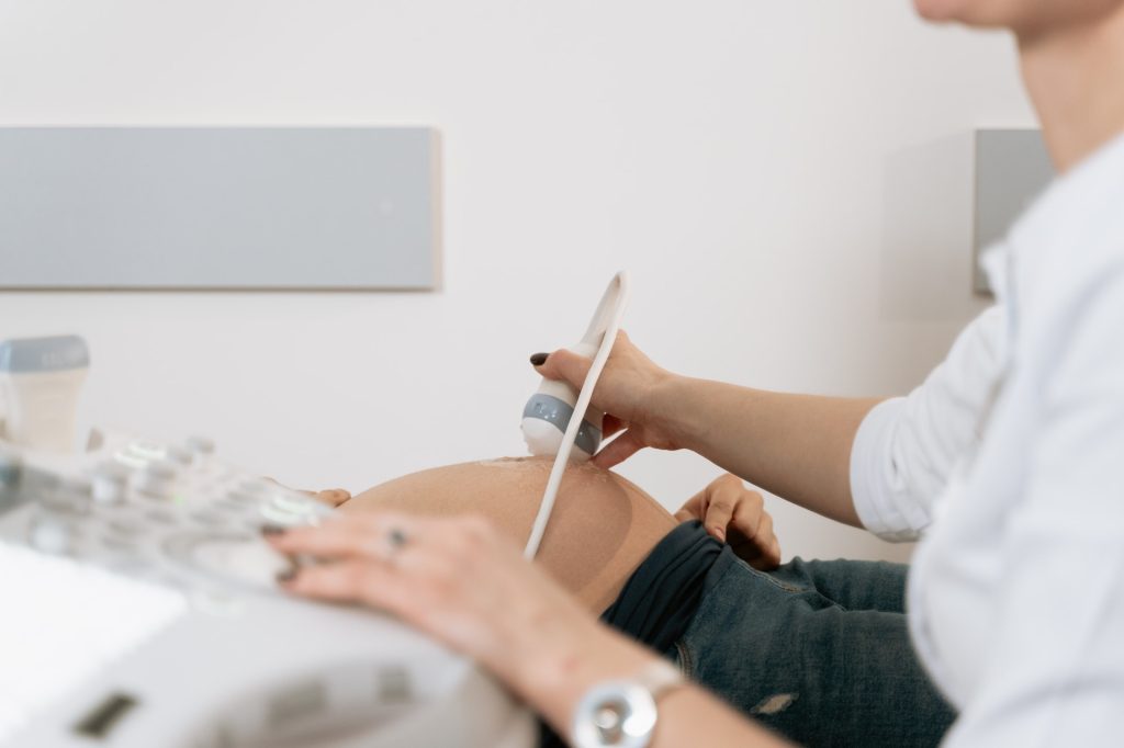
Researchers have developed a way to use ultrasound to estimate the risk of delivering a baby preterm. The new method measures microstructural changes in a woman’s cervix using quantitative ultrasound. The method works as early as 23 weeks into a pregnancy, according to the research, which is published in the American Journal of Obstetrics & Gynecology Maternal Fetal Medicine.
The current method for assessing a woman’s risk of preterm birth is based solely on whether she has previously given birth prematurely. This means there has been no way to assess risk in a first-time pregnancy.
“Today, clinicians wait for signs and symptoms of a preterm birth,” such as a ruptured membrane, explained lead author Barbara McFarlin, a professor emeritus of nursing at University of Illinois Chicago.
“Our technique would be helpful in making decisions based on the tissue and not just on symptoms.”
The new method is the result of more than 20 years of collaboration between researchers in nursing and engineering at UIC and University of Illinois Urbana-Champaign. In a study of 429 women who gave birth without induction at the University of Illinois Hospital, the new method was effective at predicting the risk of preterm births during first-time pregnancies.
And for women who were having a subsequent pregnancy, combing the data from quantitative ultrasound with the woman’s delivery history was more effective at assessing risk than just using her history.
The new approach differs from a traditional ultrasound where a picture is produced from the data received.
In quantitative ultrasound, a traditional ultrasound is performed but the radio frequency data itself is read and analysed to determine tissue characteristics.
The study is the culmination of a research partnership that began in 2001 when McFarlin was a nursing PhD student at UIC. Having previously worked as a nurse midwife and sonographer, she had noticed that there were differences in the appearance of the cervix in women who went on to deliver preterm.
She was interested in quantifying this and discovered that “no one was looking at it.”
She was put in touch with Bill O’Brien, a UIUC professor of electrical and computer engineering, who was studying ways to use quantitative ultrasound data in health research.
Together, over the past 22 years, they established that quantitative ultrasound could detect changes in the cervix and, as McFarlin had suspected long ago, that those changes help predict the risk of preterm delivery.
The preterm birth rate hovers around 10-15% of pregnancies, O’Brien said.
“That’s a very, very high percentage to not know what is happening,” he said.
If a clinician could know at 23 weeks that there was a risk of preterm birth, they would likely conduct extra appointments to keep an eye on the foetus, the researchers said.
But since there had previously been no routine way to assess preterm birth risk this early, there have been no studies to show what sort of interventions would be helpful in delaying labour.
This study, O’Brien explains, will allow other researchers to “start studying processes by which you might be able to prevent or delay preterm birth.”
A meta-analysis of studies published over the past 40 years on cannabis use during pregnancy has found an association between foetal exposure to cannabis in the womb and preterm delivery, low birth weight and the need for neonatal intensive care admission (NICU). The study was published today in the journal Addiction.
Previous research has indicated that THC, the main psychoactive component in cannabis, can cross the placenta to the foetus during pregnancy and bind to receptors in the foetal brain.
The meta-analysis examined the results of 57 studies around the world that included almost 13 million infants in total. Based on either self-reports from pregnant women, or blood and saliva testing depending on the study, just over 100 000 infants were found to be exposed to cannabis in the womb. While none of the studies found a direct causal relationship between cannabis use during pregnancy and adverse birth outcomes, the combined results indicated that newborns exposed to cannabis during pregnancy were twice as likely to require NICU admission, twice as likely to have a low birth rate and one and a half times more likely to be born early.
While there has been little research on cannabis use during pregnancy since cannabis was legalised in Canada five years ago, an American study has indicated an increase in cannabis use during pregnancy in states where it has been legalised and the perceived risk of harm from cannabis has decreased. The study states that overall cannabis use in pregnancy has doubled in the past 20 years, with approximately 10% of pregnancies associated with cannabis exposure. Some studies indicated it was being used to alleviate symptoms of nausea, poor appetite, insomnia or anxiety during pregnancy.
“This research emphasizes the importance of healthcare providers making an effort to create a safe space talking to pregnant women and women planning to be pregnant about their cannabis use and their motivations for using it to educate them about the potential risks and empower them to make informed decisions for their child,” says lead author Maryam Sorkhou, a PHD student within the addictions division at CAMH as well as the University of Toronto. Ms Sorkhou is overseen at CAMH by Senior Scientist and paper co-author Dr Tony George.
A treatment to move blood from the umbilical cord into an infant’s body may provide a safe option for preterm infants born after 28 weeks who need rapid support, suggests a study supported by the National Institutes of Health. The procedure, called umbilical cord milking, involves gently squeezing the cord between the thumb and forefinger and pushing the blood into the newborn’s abdomen.
The new findings suggest that concerns raised by a 2019 study of infants born before 28 weeks (which concluded that umbilical cord milking might increase the risk of bleeding inside the brain) do not apply to preterm infants born after 28 weeks. The current study appears in Pediatrics.
The standard procedure, delaying cord clamping while blood naturally flows into the infant’s body, takes 30 to 180 seconds. However, cord milking, takes about 20 seconds, reducing delay for infants who need immediate assistance, such as respiratory support. Both procedures allow for umbilical cord blood to reach the infant’s body before clamping, reducing the risk of anaemia and other complications seen among infants receiving immediate cord clamping and cutting.
The study was conducted by Anup Katheria, M.D., of the Sharp Mary Birch Hospital for Women & Newborns in San Diego, and colleagues in the United States, Canada and Europe. It was supported by NIH’s Eunice Kennedy Shriver National Institute of Child Health and Human Development.
More than 1000 infants were randomly assigned either to umbilical cord milking or delayed cord clamping. Rates of severe intraventricular haemorrhage and/or death did not differ significantly between the two groups (just over 1%). Moreover, the rates of overall intraventricular haemorrhage were also similar between the groups (approximately 12%). The researchers will follow all the infants in the study for two years to observe longer term outcomes.
A new study published in JAMA Pediatrics showed an association between being born preterm or large for gestational age and increased risks of atrial fibrillation later in life. Being small for gestational age at birth was only associated with an increased risk of atrial fibrillation up to the age of 18.
The incidence of atrial fibrillation in the young has increased over the past few decades, from low levels.
To date there have been little or mixed findings regarding the risk of atrial fibrillation in those with adverse birth outcomes. Atrial fibrillation increases the risk of stroke and other cardiovascular conditions, and is the most common form of cardiac arrhythmia. It mainly affects the middle-aged and the elderly. The estimated incidence in the young is low, 0.12 to 0.16%.
Low incidence in the young
A collaborative study involving researchers from Karolinska Institutet has now investigated the risk of atrial fibrillation according to preterm birth and foetal growth.
“Atrial fibrillation at a young age may involve a heavy socioeconomic burden for the affected individuals and we need to learn more about the underlying causes of the disease,” says first author Fen Yang, doctoral student at Karolinska Institutet. “Our findings may highlight the need to monitor and prevent the disease in individuals with an elevated risk of atrial fibrillation.”
“We found that individuals born preterm and those who were large for gestational age at birth had a slightly higher risk of developing atrial fibrillation up to middle-age than those with corresponding normal birth outcomes,” says principal investigator Krisztina László, associate professor at the Department of Global Public Health, Karolinska Institutet, and senior lecturer at the Department of Public Health and Caring Sciences at Uppsala University. “Individuals who were small for gestational age at birth had an increased risk of atrial fibrillation up to the age of 18, but not later in adulthood.”
The risk increase was 30% for individuals born preterm, 55% for individuals who were large at birth and 71% for individuals who were both preterm and large for gestational age at birth.
Eight million participants
The results of the study are based on statistical analyses of over eight million births from Danish (1978–2016), Finnish (1987–2014) and Swedish (1973–2014) medical birth registries who were followed for incident atrial fibrillation in the national patient and cause of death registries up to 2021. The results were compared with siblings in the same families. Since the study was observational, no causal relationships could be ascertained.
The researchers say that future studies may investigate the association between preterm birth, foetal growth, and the risk of atrial fibrillation up to old age.
The immune systems of preterm babies are especially weak, making them more vulnerable to infection. A new study published in JCI Insight suggests that this vulnerability instead stems from an immune signalling pathway being suppressed, perhaps due to a requirement for it for successful foetal development in utero.
The earlier babies are born, the higher the risk of life-threatening complications. Infections can lead to sepsis and are among the most frequent causes of death.
“In the case of very prematurely born infants, a bacterial infection can lead to death within hours,” says LMU physician Prof Markus Sperandio. The physiologist and former paediatrician and neonatologist researches the causes of this high susceptibility to infection together with his team at LMU’s Biomedical Center Munich. Now the researchers have demonstrated that an immunostimulatory signalling pathway is suppressed in the developing immune system.
In preterm infants, neutrophils ‘turned off‘
Sperandio had already shown in earlier studies that, in the foetus and in newborns, neutrophils do not work as in adults. Unlike in adults, foetal and neonatal neutrophils do not manage to sufficiently attach to the walls of blood vessels and extravasate into inflamed tissue. This is necessary, however, to trigger an inflammatory response and thus initiate immune defence.
Now the LMU researchers, working in collaboration with the Children and Women’s Clinic at University of Munich Hospital, have investigated which mechanisms are behind this immaturity. By means of a so-called transcriptomic analysis, they compared the gene activity of neutrophils in umbilical cord blood of premature and full-term babies with adult neutrophils. Compared to adults, there is a lot of gene activity in premature and full-term infants that counteracts immune defence. “In this case, these neutrophils act as if they were switched off,” says Sperandio.
Balance shift of immunoregulatory signalling pathways
This particularly affects signals transmitted via the NF-κB signalling pathway, which plays a decisive role in immune and inflammatory responses. It consists of two possible pathways for signals: one that promotes inflammation and one that can suppress it. Therefore, the activity of these two pathways must be finely balanced for proper regulation of the immune response.
“Our experiments have shown that this balance is shifted towards the anti-inflammatory pathway in foetal and neonatal neutrophils,” says Sperandio. “Whereas this regulation of neutrophil function is clearly a requirement for normal foetal growth in utero, it leads to immune defence problems in prematurely born infants who have to adapt ‘too soon’ to the world outside the uterus.” To what extent these findings will be a springboard for new therapeutic approaches in the future remains to be seen. “Due to the complex processes in the growing foetal and neonatal organism, maturation-adapted therapies are conceivable but remain a long way off at this stage,” says Sperandio.
Chemicals that accumulate in the vagina, potentially originating from personal care products, may contribute to spontaneous preterm birth, according to a new study appearing in Nature Microbiology.
Columbia University Vagelos College of Physicians and Surgeons researchers performed a study of 232 pregnant women and found that a few non-biological chemicals previously found in cosmetics and hygiene products are strongly associated with preterm birth.
“Our findings suggest that we need to look more closely at whether common environmental exposures are in fact causing preterm births and, if so, where these exposures are coming from,” says study co-leader Tal Korem, PhD, assistant professor at Columbia University. “The good news is that if these chemicals are to blame, it may be possible to limit these potentially harmful exposures.”
Preterm birth, childbirth before 37 weeks of pregnancy, is the number one cause of neonatal death and can lead to a variety of lifelong health issues. Two-thirds of preterm births occur spontaneously, but despite extensive research, there are no methods for predicting or preventing spontaneous preterm birth.
Several studies have suggested that imbalances in the vaginal microbiome play a role in preterm birth and other problems during pregnancy. However, researchers have not been able to reproducibly link specific populations of microorganisms with adverse pregnancy outcomes.
The research team opted to take a more expansive view of the vaginal microenvironment by looking at its metabolome – the complete set of small molecules found in a particular biological niche, including metabolites produced by local cells and microorganisms and molecules from external sources. “The metabolome can be seen as a functional readout of the ecosystem as a whole,” Korem says. “Microbiome profiling can tell us who the microbes are; metabolomics gets us close to understanding what the microbes are doing.”
In the current study, the researchers measured over 700 different metabolites in the second-trimester metabolome of 232 pregnant women, including 80 pregnancies that ended prematurely.
The study found multiple metabolites that were significantly higher in women who had delivered early than in those who delivered at full term.
“Several of these metabolites are chemicals that are not produced by humans or microbes – what we call xenobiotics,” says Korem. “These include diethanolamine, ethyl-beta glucoside, tartrate, and ethylenediaminetetraacetic acid. While we did not identify the source of these xenobiotics in our participants, all could be found in cosmetics and hygiene products.”
Algorithm predicts preterm birth
Using machine learning models, the team also developed an algorithm based on metabolite levels that can predict preterm birth with good accuracy, potentially paving the way for early diagnostics.
Though the predictions were more accurate than models based on microbiome data and maternal characteristics (such as age, BMI, race, preterm birth history, and prior births), the new model still needs improvement and further validation before it could be used in the clinic.
Despite the current limitations, Korem says, “our results demonstrate that vaginal metabolites have the potential to predict, months in advance, which women are likely to deliver early.”
WHO today launched new guidelines to improve survival and health outcomes for babies born preterm (< 37 weeks) or small (< 2.5kg). In a significant departure from common clinical practice, the guidelines advise that caregiver skin to skin contact with a caregiver – aka kangaroo mother care – should start immediately after birth, without incubator stabilisation. This reflects the immense health benefits of ensuring caregivers and their preterm babies can stay close, without being separated, after birth.
The guidelines also provide recommendations to ensure emotional, financial and workplace support for families of very small and preterm babies, who can face extraordinary stress and hardship because of intensive caregiving demands and anxieties around their babies’ health.
“Preterm babies can survive, thrive, and change the world – but each baby must be given that chance,” said Dr Tedros Adhanom Ghebreyesus, WHO Director-General. “These guidelines show that improving outcomes for these tiny babies is not always about providing the most high-tech solutions, but rather ensuring access to essential healthcare that is centred around the needs of families.”
Depending on where they are born, there remain significant disparities in a preterm baby’s chances of surviving. While most born at or after 28 weeks in high-income countries go on to survive, in poorer countries survival rates can be as low as 10%.
Most preterm babies can be saved through feasible, cost-effective measures including quality care before, during and after childbirth, prevention and management of common infections, and kangaroo mother care – combining skin to skin contact in a special sling or wrap for as many hours as possible with a primary caregiver, usually the mother, and exclusive breastfeeding.
Previous recommendations for preterm babies were for an initial period of separation from their primary caregiver, with 3–7 days of initial stabilisation in an incubator or warmer. However, research has now shown that starting kangaroo mother care immediately after birth reduces mortality, infections and hypothermia, and improves feeding.
Breastfeeding is also strongly recommended to improve health outcomes for preterm and low birthweight babies, with evidence showing it reduces infection risks compared to infant formula. Where mother’s milk is not available, donor human milk is the best alternative, though fortified ‘preterm formula’ may be used if there are no donor milk banks.
Integrating feedback from families gathered through over 200 studies, the guidelines also advocate for increased emotional and financial support for caregivers. Parental leave is needed to help families care for the infant, the guidelines state, while government and regulatory policies and entitlements should ensure families of preterm and low birthweight babies receive sufficient financial and workplace support.
Earlier this year, WHO released related recommendations on antenatal treatments for women with a high likelihood of a preterm birth. These include antenatal corticosteroids, which can prevent breathing difficulties and reduce health risks for preterm babies, as well as tocolytic treatments to delay labour and allow time for a course of corticosteroids to be completed. Together, these are the first updates to WHO’s preterm and low birth weight guidelines since 2015.
The guidelines were released ahead of World Prematurity Day, which is marked every year on 17th November.
Inflammatory bowel disease is a risk factor for giving birth preterm even when in apparent disease remission, according to a study published in the journal eClinicalMedicine. If corroborated, the results may eventually affect recommendations for women with ulcerative colitis wishing to conceive.
Inflammatory bowel disease (IBD) is chronic inflammatory disease with a prevalence of approximately 0.5%. IBD, which includes ulcerative colitis and Crohn’s disease, and – unlike irritable bowel syndrome (IBS) – causes visible damage to the mucosa lining the intestines. IBD is characteristic for its recurrent tendency for symptoms to relapse, followed by periods of remission.
Onset of IBD commonly occurs at age 15–30, so questions about its impact on pregnancy and the foetus are common. IBD has previously been linked to negative birth outcomes, such as preterm birth (< 37 weeks of pregnancy), mainly in women showing signs of active disease.
Also, women without obvious IBD activity often have microscopic inflammation in the intestinal mucosa. Until now, however, it has been unknown whether even microscopic inflammation may be associated with risks in pregnancy.
Higher risk of preterm birth
The present study, shows that microscopic inflammation in IBD, especially ulcerative colitis, is linked to an elevated risk of giving birth prematurely.
Among babies born to women with microscopic inflammation due to IBD, 9.6% were preterm, while 6.5% of children were born preterm to women without microscopic inflammation of IBD. This corresponds to a relative risk increase of 46 percent. Microscopic inflammation was not clearly associated with other adverse pregnancy outcomes, such as growth restriction.
The results are based on register data on women in Sweden, diagnosed with IBD in 1990–2016, in whom information was available on the microscopic appearance of the intestine shortly before pregnancy. The study included 1223 children of women with microscopic IBD inflammation of the intestine and 630 children of women with IBD but with microscopically healed intestinal mucosa.
Through register linkages, data were also retrieved from several national health registers, such as the Swedish Medical Birth Register and the Swedish Quality Register for Inflammatory Bowel Desiease (SWIBREG).
Prospectof new treatment targets
“Our results suggest that IBD treatment aimed at not merely alleviate symptoms of IBD, but also microscopically heal the intestine, can reduce the risk of giving birth preterm,” sayd first author and corresponding author is Karl Mårild, associate professor of paediatrics at Sahlgrenska Academy, University of Gothenburg. “If our results hold up in future studies, they may therefore be the basis for recommendations to confirm microscopic healing before pregnancy, to reduce such risks.”
“Even a modestly increased relative risk of preterm birth is important, given that preterm birth can greatly affect the child’s health in both the short and the long term. Preterm birth is still one of the most common causes of death for children under the age of five in Sweden,” Karl Mårild concludes.
A serious neonatal health threat, preterm labour has long mystified researchers – and how does the uterus normally stay dormant, letting it stretch and expand during the 40 weeks it takes a foetus to grow? New research published in The Journal of Physiology suggests that a protein called Piezo1 keeps the uterus relaxed throughout gestation.
Preterm birth is a major cause of neonatal mortality and morbidity. The identification of Piezo1 in the uterus, and its role to maintain relaxation of uterus through stretch-activation during pregnancy, paves the way for drugs and therapies to be developed that could one day treat or delay preterm labour.
The muscular outer layer of the uterus is peculiar because it is the only muscle that it is not regulated by nerves and it must remain dormant for the 40 weeks despite significant expansion and stretch as the foetus develops into a baby. The researchers from University of Nevada USA studied tissue samples of the smooth muscle of the uterus to explore the mechanistic pathways to better understand the dynamics controlling the uterus, how pregnancy is maintained and what maintains quiescence until labour.
Stretching the uterus tissue, to mimic what happens during pregnancy, activates Piezo1 channels. This drives the flow of calcium molecules generating a signalling cascade that activates the enzyme nitric oxide synthase to produce the molecule nitric oxide. This Piezo1 cascade promotes and maintains the dormant state of the uterus.
Piezo1 controls the uterus by working in a dose-dependent manner, where channel activity is stimulated by the chemical Yoda1 and inhibited by a chemical called Dooku1 (Star Wars fans will no doubt recognise the inspiration behind these two names). When Piezo1 is upregulated, the uterus remains in a relaxed state. However, in preterm tissue, the expression of Piezo1 is significantly downregulated, ‘switching off’ the dormant signalling to the muscle, so the uterus contracts and initiates labour.
Professor Iain Buxton at the Myometrial Research Group at the University of Nevada said: “Pregnancy is the most impressive example of a human muscle enduring mechanical stress for a prolonged period. Finding Piezo1 in the muscular layer of the uterus means the uterus is controlled locally and is coordinated by a stretch-activated mechanism rather than hormonal influence from the ovaries or the placenta, which has been the assumption.
“It is troubling that there are still no drugs available to stop preterm labour. Thanks to the Nobel Prize winning discovery of Piezo proteins, which are responsible for how the body responds to mechanical force, and our investigation we are now closer to developing a treatment. Piezo1 and its relaxation mechanism provide a target for us which we could potentially activate with drugs. We need to test this with further studies and we hope to carry out clinical trials in the future.”
Contraction and relaxation were assessed in tissue samples compared for the following gestational periods: non-pregnant, term non-labouring, term labouring, preterm non-labouring and preterm labouring. The presence of Piezo1 channels was discovered using molecular tools while pregnant tissues contracting in a muscle bath were stimulated with Piezo1 channel activator and inhibitor to characterize the regulation of quiescence.
More research is needed to understand just how all the molecular signals and steps involved in the Piezo1 channel regulate uterus relaxation, and to identify other chemicals that may be involved.