Tag: platelets

Using Chilled Platelets Could Safely Expand Life-Saving Supply to Bleeding Patients
Photo by Marcelo Leal on Unsplash

In a thoughtfully designed, adaptive clinical trial, University of Pittsburgh School of Medicine physician-scientists demonstrated that the United States could triple to quadruple the supply of platelets available to help save bleeding patients of all ages and extend the life-saving therapy into rural regions and community hospitals. Platelets are the component of blood that encourage clotting, plugging cuts and tears in blood vessels and stemming blood loss. 

Results from the Chilled Platelet Study (CHIPS) trial were announced Aug 17 in JAMA and under consideration by the US Food and Drug Administration and multiple other countries to revise regulations for use of refrigerated platelets stored for up to 21 days in actively bleeding patients. 

Of the 2.5 million room-temperature platelet units collected per year in the United States, roughly 10% to 20% are wasted due to the short shelf life, costing hospitals $300 million, according to recent research in the journal Hematology

“Our findings could result in major international public health benefits,” said lead author Philip Spinella, professor of surgery and of critical care medicine in Pitt’s School of Medicine and codirector of Pitt’s Trauma and Transfusion Medicine Research Center. “This is incredibly transformative and will dramatically improve the availability of platelets while also reducing waste, allowing hospitals that aren’t in major cities to afford to keep this life-saving blood product on hand for bleeding patients.” 

Currently, donated platelets are conventionally stored at room temperature with a five- to seven-day shelf life. Early studies conducted half a century ago in healthy volunteers led regulators to believe that refrigerating platelets to extend their shelf life made them less effective.  

Recent preclinical studies indicated the opposite, though, so Spinella and colleagues designed CHIPS to safely test using refrigerated platelets stored for increasing amounts of time up to 21 days. The trial enrolled 1000 paediatric and adult patients undergoing cardiac surgery at 27 sites in the United States and Australia from December 2021 to March 2025.  

The patients were randomised to receive either standard, room-temperature platelets stored for less than seven days or chilled platelets. At every 200 participants treated, independent data analysts looked at the results. If the chilled platelets were performing just as well as room temperature, they’d add up to another five days to the age of the chilled platelets used and enrol another 200 participants, up to a maximum of 21 days.  

The research team discovered that platelets refrigerated for up to three weeks were just as effective at treating bleeding as room-temperature platelets stored for up to one week. Since it can take two days for blood banks to process donations, the finding holds the potential to extend the lifespan of platelets for use in controlling bleeding by almost four-fold. 

“Rural and community hospitals, for the most part, simply cannot justify keeping room temperature platelets in stock – they don’t see enough severely bleeding patients, so the waste would far exceed the benefit, something our nation’s fragile blood supply chain cannot accommodate,” said coauthor Michael Boisen, cardiothoracic anaesthesiologist and medical director of the UPMC Patient Blood Management Program, who served as principal investigator for UPMC’s CHIPS trial site. “So, if we can safely extend the shelf life of platelets and reduce the waste, imagine how many more people we could help.” 

More than 97% of platelets are donated through apheresis, where a donor’s blood runs from a tube in their arm to a machine that collects the platelets and returns the rest of the blood back to the donor–a time-consuming process. The age of platelet donors has steadily increased over the past decade and younger donors are not replacing the aging donor base fast enough to keep up with demand, according to data from Vitalant, which supplied the platelets for the study and is one of the nation’s largest nonprofit blood and biotherapies services providers.  

“About 15% of hospitals can experience one or more delayed platelet transfusions within any given month due to supply shortages,” said Ralph Vassallo, Vitalant’s chief medical and scientific officer. “A 21-day shelf life for cold-stored platelets will reduce outdate rates and ensure platelets are available on hospital shelves when a surge in need occurs.” 

In addition to the civilian applications, the findings could also benefit military personnel by making it more feasible to have platelets available in conflict areas, said Spinella, who is also associate medical director of Pitt’s Center for Military Medicine Research

A unique facet of the trial is that it included children and babies from the start. Usually, clinical trials that aren’t specific to paediatric populations will exclude children. 

“I believe it is unethical not to include children in a trial when there isn’t a rational biological reason to exclude them,” Spinella said. “Of the 1,000 participants in our trial, roughly a third were children—which makes sense because it is the very young who bleed the most from cardiac surgery and most frequently benefit from platelets. Our trial will hopefully motivate others to include children in their trials.” 

Additional authors are listed in the JAMA article.  
Source: University of Pittsburgh School of Medicine

Synthetic Platelets Stop Bleeding and Promote Healing in Animal Models

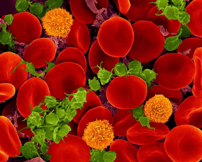
Scanning electron micrograph of red blood cells, T cells (orange) and platelets (green). Source: Wikimedia CC0

Researchers have developed hydrogel-based synthetic platelets that can be used to stop bleeding and, in animal models, has been shown to enhance healing at an injury site. The research is presented in Science Translational Medicine.

A number of medical situations require platelet transfusions – such as cases of severe bleeding, or for patients who are going into surgery or receiving chemotherapy. Currently, patients in any of those situations receive platelets harvested from blood donors, ideally from donors with a compatible blood type. This is challenging, because there is a very limited supply of platelets available, those platelets have a limited shelf life, and the platelets must be stored under controlled conditions.

“We’ve developed synthetic platelets that can be used with patients of any blood type and are engineered to go directly to the site of injury and promote healing,” says Ashley Brown, corresponding author of a paper on the synthetic platelets and biomedical engineering programme. “The synthetic platelets are also easy to store and transport, making it possible to give the synthetic platelets to patients in clinical situations sooner – such as in an ambulance or on the battlefield.”

The synthetic platelets are made of hydrogel nanoparticles that mimic the size, shape and mechanical properties of human platelets. Hydrogels are water-based gels that are composed of water and a small proportion of polymer molecules.

“Our synthetic platelets are deformable – meaning they can change shape – in the same way that normal platelets are,” Brown explains.

The researchers engineered the surface of the synthetic platelets to incorporate antibody fragments that bind to a protein called fibrin. When a body is injured, it synthesises fibrin at the site of the wound. The fibrin then forms a mesh-like substance to promote clotting.

“Because the synthetic platelets are coated with these antibody fragments, the synthetic platelets travel freely through the blood stream until they reach the wound site,” Brown says. “Once there, the antibody fragments bind to the fibrin, and the synthetic platelets expedite the clotting process.”

In addition to forming a clot within the fibrin network, the synthetic platelets act to contract the clot over time – just like normal platelets.

“This expedites the process of healing, allowing the body to move forward with tissue repair and recovery,” Brown says.

The researchers initially demonstrated the efficacy of the antibody fragments via in vitro testing, as well as demonstrating that the antibody fragments and synthetic platelets could be produced at scales that would make them viable for large-scale manufacturing.

The researchers then used a mouse model to determine the optimal dose of synthetic platelets necessary to stop bleeding.

Subsequent research in both mouse and pig models demonstrated that the synthetic platelets travelled to the site of a wound, expedited clotting, did not cause any clotting problems in areas outside of the wound, and accelerated healing.

“In the mouse and pig models, healing rates were comparable in animals that received platelet transfusions and synthetic platelet transfusions,” Brown says. “And both groups fared better than animals that did not receive either transfusion. We also found that the animals in both mouse and pig models were able to safely clear the synthetic platelets over time through normal kidney function. We didn’t see any adverse health effects associated with the use of the synthetic platelets.

“In addition, based on our preliminary estimates, we anticipate that the cost of the synthetic platelets – if they are approved for clinical use – would be comparable to the current cost of platelets,” Brown says.

“We are wrapping up preclinical efficacy testing and are in the process of securing funding for preclinical safety work that should allow us to obtain FDA approval to begin clinical trials within two years.”

Source: North Carolina State University

Could a Simple Dietary Change Increase Platelet Counts?

Scanning electron micrograph of red blood cells, T cells (orange) and platelets (green). Source: Wikimedia CC0

Aside from transfusions, there currently is no way to boost people’s platelet counts, which can drop for reasons such as chemotherapy, leaving them at risk for uncontrolled bleeding. But new research published in Nature Cardiovascular Research suggests that there could be a simple alternative: a dietary change in type of fat intake could raise platelet counts in people with low levels.

A study led by Kellie Machlus, PhD, and Maria Barrachina, PhD at Boston Children’s Hospital found that they could raise platelet counts in mice by feeding them polyunsaturated fatty acids (PUFAs) like those found in the Mediterranean diet. In contrast, mice fed a diet high in saturated fatty acids had decreased platelet counts.

“We were honestly surprised at how profound the effects were,” says Machlus, whose lab focuses on studying platelets and their precursor cells, megakaryocytes, and ways to get the body to increase platelet production.

But equally interesting is the apparent reason for the dietary effect.

“What brought me to the idea of diet is that megakaryocytes make these long extensions from their membrane when they form platelets,” Machlus says. “We thought the membrane must have an unusual composition to make it so fluid.”

A fluid megakaryocyte membrane

No one had studied megakaryocyte membranes before, perhaps because megakaryocytes are in the bone marrow and hard to access. Machlus, Barrachina, and their colleagues decided to comprehensively assess the membranes’ fat content with lipidomics.

“We found that PUFAs are enriched in megakaryocytes, especially right before they begin making platelets,” says Machlus. “We think they provide the fluidity necessary for the membrane to move and reshape.”

In culture, the megakaryocytes with higher amounts of PUFAs in their membrane made more platelets. When the cells were instead supplied with saturated fats as their lipid source, platelet production declined. The same thing happened when the team added compounds to inhibit uptake of PUFAs from the blood.

The researchers also identified one of the receptors on megakaryocytes that’s responsible for taking up PUFAs from blood: CD36. When they deleted the gene for CD36 in their mouse model, the animals had low platelet counts.

Serendipitously, the researchers were able to connect the dots to humans. Through a colleague in the U.K., they identified a family in which several members had a mutation in the CD36 gene. Those affected had low platelet counts and, in the mother’s case, bleeding episodes.

An olive oil intervention?

Intrigued by their findings, Barrachina hopes to extend the study by collaborating with a team in her native Spain. The team is studying dietary interventions for cardiovascular disease, including the Mediterranean diet.

“We want to look at platelets from these patients,” she says. She thinks that platelets with more saturated fatty acids in their membranes might be in a more activated state that could lead them to aggregate and form blood clots.

While Machlus thinks it may be worth encouraging patients with thrombocytopenia to consume more olive oil to increase PUFA levels, she recognises that a drug treatment may be more practical.

“Our next steps are to find out the enzymes that create PUFAs,” she says. “Maybe we can target them to make more platelets.”

Source: Boston Children’s Hospital

Platelets Get to Work Right Away on Wound Sites

Scanning electron microscope image of red blood cells, platelets (green) and T cells (orange). . Image courtesy of Dennis Kunkel, Dennis Kunkel Microscopy, Inc. Part of the exhibit Life:Magnified by ASCB and NIGMS.

New research shows that platelets at a wound site can sense where they are within a blood clot they are and that they can remodel their surroundings accordingly.

Platelets are key to initiating wound healing and the formation of blood clots (thrombus). Fibroblasts are connective tissue cells that are essential for the later stages of wound healing. Fibroblasts invade the clot that has been formed and produce vital proteins, including fibronectin, that then form a structural framework to build the new tissue needed to heal.

This new study, published in Science Advances, indicates that platelets can also form a provisional fibronectin matrix in their surroundings, similar to what fibroblasts do in the later stages of wound healing. This has potential implications for how the integrity of blood clots might be maintained during vascular repair.

Commenting on the discovery, lead author Dr Ingmar Schoen said: “We have identified an additional unexpected role for the most prominent platelet adhesion receptor. Our results show that platelets not only form the clot but also can initiate its remodelling by erecting a fibrous scaffold. This finding challenges some existing paradigms in the field of wound healing, which is dominated by research on fibroblasts.”

The researchers made use of super-resolution microscopy, a powerful imaging technique which enables much finer resolution of structures inside or around cells in vitro. To develop this finding further, in vivo observation of this platelet behaviour will be necessary.

“Without super-resolution microscopy, this discovery would not have been possible,” Dr Schoen noted.

Source: RCSI University

Platelet Signalling Implicated in COVID Organ Damage

Source: Fusion Medical Animation on Unsplash

New research finds that one cause of organ damage in COVID patients is abnormal crosstalk between blood platelets and cells lining blood vessels.

The study published in Science Advances, revealed the protein signals released by platelets cause inflammation, abnormal clotting, and damage to vessels when exposed to SARS-CoV-2.

The work identified two related genes, S1000A8 and S1000A9, which are turned up in the platelets of COVID patients, causing them to make more of myeloid-related proteins (MRP) 8 and 14. Higher levels of the dual proteins were linked in the study to higher levels of clotting and inflammation in vessels and worse outcomes.

In support of the theory that platelets are at the core of blood vessel damage in COVID, the research team also presented evidence that approved medications known to block platelet activation via the platelet surface protein P2Y12 (clopidogrel or ticagrelor) reduced COVID-related inflammation in vessels. The study also found that COVID-exposed platelets change cells lining blood vessels (endothelial cells) largely through a protein called p-selectin, which makes platelets stickier and more likely to form clots.

“Our findings reveal a new role for platelets in COVID blood vessel damage, and may explain in large part what makes the COVID virus so much more deadly than its relatives that cause the common cold,” said corresponding author Tessa Barrett, PhD,.

Abnormal, body-wide inflammation and blood clotting were identified early in the pandemic as central features of severe COVID-19, with the two thought to be interrelated, say the study authors. As blood components that react to injuries in vessels by triggering inflammation, and by becoming sticky to clump together in clots, platelets are a possible culprit. Increasing evidence shows that interplay between platelets and endothelial cells may be important to these disease mechanisms.

For the current study, endothelial cells from small blood vessels were exposed to fluid released from the platelets of either COVID patients or healthy controls. RNA was then sequenced, In the presence of COVID-activated platelets, changes were observed in the activity of the exposed endothelial cells. Genes expressed differently in COVID-19 were linked to clotting, inflammation, and the weakening of junctions between endothelial cells, which lets blood serum seep into tissue to cause the pulmonary oedema seen in severe cases, where patients’ lungs fill with fluid.

The large list was narrowed down to S100A8 and S100A9, which coded for the building of MRP 8 and 14. COVID in patients was found to increase the amount of MRP8/14 produced by platelets and other cells by 166 percent compared to controls. Higher levels of these proteins were linked to abnormal thrombosis, inflammation, and critical illness among hospitalised COVID patients. Curiously S100A8/A9 were not upregulated after exposure of platelets to a coronavirus relative, CoV-OC43, which causes the common cold.

Additionally, damage and abnormal clotting could arise from p-selectin, which promotes platelet clumping and immune-boosting signals. The researchers also found that the anti-clotting P2Y12 inhibitors reduced the expression of S100A8 and S100A9 in platelets by 18 percent over four weeks, and in lab tests prevented COVID platelets from inducing blood vessel damage.

“The current study supports the theory that platelets are activating endothelial cells through P-selectin, and that both p-selectin and MRP8/14 contribute to vessel damage and an increased risk of dying,” said senior study author Jeffrey S. Berger, MD. “As our team also leads ACTIV4a, a large, ongoing NIH-funded, anti-clotting trial in COVID, we are currently testing in patients whether P2Y12 inhibitors can better prevent severe disease, with the results to be presented at the American Heart Association annual meeting in November.”

Source: NYU Langone Health

Degree of Platelet Drop, Not Count, Important in Sepsis Mortality

Photo by National Cancer Institute on Unsplash

Mortality risk in sepsis is linked to the degree of platelet reduction, rather than absolute platelet count, according to new Japanese research.

Sepsis, a potentially life-threatening condition, arises from tissue and organ damage from an overactive infection response. Sepsis is commonly characterised by abnormally low platelet counts, which is believed to be associated with its high mortality rate.

Recently, Nagoya University researchers and colleagues have shown that a high degree of platelet reduction, rather than an abnormally low platelet count, raises mortality risks in sepsis. The findings, recently presented in the journal Scientific Reports, could lead to the development of precise and preventive treatments for sepsis-associated coagulopathy.

It is known that during sepsis, disseminated intravascular coagulation (DIC) forms tiny blood clots throughout the bloodstream, depleting platelets. Based on this, the international criterion for the diagnosis of sepsis-associated DIC uses platelet count and trials have been done using this criterion. However, very few trials have led to the development of effective treatments for sepsis-associated DIC.

There is however a different theory, that degree of platelet depletion (a rapid drop), rather than the absolute platelet count, accounts for much mortality risk in sepsis-associated DIC. But since there is little evidence for this theory, it has not been considered an international criterion for the disease prognosis.

With this in mind, researchers conducted a study to examine the significance of the degree of platelet reduction on sepsis mortality rate, using data from 200 859 sepsis patients staying in intensive care units of 208 US hospitals.

Corresponding author Dr Daisuke Kasugai of the Nagoya University Hospital, said: “To our knowledge, it was the largest study to evaluate the prognostic impact of both the degree of platelet depletion and absolute platelet counts in patients with sepsis.”

The degree of platelet reductions was found to be associated with the mortality risk associated with sepsis, regardless of absolute platelet count, indicating higher mortality risk with a fast decrease in platelet count. Dr Kasugai said:  “Surprisingly, we also found that if the platelet count decreases by 11% or more, the risks of bleeding, as well as thrombosis development (a serious condition caused by the formation of blood clots in blood vessels or the heart), increases.”

The researchers therefore concluded that, compared to the absolute platelet count, the degree of platelet reduction could be a more plausible criterion for assessing the mortality risk of the sepsis-associated DIC. They hope that this study will lead to effective treatments for sepsis-associated DIC.

Source: Nagoya University