Tag: placenta

Microplastics Found in Every Human Placenta Tested in Study
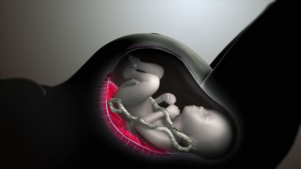
Image by Scientific Animations CC4.0

A flurry of recent studies has found that microplastics are present in virtually everything we consume, from bottled water to meat and plant-based food. Now, University of New Mexico Health Sciences researchers have used a new analytical tool to measure the microplastics present in human placentas.

In a study published in the journalĀ Toxicological Sciences, a team led by Matthew Campen, PhD, Regents’ Professor in the UNM Department of Pharmaceutical Sciences, reported finding microplastics in all 62 of the placenta samples tested, with concentrations ranging from 6.5 to 790 micrograms per gram of tissue.

Although those numbers may seem small, Campen is worried about the health effects of a steadily rising volume of microplastics in the environment.

For toxicologists, “dose makes the poison,” he said. “If the dose keeps going up, we start to worry. If we’re seeing effects on placentas, then all mammalian life on this plant could be impacted. That’s not good.”

In the study, Campen and his team, partnering with colleagues at the Baylor College of Medicine and Oklahoma State University, analyzed donated placenta tissue. In a process called saponification, they chemically treated the samples to “digest” the fat and proteins into a kind of soap.

Then, they spun each sample in an ultracentrifuge, which left a small nugget of plastic at the bottom of a tube. Next, using a technique called pyrolysis, they put the plastic pellet in a metal cup and heated it to 600 degrees Celsius, then captured gas emissions as different types of plastic combusted at specific temperatures.

“The gas emission goes into a mass spectrometer and gives you a specific fingerprint,” Campen said. “It’s really cool.”

The researchers found the most prevalent polymer in placental tissue was polyethylene, which is used to make plastic bags and bottles. It accounted for 54% of the total plastics. Polyvinyl chloride (better known as PVC) and nylon each represented about 10% of the total, with the remainder consisting of nine other polymers.

Marcus Garcia, PharmD, a postdoctoral fellow in Campen’s lab who performed many of the experiments, said that until now, it has been difficult to quantify how much microplastic was present in human tissue. Typically, researchers would simply count the number of particles visible under a microscope, even though some particles are too small to be seen.

With the new analytical method, he said, “We can take it to that next step to be able to adequately quantify it and say, ‘This is how many micrograms or milligrams,’ depending on the plastics that we have.”

Plastic use worldwide has grown exponentially since the early 1950s, producing a metric ton of plastic waste for every person on the planet. About a third of the plastic that has been produced is still in use, but most of the rest has been discarded or sent to landfills, where it starts to break down from exposure to ultraviolet radiation present in sunlight.

“That ends up in groundwater, and sometimes it aerosolizes and ends up in our environment,” Garcia said. “We’re not only getting it from ingestion but also through inhalation as well. It not only affects us as humans, but all off our animals — chickens, livestock — and all of our plants. We’re seeing it in everything.”

Campen points out that many plastics have a long half-life — the amount of time needed for half of a sample to degrade. “So, the half-life of some things is 300 years and the half-life of others is 50 years, but between now and 300 years some of that plastic gets degraded,” he said. “Those microplastics that we’re seeing in the environment are probably 40 or 50 years old.”

While microplastics are already present in our bodies, it is unclear what health effects they might have, if any. Traditionally, plastics have been assumed to be biologically inert, but some microplastics are nanometres in size and are capable of crossing cell membranes, he said.

Campen said the growing concentration of microplastics in human tissue might explain puzzling increases in some types of health problems, such as inflammatory bowel disease and colon cancer in people under 50, as well as declining sperm counts.

The concentration of microplastics in placentas is particularly troubling, he said, because the tissue has only been growing for eight months (it starts to form about a month into a pregnancy). “Other organs of your body are accumulating over much longer periods of time.”

Campen and his colleagues are planning further research to answer some of these questions, but in the meantime he is deeply concerned by the growing production of plastics worldwide.

“It’s only getting worse, and the trajectory is it will double every 10 to 15 years,” he said. “So, even if we were to stop it today, in 2050 there will be three times as much plastic in the background as there is now. And we’re not going to stop it today.”

Source: University of New Mexico Health Sciences Center

New Study Links Placental Oxygen Levels to Foetal Brain Development

Image by Scientific Animations CC4.0

A new study published in JAMA Network Open shows oxygenation levels in the placenta, formed during the last three months of foetal development, are an important predictor of cortical growth and is likely a predictor of childhood cognition and behaviour.

“Many factors can disrupt healthy brain developmentĀ in utero, and this study demonstrates the placenta is a crucial mediator between maternal health and foetal brain health,” said Emma Duerden, Canada Research Chair in Neuroscience & Learning Disorders at Western University, Lawson Health Research Institute scientist and senior author of the study.

The connection between placental health and childhood cognition was demonstrated in previous research using ultrasound, but for this study, Duerden, research scientist Emily Nichols and an interdisciplinary team of Western and Lawson researchers used magnetic resonance imaging (MRI), a far superior and more holistic imaging technique. This novel approach to imaging placental growth allows researchers to study neurodevelopmental disorders very early on in life, which could lead to the development of therapies and treatments.

“While ultrasound provides some measure of placental function, it is imprecise and prone to error, so MRI is just a bit more specific and precise,” said Nichols, lead author of the study. “You wouldn’t use MRI necessarily to diagnose placental growth restriction, you would use ultrasound, but MRI gives us a much better way to understand the mechanisms of the placenta and how placental function is affecting the foetal brain.”

The study was led by Duerden and Nichols and co-authored by researchers from the Faculty of Education, Schulich School of Medicine & Dentistry, Western Engineering and Lawson Health Research Institute.

The placenta, an organ that develops in the uterus during pregnancy, is the main conduit for oxygenation and nutrients to a fetus, and a vital endocrine organ during pregnancy.

“Anything a foetus needs to grow and thrive is mostly delivered through the placenta so if there is anything wrong with the placenta, the foetus might not be receiving the nutrients or the levels of oxygenation it needs to thrive,” said Nichols.

Poor nutrition, smoking, cocaine use, chronic hypertension, anaemia, and diabetes may result in foetal growth restriction and may cause problems for the development of the placenta. Foetal growth restriction is relatively common and happens in about six per cent of all pregnancies and globally impacts 30 million pregnancies each year.

“There can be many issues related to the healthy development of the placenta,” said Duerden. “If it does not develop properly, the foetal brain may not get enough oxygen and nutrients, which may affect childhood cognition and behaviour.”

Impact, affect and change

The study revealed that a healthy placenta in the third trimester particularly impacts the cortex and the prefrontal cortex, regions of the child’s brain that are important for learning and memory.

“An unhealthy placenta can place babies at risk for later life learning difficulties, or even something more serious, like a neurodevelopmental disorder,” said Duerden. “This research can open a lot of doors as we still don’t really understand everything there is to know about the placenta. We are just scratching the surface.”

The study, funded by grants from Brain Canada, The Children’s Health Research Institute, Canadian Institutes of Health Research, BrainsCAN and the Molly Towell Perinatal Research Foundation, is also an important first step in biomarking the impact of oxygenation levels in the placenta and considering changes for expectant mothers to deal with less-than-ideal placental conditions.

While oxygenation in the placenta in the third trimester predicts foetal cortical growth (development of the outermost layer of the brain ā€“ the cerebral cortex), results of the study indicate it may not affect subcortical maturation, or the deep grey and white matter structures of the brain.

Subcortical structures in the brain, responsible for children’s temperament or motor functions such as the amygdala and basal ganglia, may be more vulnerable to factors affecting the placenta in the second trimester.

“We now have a better understanding of how the placenta affects the cortex. With this basic knowledge, we now have an idea of how these two things are related and we can identify or benchmark healthy levels that lead to brain cortical growth,” said Nichols. “The subcortical regions of the brain appear to be unaffected by placental growth, at least in the healthy samples from our study.”

Duerden, Nichols, and the team scanned pregnant women twice (during their third trimester) for the study at Western’s Translational Imaging Research Facility.

“This is one of the few datasets in the world where there are two scans collected in utero during the third trimester. There are not many groups in the world doing foetal MRI, so it is a super-rich data set that allows us to look at growth over time,” said Duerden. “Western is probably one of the few places where we can do the research because we have the expertise and the facilities to do it.”

Source: University of Western Ontario

New Study Sheds Light on Placenta Accreta Spectrum Disorder

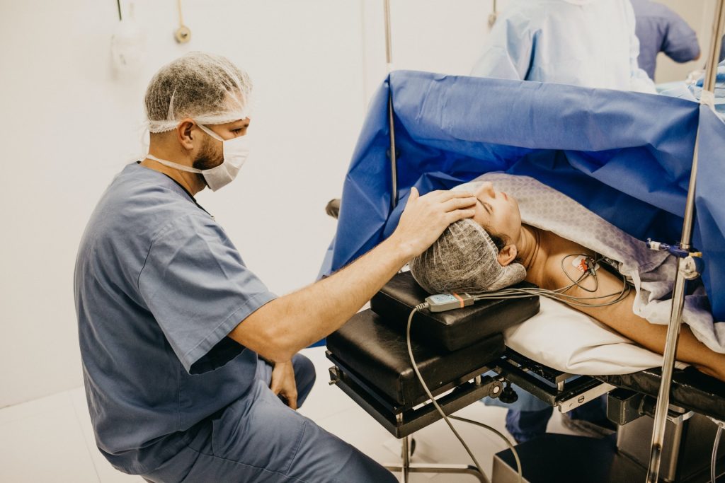
Photo by Jonathan Borba on Unsplash

A new UCLA-led study published in American Journal of Obstetrics & Gynecology may change the way clinicians and scientists understand, diagnose and treat placenta accreta spectrum disorder, a serious condition in which the placenta fails to separate from the uterus at birth. Researchers previously believed that certain overly invasive placental cells, called trophoblasts, were responsible for keeping the connection intact.

But this new research, which identifies genetic and cellular changes within single cells where the placenta and uterus join, shifts the focus to how the structural support of tissues, and the blood vessels of the uterus, can cause a “loss of normal boundary limits” between the placenta and the uterus.

“We utilized two new techniques in single-cell analysis to create an atlas of cells involved in placenta accreta to better understand this increasingly prevalent disorder that can have devastating implications for maternal and neonatal health,” said Dr Yalda Afshar, a maternal-foetal medicine specialist and researcher at the David Geffen School of Medicine at UCLA, and the first and corresponding author.

“This work revealed a subset of genes differentially expressed in placenta accreta spectrum disorder, which provides the basis for the ‘permissive environment’ for the placenta to attach to the uterine lining,” said Dr Deborah Krakow, a maternal-foetal medicine specialist and researcher, chair of the Department of Obstetrics and Gynecology at the David Geffen School of Medicine at UCLA, and the paper’s senior author.

The research showed that the decidua, the layer of the uterine lining that forms during pregnancy, and blood vessels, are sending different signals to the placenta when a pregnant person has placenta accreta.

In placenta accreta, the placenta is stuck on too tight, which becomes the reason for many of the maternal complications of placenta accreta.

“Our goal was to characterize the intimate relationship between the maternal and fetal tissue at the site of accreta or malfunction,” Afshar said.

“The genes and signaling pathways we identified go beyond providing a better understanding of the mechanism of the disease; they may be used as targets to help us refine diagnostic tests, track disease progression over time, and discover new, more effective therapies.”

The incidence of placenta accreta spectrum (PAS) disorders has increased dramatically in recent decades, the cause of which is not certain, though cesarean deliveries, is one of several risk factors.

Today, incidence is estimated at 1 in 272 births in the U.S., up from 1 in about 30 000 pregnancies in the 1960s, researchers say.

For this study, the research team performed multiple placental biopsies on 12 placentas, six with PAS disorder and six controls, conducting single-cell RNA analysis on 31 406 individual cells.

The researchers also applied spatial transcriptomics to 36 regions of interest: 12 in PAS-adherent, 12 in PAS-nonadherent, and 12 in controls.

Spatial transcriptomics allow researchers to precisely measure and map the gene activity within a single tissue sample.

“At the end of the day, understanding the biology of pregnancy and pregnancy-related diseases, like accreta, is inspired by only one thing ā€“ finding ways to improve the care we can provide to pregnant people and their families,” said Afshar, a physician-scientist who manages the care of many patients with placenta accreta spectrum disorders at UCLA Health.

Source: University of California – Los Angeles Health Sciences

Placental Cells Could Help Growth-restricted Babies

Photo by Jonathan Borba on Unsplash

Healthy placenta from mothers of healthy newborns could potentially reduce brain injury in growth-restricted babies, University of Queensland researchers suggest. UQ’s Dr Julie Wixey said the study found stem cells sourced from a healthy placenta may reduce damaging inflammation in these babies after only three days.

ā€œThere is currently no treatment to protect the brains of a growth-restricted baby,ā€ Dr Wixey said.Ā ā€œUp to 50% of them have long term issues ranging from mild learning and behavioural disorders all the way through to cerebral palsy. We know thereā€™s inflammation in the brain and it doesnā€™t cease once these babies are born. Ā 

ā€œOur study has shown we could reduce inflammation and ongoing brain injury by treating these newborns on the day theyā€™re born using a combination of two types of stem cells ā€“ endothelial colony forming cells and mesenchymal stromal cells ā€“ isolated from a healthy human placenta.ā€

About 32 million growth-restricted babies are born around the world each year. Many of them did not receive enough nutrients and oxygen from the placenta.

ā€œOur research has found after just three days, the combination stem cell therapy not only reduced inflammation but also, importantly, appeared to repair damaged blood vessels in the brain in animal models,ā€ Dr Wixey said. ā€œWeā€™re really excited by the outcomes of this study and we hope itā€™ll improve these babiesā€™ lives long term.ā€

Dr Jatin Patel, who co-invented the stem cell harvesting technology, said: ā€œThis has been a fantastic collaborative study and demonstrates the exciting potential of stem cell therapy in the near future in treating unwell babies.

ā€œWe are now working towards scaling up our patented stem cell technology, that will result in greater quantities of cells to drive and expand the preclinical animal studies with the aim of progressing towards a human trial.ā€

The study was published in npj Regenerative Medicine. The researchers will now investigate the longer-term outcomes of the combination stem cell treatment.

Source: University of Queensland