Tag: physical rehabilitation

Meet Kamogelo – The Teen with the Can-do Attitude

Spinal cord injury survivor is a capable and helpful big brother
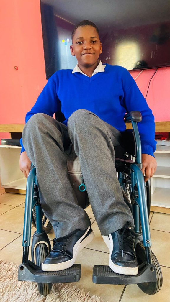
Kamogelo Sodi, who was injured in a car crash when he was just six years old, says he learned valuable skills on how to regain his independence at the Netcare Rehabilitation Hospital. The teenager enjoys cooking for himself, taking care of his three younger brothers, and playing basketball when he’s not studying hard to achieve his dream of being a medical practitioner one day.

5 September 2024: At 14 years old, Kamogelo Sodi of Alberton enjoys listening to music, chatting with his friends on social media and working hard at school towards his dream of becoming a neurosurgeon one day. He cooks for himself when he’s hungry and loves looking after his three little brothers. He also likes playing basketball. The difference between him and most other teenagers is that he does all this from his wheelchair.

“Since I’ve been in a wheelchair, I’ve become more confident,” says the vivacious teenager. “I was extremely shy, and I didn’t have a lot of friends, but now I have loads of friends.”

In 2016, when he was just six years old, Kamogelo’s life changed forever. He was in a devastating car crash, which left him with fractures in the lumbar region of his spine, resulting in complete paraplegia.

Once discharged from the hospital, where he had emergency surgery, Kamogelo was sent to the Netcare Rehabilitation Hospital to learn how to cope with, as his mother Reshoketswe Sodi calls it, his new normal. He was to stay there for almost six months.

Mrs Sodi, a radiation therapist, says the enduring care of the doctors, occupational therapists and physiotherapists there helped support Kamogelo and their family on their journey towards accepting and learning to cope with this difficult transition in his life. “It was important for me that he continued his schoolwork while there. When the social worker asked me what I wanted to happen, the first thing I said was that I didn’t want to break the routine of what he had been doing and that I wanted him to continue with school.

“It’s been a struggle, but with the help of the occupational therapists and physiotherapists, it has been an easier journey. We saw real progress when they taught Kamogelo something, and he grasped it, putting all his energy into it by thinking positively about it. It’s been hard, but with the support of the team from Netcare Rehabilitation Hospital, we managed it,” she says.

“After he was discharged, initially, we lived in a flat on the seventh floor. When the lifts weren’t working, like during load shedding, I’d have to carry him upstairs on my back – there was no other way to take him up. I’m so fortunate that I had a lot of support from my family and friends who’ve been pillars of strength for us.”

Kamogelo remembers his first visit to the Netcare Rehabilitation Hospital in Auckland Park. “When I first got to the hospital, I was lost. I didn’t know how to use a wheelchair. I was still so young. But they were so kind and taught me everything I needed to know. 

“At first, I struggled to move around. I battled to transfer myself from place to place, but they showed me what to do, and over time, I started getting used to it. I managed to start moving myself around, and I began to enjoy it. From that day forward, I didn’t like people pushing me around. The staff also taught me how to transfer myself from my wheelchair to the car. It was a bit difficult at first, but I learned to push myself up properly so my bottom wouldn’t scrape on the wheelchair.

“It does help you become more independent, but you must be consistent. You don’t need to complain about things,  you just need to listen to the people who want to help you learn to be independent.”

Later, in 2022, when he was 12 years old, Kamogelo returned to the Netcare Rehabilitation Hospital after he developed a severe pressure sore.

Dr Anrie Carstens, a doctor at the Netcare Rehabilitation Hospital, said Kamogelo was operated on at Netcare Milpark Hospital under the care of a plastic surgeon who did a flap to close the wound. “When the doctor was happy with his progress, Kamogelo came to us to help him because you get weak after surgery. The wound had healed, but the skin was delicate, so we had a graded seating approach for him to build up his strength and so that the areas of the skin didn’t break down. Another area of focus for Kamogelo was spasticity at the ankles. We worked on relaxing the ankles to get to a ninety-degree angle so he could sit better in his chair with his feet positioned well in the footrest.”

When homesickness inevitably struck, the staff comforted Kamogelo. “I began to miss home, and I cried and said I wanted to go home. They spoke nicely to me and said they first had to help me so I could go back home with no problems so my parents wouldn’t have to worry about me because of the pressure sore.”

Kamogelo said the staff also taught him valuable techniques to help him empty his bladder and bowels and assisted him in his journey to independence. “I was worried it would be painful and was a bit hesitant to try them out. But, doing it daily helped my routine and helped me become independent.”

Charne Cox, a physiotherapist at Netcare Rehabilitation Hospital, describes Kamogelo as bubbly, intelligent and with lovely manners. “He’s so motivated and tried so hard in therapy. He manages to go to school each day, not because of us, but because of his character.”

She says as children grow, their needs change. “The pressure sore developed because his seating in his wheelchair was not adequate because he had grown so much. We collaborated with the wheelchair manufacturer to re-evaluate and reassess the wheelchair seating, and they made him a new wheelchair. He was getting heavier, and his feet weren’t in alignment, so it was trickier for him to safely transfer from the wheelchair to the bed, for instance. It was good to re-educate him on pressure relief and pressure sores. It’s vital that adolescents are taught to take responsibility for themselves.”

Cox also helped Kamogelo work towards getting his feet in a better position.

“Children are so good about learning to use a wheelchair. Kamogelo was so motivated to move and be independent. He absorbed the information we gave him to enable him to go up ramps, turn and even do wheelies because he liked to explore.

“Children want to learn and have fun. They want to be independent. It’s amazing to help give them the tools to be the best new person they can be. Unfortunately, sometimes we can’t fix the injury, but we can give them the best opportunity to be as independent as possible. It’s so satisfying to know that Kamogelo is going to school and playing basketball.”

Kamogelo is determined to pursue a career as a neurosurgeon. “As long as I follow the path that I want to do and enjoy it, I will continue pursuing that path.  Academically, I was the top achiever from grade four to grade six at my school.”

When he’s not at school, he loves going around the estate he lives in, getting fresh air, and being a good big brother to his three younger brothers. “They’re a handful, but what can I say – they’re my brothers, and I love them,” he says with a laugh.

Asked who his hero is, Kamogelo is quick to say his mother and father are both his heroes. His mom clearly thinks he’s a hero too. She’s smiling as she speaks about her son. “He’s playful and has a great sense of humour. He’s helpful in the house. Instead of wanting us to help him, thanks to the skills he learned at Netcare Rehabilitation Hospital, Kamogelo always says, ‘Let me give you a hand. Let me help you.’”

More than Mobility: Gaining Independence after a Spinal Cord Injury

International Spinal Cord Injury Day marked on 5 September 2024

Photo by Elevate on Unsplash

5 September 2024, International Spinal Cord Injury Day is commemorated on Thursday 5 September, drawing attention to the many ways people can be affected by spinal cord injury, creating awareness of prevention, and highlighting the possibilities for a fulfilling life after injury.

According to the World Health Organization, globally, over 15 million people are living with spinal cord injuries. Most of these cases are due to trauma, including falls, road traffic injuries or violence.

Jessica Morris, an occupational therapist at the Netcare Rehabilitation Hospital in Auckland Park, says one of the most critical aspects of care for those who’ve been impacted by spinal cord injuries is the importance of successful rehabilitation through a holistic, integrated approach from a multidisciplinary team.

“Many people just think it’s just about mobility. It’s so much more than that. Rehabilitation is complex because many different areas of our patients’ lives are affected.” Morris says they are fortunate that their team has so many different practitioners who can contribute to treating spinal cord injury patients, helping them regain a level of independence, which is vital to their self-confidence and sense of empowerment.

Dr Anrie Carstens,  a general practitioner with a particular interest in physical medicine and rehabilitation who practises at the Netcare Rehabilitation Hospital, says the message of Spinal Cord Injury Awareness Day has relevance all year round, as people with spinal cord injuries need to be incorporated into society.

“It’s an opportunity to tell people not to be nervous to talk to someone in a wheelchair. They’re just like you or me, and they just have special ways of moving around and managing their pain and different aspects of their bodies. With the help of proper rehabilitation, the person can be better integrated as a functional, contributing member of society.”

Dr Carstens says people should also be aware that if they or their loved ones are ever impacted by a spinal cord injury, professional support is available. “Don’t just go straight home after your hospital stay and try to do everything on your own. Instead, come to a specialised spinal cord injury unit like ours, with therapists, doctors and nursing staff who are well versed in spinal cord injury and know the finer nuances necessary to optimally treat the person and show them how best to cope with their injury.

“In the multidisciplinary approach, every practitioner has a role in getting the person back into the real world, whether it means going back home, back to school, back to work or wherever they were before their injury occurred.”

From doctors and nurses with specialised skills to physiotherapists, occupational therapists, social workers and psychologists, speech therapists, a prosthetist and dieticians, the team provides a broad person focussed rehabilitation service to both adults and children. Their aim is to optimise their patients’ independence level using specialised equipment and teaching specific techniques to help overcome the obstacles a person may face.

Dr Carstens says it’s rewarding work for the staff at the hospital, who build up enduring relationships with those they care for. “One of the highlights is to compare and see what the patient was like when you admitted them and then see on discharge how much they’ve grown, how they’ve gained confidence and become more independent. What’s even better is to see them after they’ve been discharged and observe how well they’ve coped and how they’ve integrated and adjusted to their environment. We build a relationship with our patients because they stay with us for quite a while, and we usually have checkups every year after the person is discharged, often for life. We get to see them grow and thrive outside the healthcare setting, and we need more awareness about how much it is possible for people with spinal cord injuries to achieve.” 

Restoring Muscle Strength Lost to Aging or Injury

Photo by Barbara Olsen on Pexels

A small molecule previously shown to enhance strength in injured or old laboratory mice does so by restoring lost connections between nerves and muscle fibres, Stanford Medicine researchers have found.

The molecule blocks the activity of an aging-associated enzyme, or gerozyme, called 15-PGDH that naturally increases in muscles as they age. The study, which was published in Science Translational Medicine, showed that levels of the gerozyme increase in muscles after nerve damage and that it is prevalent in muscle fibres of people with neuromuscular diseases.

The research is the first to show that damaged motor neurons can be induced to regenerate in response to a drug treatment and that lost strength and muscle mass can be at least partially regained. It suggests that, if similar results are seen in humans, the drug may one day be used to prevent muscle loss of muscle strength due to aging or disease or to hasten recovery from injury.

It’s estimated that sarcopenia, or debilitating muscle frailty, affects about 30% of people over 80 and costs the United States around $380 billion each year.

“There is an urgent, unmet need for drug treatments that can increase muscle strength due to aging, injury or disease,” said Helen Blau, PhD, professor of microbiology and immunology. “This is the first time a drug treatment has been shown to affect both muscle fibres and the motor neurons that stimulate them to contract in order to speed healing and restore strength and muscle mass. It’s unique.”

Blau, the Donald E. and Delia B. Baxter Foundation Professor and director of the Baxter Laboratory for Stem Cell Biology, is the senior author of the study. Postdoctoral scholar Mohsen Bakooshli, PhD, and former postdoctoral scholar Yu Xin Wang, PhD, are the lead authors of the study. Wang is now an assistant professor at the Sanford Burnham Prebys Medical Discovery Institute in San Diego.

Addressing loss of strength

The finding is the latest from the Blau laboratory focused on understanding how muscles weaken from aging or disease, and whether it’s possible to combat this decline. In 2021, the group showed that blocking the activity of 15-PGDH in 24-month-old laboratory mice significantly enhances the animals’ leg strength and endurance when running on a treadmill. (Laboratory mice typically live about 26 to 30 months.) But it wasn’t clear exactly how.

The new research shows that the effect is due to the restoration of lost connections between the nerves and the muscle. These connections, called neuromuscular junctions, are how the brain signals muscles to contract. In aging, some of these connections are lost, causing muscle contractions to become less powerful and muscles to atrophy. People typically lose muscle mass and strength, up to 10% per decade, after the age of 50.

Conditions other than aging can also destabilise these connections, including the disuse of muscles due to bedrest after illness or injury, or muscle-wasting diseases like spinal muscular atrophy or amyotrophic lateral sclerosis (also known as ALS).

Blau’s previous research showed that a molecule called PGE2 is critical to the function of stem cells in muscle fibres that repair damage – including the microtears from exercise that lead to an increase in muscle mass and strength. They subsequently showed that levels of 15-PGDH, which breaks down PGE2, increase in the muscles with age and that the loss of strength with aging could be overcome by inhibiting the activity of this PGE2-degrading enzyme.

“PGE2 is part of the body’s natural healing mechanism, and its levels increase in muscle after injury,” Blau said. “We wanted to learn how age triggers an increase in 15-PGDH, and therefore the degradation and loss of PGE2.”

A lack of nerves

The researchers knew that muscles become less innervated, or infiltrated with nerves, as people and animals age. They wondered if that loss could be what triggers the rising levels of 15-PGDH.

“We found that when you cut the nerve that innervates the leg muscles of mice, the amount of 15-PGDH in the muscle increases rapidly and dramatically,” Blau said. “This was an exciting new insight. But what surprised us most was that when these mice are treated with a drug that inhibits 15-PGDH activity, the nerve grows back and makes contact with the muscle more quickly than in control animals, and that this leads to a faster recovery of strength and function.”

Additional experiments showed that treatment with the drug restored neuromuscular junctions lost during aging and increased muscle strength and function in old laboratory mice. The researchers also identified discrete clumps of 15-PGDH in the muscle fibres of people with several types of neuromuscular disorders suggesting that the gerozyme may have a role in causing these human disorders.

Blau and her colleagues plan to investigate at a molecular level how neural growth is stimulated by blocking 15-PGDH activity. Blau has also co-founded a company, Epirium Bio, to develop similar drugs for use in humans. Although her lab is still conducting animal studies, the company hopes to launch a clinical trial within the next year or so.

“Our next steps will be to examine whether blocking 15-PGDH function in people with spinal muscular atrophy can increase lost muscle strength in combination with gene therapy or other treatments,” Blau said. “We are also looking at ALS to see if something like this might help these patients. It’s really exciting that we are able to affect both muscle function and motor neuron growth.”

Source: Stanford Medicine

Vigorous Exercise Improves Walking in Chronic Stroke Patients

Photo by Kampus Production on Pexels

When 67-year-old Larry Christian suffered a sudden loss of balance, he was diagnosed with a haemorrhagic stroke, and referred to the University of Delaware’s Physical Therapy Clinic for rehabilitation. 

“Initially, I had a lot of balance problems that we worked pretty intensely to correct,” Christian said. 

He enrolled in a clinical trial at UD, led by co-investigator Darcy Reisman, professor and chair of the Department of Physical Therapy, that sought to explore whether high-intensity interval training (HIIT) aids in improved gait post-stroke. UD was one of three sites selected for the clinical trial led by primary investigator and associate professor Pierce Boyne of the University of Cincinnati. Sandra Billinger, professor and vice chair of stroke translation research at the University of Kansas Medical Center, is also a co-investigator and represents the third site involved in the clinical trial. 

Now, seven years later, Christian is walking better. 

“Participating in this study got me to a point where I could walk better and even take a walk outside,” Christian said. “I’ve been pretty healthy all my life, and while I can’t play volleyball anymore, walking again made me feel great.”

Christian is among the lucky ones. Among 7 million stroke survivors in the US, fewer than 10% have adequate walking speed and endurance to complete normal daily activities like grocery shopping. 

Reisman said the results of the multi-million-dollar, five-year clinical trial showed HIIT helped more people than just Christian. The results, published in JAMA Neurology, show that chronic stroke survivors who engaged in high-intensity exercise with bursts of maximum-speed walking alternated with recovery periods saw a significant difference in their walking capacity over 12 weeks. The improvements were so dramatic Boyne and Reisman have secured a clinical trial grant renewal to triple the size of their study to 165 participants. 

She added HIIT looks different for each stroke survivor, and the optimal exercise program for each person with stroke remains unknown. 

“We want them to train at the fastest possible speed, which varies from person to person,” Reisman said. “But we don’t want them running.”

For those already walking at a reasonably fast pace, research associate Henry Wright in Reisman’s lab will add an incline or a weighted vest or wrap a bungee cord around their waist to create resistance. 

“It’s self-reported data, but participants tell me they have more energy, or they’re able to do more around the house, or they’re not winded when they go shopping,” Wright said. “By the end of the training, I can see their walking is smoother, they’re getting farther on clinical testing, and it’s rewarding to see their gains.”  

The results from the initial clinical trial showed Reisman and collaborators that HIIT was feasible and safe in a small group of stroke survivors, who saw sustained gains in walking capacity, more so than patients engaged in moderate-intensity exercise. 

However, further study of the intervention in larger populations is crucial to change the standard of care.

“Many physical therapists were trained during a time when patients with neurologic conditions, particularly stroke, were treated with kid gloves, partly because they say stroke is the heart attack of the brain,” Reisman said. “It’s common they also have cardiovascular conditions, so people tend to be extra careful with those patients in terms of pushing them.

“But what we know now is at least moderate-intensity, and likely high-intensity interval training, is essential not only for stroke survivors’ cardiovascular system but also for their brain,” Reisman said. “The evidence shows that intensity is linked to the release of neurotrophins in the brain that help the brain remodel after a stroke.” 

Kiersten McCartney, a physical therapist obtaining her doctorate in biomechanics and movement science, worked on the clinical trial with Reisman. She spent the 2022 Winter Session at Magee Rehabilitation Hospital in Philadelphia, helping them implement moderate-to-high-intensity exercise and saw the benefits first-hand. 

“I’ll never be able to say there’s no risk of heart attack. Even the fittest people can have a heart attack when exercising,” McCartney said. “Still, the data points to the idea that you’re doing more harm than good by not engaging your patients with stroke in high-intensity exercise when we talk about those longer-term outcomes.”

The HIIT-Stroke Trial 2 will continue to examine dosing to confirm whether a full 12 weeks of vigorous exercise is needed to see significant improvements in walking. Reisman and collaborators will identify whether differences in sex and other factors played a role in rehabilitation. If the five-year study results are similar and show significant gains from high-intensity interval exercise in a larger population, investigators would next work with NIH Strokenet to launch a nationwide clinical trial in people with stroke.  

“We’ve known about the value of moderate-intensity exercise for more than a decade, and it’s still not the standard of care,” Reisman said. “If we find that HIIT is the optimal intervention, the next phase would be the knowledge translation phase, where we’d systematically develop a methodology to get HIIT into clinics.” 

For HIIT to work as an intervention, Reisman said therapists will need the proper tools. She’s been pushing for commercially available heart rate monitors, placed around the chest during exercise, to be the standard of care in clinics for years.

“They’re already a standard of care for people in the community,” Reisman said. “Getting them into clinics is imperative so PTs can monitor patients’ heart rate the entire time they exercise. That constant monitoring gives therapists data on how a person is responding beyond visible signs and symptoms, and in turn, more peace of mind.” 

But beyond tools and training, Reisman said, it comes down to evidence and education. 

“If we have hundreds and hundreds of stroke survivors who’ve gone through our high-intensity exercise intervention, and we’ve seen no major adverse events – that will help,” Reisman said. “The more data we have to show therapists, the better we can implement this intervention that will change lives.”

Source: University of Delaware

For Stroke Recovery, Deep Brain Stimulation may Aid Rehabilitation

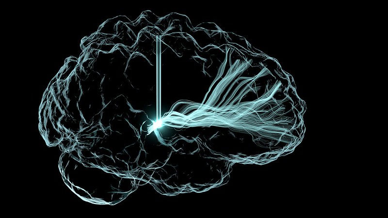
Deep brain stimulation illustration. Credit: NIH

A first-in-human trial of deep brain stimulation (DBS) for post-stroke rehabilitation patients has shown that using DBS to target the dentate nucleus – which regulates fine-control of voluntary movements, cognition, language, and sensory functions in the brain – is safe and feasible.

The EDEN trial (Electrical Stimulation of the Dentate Nucleus for Upper Extremity Hemiparesis Due to Ischemic Stroke) also shows that the majority of participants (9 of 12) demonstrated improvements in both motor impairment and function. Importantly, the study found that participants with at least minimal preservation of distal motor function at enrolment showed gains that almost tripled their initial scores.

Published in Nature Medicine, these findings build on more than a decade of preclinical work led by principal investigators Andre Machado, MD, PhD, and Kenneth Baker, PhD, at Cleveland Clinic.

“These are reassuring for patients as the participants in the study had been disabled for more than a year and, in some cases, three years after stroke. This gives us a potential opportunity for much needed improvements in rehabilitation in the chronic phases of stroke recovery,” said Dr Machado, patented the DBS method in stroke recovery. “The quality-of-life implications for study participants who responded to therapy have been significant.”

“We saw patients in the study regain levels of function and independence they did not have before enrolling in the research,” Dr Machado said. “This was a smaller study and we look forward to expanding as we have begun the next phase.”

The completed EDEN trial enrolled 12 individuals with chronic, moderate-to-severe hemiparesis of the upper extremity as a result of a unilateral middle cerebral artery stroke 12-to-36 months prior. There were no major complications throughout the study. Nine of the 12 participants improved to a degree that is considered meaningful in stroke rehabilitation.

Source: Cleveland Clinic

Simple Oxygen Therapy can Boost Motor Skills Rehabilitation

Photo by Samuel Ramos on Unsplash

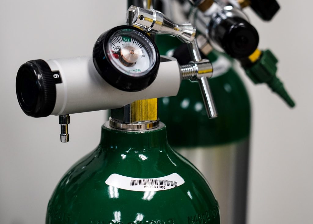
Scientists studying the impact of oxygen supplementation on motor learning have found a promising treatment that could help patients who have experienced neurological trauma recover lost motor skills.

“A simple and easy to administer treatment with 100% oxygen can drastically improve human motor learning processes,” said Dr Marc Dalecki, now at the German University of Health and Sports in Berlin, senior author of the study in Frontiers in Neuroscience.

Repurposing a frontline treatment

Brains have a high oxygen demand, and hypoxia causes cognitive function to decrease, while in high-oxygen contexts it recovers, and the delivery of 100% oxygen is already used to help preserve as much of the brain as possible in patients with neurological injuries.

Motor learning is particularly dependent on oxygen-reliant information processing and memory functions: humans learn by trial and error, so the ability to remember and integrate information from previous trials is critical to efficient and effective motor learning. So could supplementing oxygen while learning a motor task help people learn faster and more effectively, offering hope for neurorehabilitation patients?

“I had this idea in my mind for almost a decade and promised myself to investigate it once I got my own research lab,” said Dalecki, who led the experimental research at the School of Kinesiology at Louisiana State University. “And with Zheng Wang, now Dr Zheng Wang, I had the perfect doctoral student to run it – a keen physiotherapist with a clinical background and stroke patient experience.”

Hand-eye coordination

Dalecki and Wang recruited 40 participants, 20 of whom received 100% oxygen at normobaric pressure and 20 of whom received medical air (21% oxygen) through a nasal cannula during the “adaptation” or learning phase of a task.

Dalecki and Wang selected a simple visuomotor task which involved drawing lines between different targets on a digital tablet with a stylus. The task was designed to test how quickly the participants were able to integrate information from the eye and hand, a crucial part of motor learning. After the task had been learned, the alignment of the cursor and the stylus was altered to see how effectively the participants adapted to the inconsistency, and then realigned for a final session to see how they adapted to the realignment.

“The oxygen treatment led to substantially faster and about 30% better learning in a typical visuomotor adaptation task,” said Wang, first author of the study and now at the Mayo Clinic in Rochester. “We also demonstrate that the participants were able to consolidate these improvements after the termination of the oxygen treatment.”

Oxygen improved learning by 30%

The scientists found that the participants who had received oxygen learned faster and performed better, improvements which extended into later sessions of the task when oxygen was not administered.

The oxygen group moved the pen more smoothly and more accurately, and when the cursor was adjusted in a deliberate attempt to throw them off, they adapted more quickly. They also made bigger mistakes when the alignment of the stylus was corrected, suggesting they had integrated the previous alignment more thoroughly than the other group.

Dalecki and Wang plan to investigate the long-term effects of this supplementation on learning and test the intervention with other motor learning tasks: it is possible that the relevant brain functions for this task in particular benefit from high ambient oxygen levels, leading to the observed advantages in performance. They also hope to bring the oxygen treatment to elderly and injured people, in the hope that it will help them re-learn motor skills.

“Our future plan is to investigate whether this treatment can also improve motor recovery processes following brain trauma,” said Dalecki. “Since it worked in the young healthy brain, we expect that the effects may even be larger in the neurologically impaired, more vulnerable brain.”

Source: Medical Xpress

Fixing Spinal Cord Injuries with Stem Cell Grafts and Rehabilitation

Photo by Cottonbro on Pexels

In recent years, researchers have made strides in promoting tissue regeneration in spinal cord injuries (SCI) through implanted neural stem cells or grafts in animal models. Separate efforts have shown that intensive physical rehabilitation can improve function after SCI by promoting greater or new roles for undamaged cells and neural circuits.

University of California San Diego researchers tested whether rehabilitation can pair with pro-regenerative therapies, such as stem cell grafting. They published their findings in in JCI Insight, 

The researchers induced a cervical lesion in rats that impaired the animals’ ability to grasp with its forelimbs. The animals were divided into four groups: animals who underwent the lesion alone; animals who received a subsequent grafting of neural stem cells designed to grow and connect with existing nerves; animals who received rehabilitation only; and animals who received both stem cell therapy and rehabilitation.

Rehabilitation therapy for some animals began one month after initial injury, a time point that approximates when most human patients are admitted to SCI rehabilitation centers. Rehabilitation consisted of daily activities that rewarded them with food pellets if they performed grasping skills.

The researchers found that rehabilitation enhanced regeneration of injured corticospinal axons at the lesion site in rats, and that a combination of rehabilitation and grafting produced significant recovery in forelimb grasping when both treatments occurred one month after injury.

“These new findings indicate that rehabilitation plays a critically important role in amplifying functional recovery when combined with a pro-regenerative therapy, such as a neural stem cell transplant,” said first author Paul Lu, PhD, associate adjunct professor of neuroscience at UC San Diego School of Medicine and research health science specialist at the Veterans Administration San Diego Healthcare System.

“Indeed, we found a surprisingly potent benefit of intensive physical rehabilitation when administered as a daily regimen that substantially exceeds what humans are now provided after SCI.”

Senior author Mark H. Tuszynski, MD, PhD, professor of neurosciences and director of the Translational Neuroscience Institute at UC San Diego School of Medicine, and colleagues have long worked to address the complex challenges of repairing SCIs and restoring function.

In 2020, for example, they reported on the observed benefits of neural stem cell grafts in mice and in 2019, described 3D-printed implantable scaffolding that would promote nerve cell growth.

“There is a great unmet need to improve regenerative therapies after SCI,” said Tuszynski. “We hope that our findings point the way to a new potential combination treatment consisting of neural stem cell grafts plus rehabilitation, a strategy that we hope to move to human clinical trials over the next two years.”

Source: University of California – San Diego

Progressive Exercise Programme Improves Outcomes after Hip Surgery

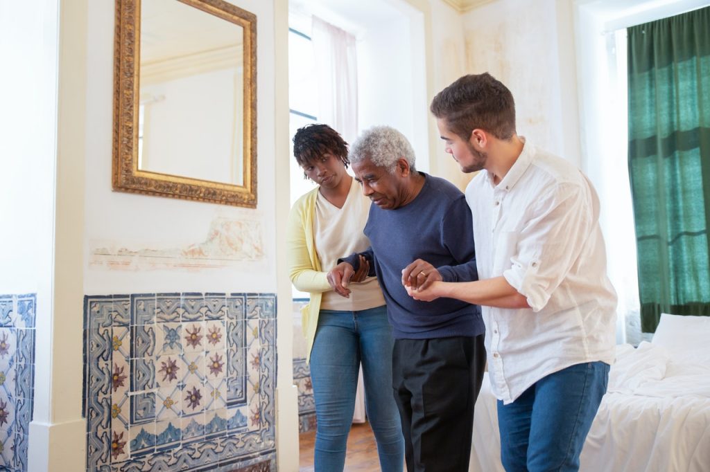
Carers help an old man to walk
Photo by Kampus Productions on Pexels

A study published in the Journal of the American Geriatrics Society shows that a 12-month home-based supervised exercise programme can help to improve physical performance and functioning after patients undergo hip fracture surgery.

Hip fracture is a major health problem among older people, often resulting in long-term, sometimes persistent, functional impairments such as poor mobility and reduced independence in daily activities. Sedentary behaviour and low level of physical activity are also common among patients recovering from surgical repair of a hip fracture.

Standard care post-discharge care does not seem to meet the requirements of effective rehabilitation, as many patients with hip fractures do not reach their pre-fracture level of functioning. Growing evidence shows that multidisciplinary and well-coordinated rehabilitation started at the hospital and continued after discharge enhances the recovery of patients with hip fractures. Multicomponent rehabilitation in particular, which includes individualised and progressive resistance training, has improved functioning and mobility and decreased dependency in everyday activities. Longer lasting exercise programs of 6 to 12 months duration have reduced or reversed incident disability after hip fractures.

For the study, 121 patients aged 60 years and older were randomised to either an exercise group or a usual care group as a control. Home-based exercise sessions were delivered by physiotherapists twice a week and included strength, balance, mobility, and functional components as well as brief counselling on physical activity and nutrition.

Compared with patients in the usual care group, patients in the exercise group saw more improvements over the course of a year in their physical performance, their handgrip strength, and their ability to complete certain activities of daily living.

“It is worthwhile to invest in rehabilitation exercise for older people after hip fracture. Better functioning benefits the individual and also society,” said lead author Paula K. Soukkio, MSc, of the South Karelia Social and Health Care District (Eksote), in Finland.

Source: Wiley

After a Stroke, Muscles Lose Basic ‘Building Blocks’

Muscle sarcomeres (consecutive green lines), the smallest functional unit of muscle, from inside a living human. Credit: Northwestern University

In a new study of stroke patients, researchers have discovered that, in an attempt to adapt for an unusable arm, muscles actually lose sarcomeres — their smallest, most basic building blocks.

Patients that have suffered a stroke are often unable to use the arm on their affected side. Sometimes, they end up holding it close to their body, with the elbow flexed. Northwestern University and Shirley Ryan AbilityLab researchers found out why this happens.

Stacked end to end (in series) and side to side (in parallel), sarcomeres form the length and width of muscle fibres. By imaging biceps muscles with three noninvasive methods, the researchers found that stroke patients had fewer sarcomeres along the length of the muscle fibre, resulting in the muscle structure being shorter overall.

This finding is consistent with the common patient experience of abnormally tight, stiff muscles that resist stretching, and it suggests that changes in the muscle potentially amplify existing issues caused by stroke, which is a brain injury. The team hopes this discovery can help improve rehabilitation techniques to rebuild sarcomeres, ultimately helping to ease muscle tightening and shortening.

“This is the most direct evidence yet that chronic impairments, which place a muscle in a shortened position, are associated with the loss of serial sarcomeres in humans,” said senior author Wendy Murray. “Understanding how muscles adapt following impairments is critical to designing more effective clinical interventions to mitigate such adaptations and to improve function following motor impairments.”

Murray is a professor of biomedical engineering at Northwestern’s McCormick School of Engineering, a professor of physical medicine and rehabilitation at the Northwestern University Feinberg School of Medicine and research scientist at the Shirley Ryan AbilityLab. The research was completed in collaboration with Julius Dewald, professor of physical therapy and human movement sciences and of physical medicine and rehabilitation at Feinberg, professor of biomedical engineering at McCormick, and research scientist at Shirley Ryan AbilityLab.

Measuring just 1.5 to 4.0 micrometres in length, sarcomeres are made up of two main proteins: actin and myosin. When these proteins work together, they enable a muscle to contract and produce force. Even though previous animal studies have found that serial sarcomeres are lost from muscles after a limb is immobilised in a cast, the phenomenon had never before been demonstrated in humans. The animal studies found that the shorter muscles due to lost serial sarcomeres also became stiffer.

There is a classic relationship between force and length,” explained first author Amy Adkins, a PhD student in Murray’s laboratory. “Given that the whole muscle is composed of these building blocks, losing some of them affects how much force the muscle can generate.”

To conduct the study in humans, the researchers combined three non-invasive medical imaging techniques: MRI to measure muscle volume, ultrasound to measure bundles of muscle fibers and two-photon microendoscopy to measure the microscopic sarcomeres.

Imaging opens new possibilities
Combining these technologies, the researchers imaged biceps from seven stroke patients and four healthy participants. As stroke patients are more affected on one side of their body, the researchers compared imaging from the patients’ affected side to their unaffected side as well as to images from the healthy participants.

In the stroke patients’ affected biceps, researchers found less volume, shorter muscle fibres and comparable sarcomere lengths. After combining data across scales, they found that affected biceps had fewer sarcomeres in series compared to the unaffected biceps. Greater differences between stroke patients’ arms than healthy participants’ arms were seen, indicating that stroke was the cause.

By combining medical imaging to better view muscle structure, the study also establishes that it is possible to study muscle adaptations in sarcomere number in humans. Prior to two-photon microendoscopy, human studies were limited either to examining dissected tissues in anatomy labs, which give imperfect insight into how muscles adapt to injury and impairment, measuring sarcomere lengths during surgery or from a muscle biopsy, which restricts who can participate in the study.

“In almost every facet of our world, there is an important relationship between how something is put together (its structure) and how it works (its function),” the researchers said. “Part of the reason medical imaging is such a valuable resource and clinical tool is that this is also true for the human body, and imaging gives us an opportunity to measure structure.”

Source: Northwestern University

Journal information: Adkins, A.N., et al. (2021) Serial sarcomere number is substantially decreased within the paretic biceps brachii in individuals with chronic hemiparetic stroke. PNAS. doi.org/10.1073/pnas.2008597118.

Using a Gaming Console can Improve Stroke Patient Rehabilitation

Photo by Tima Miroshnichenko from Pexels

A study by the Faculty of Physiotherapy of the University of Valencia (UV) has shown that a physiotherapy programme using the Nintendo Wii console improves the functionality, balance and daily activities of patients who have suffered a cerebrovascular accident or stroke.
Wii research group

The study found that when the Wii is added to conventional physiotherapy techniques, the benefits are significant in stroke patients. Besides improvements in functionality and balance, the physiotherapy programme using the Wii also helps to improve daily activities.

The use of game consoles in medicine has focused on aspects such as helping build motor skills and pain management with virtual reality immersion. They are relatively cheap and available, and simple to use. One study looked at using a Microsoft Xbox to help rehabilitation of patients with Parkinson’s disease.

Previous research had already shown that the Wii can help normal treatments in restoring functionality in some chronic diseases. However, until now, there was little evidence of its use in people who had suffered strokes. “Before conducting the study we realised that not much research had been done with stroke patients, so we wanted to know if console games could promote mobility, balance and the day-to-day life of people with this pathology”, explained Elena Marqués, one of the researchers and professor of Physiotherapy at the UV.

The study recruited 29 participants into two groups, one using the Wii and conventional techniques, and the other performing traditional physiotherapy exercises. “The sample is relatively large considering it is comprised by patients who suffered strokes, as because they have many physical limitations, their treatment is usually much more individualised than that of other pathologies”, said the author.

These video games allow therapists to design rehabilitation programs that improve the principles of brain plasticity. An additional advantage is that the console provides real-time feedback on performance and progress, which can increase patient motivation, fun, and treatment adherence. “It should be taken into account that some patients have not performed any exercise before, regardless of the pathology they have, so being presented as a game can be an incentive”, said Prof Marqués.

She pointed out that other benefits include the Wii being easy to use, relatively affordable and, most importantly, can be used individually and at home, without needing to visit a rehab centre. This is particularly useful with COVID lockdowns.

This is one of the first studies using consoles as a therapeutic option, but it can be extended to patients with other pathologies, “because it allows you to work the balance with the console table, both in the chronic phase and in the subacute phase”, said Marqués.

Strengthening mirror neurons

Many Wii games use the remote control, but the console also offers a balance table that detects weight transfer by reflecting it in an avatar on the screen, letting the patient observe his/her own movements and generate positive feedback.

Thus, when the person observes his/her movements, the plasticity changes that depend on the use of sensory areas belonging to the mirror neuron system are strengthened. This exemplifies, among other factors, the improvements the Wii can provide in such patients. This feedback could result in a strengthening of the learning mechanisms of different motor and sensory activities and ultimately improving quality of life.

Source: Asociación RUVID