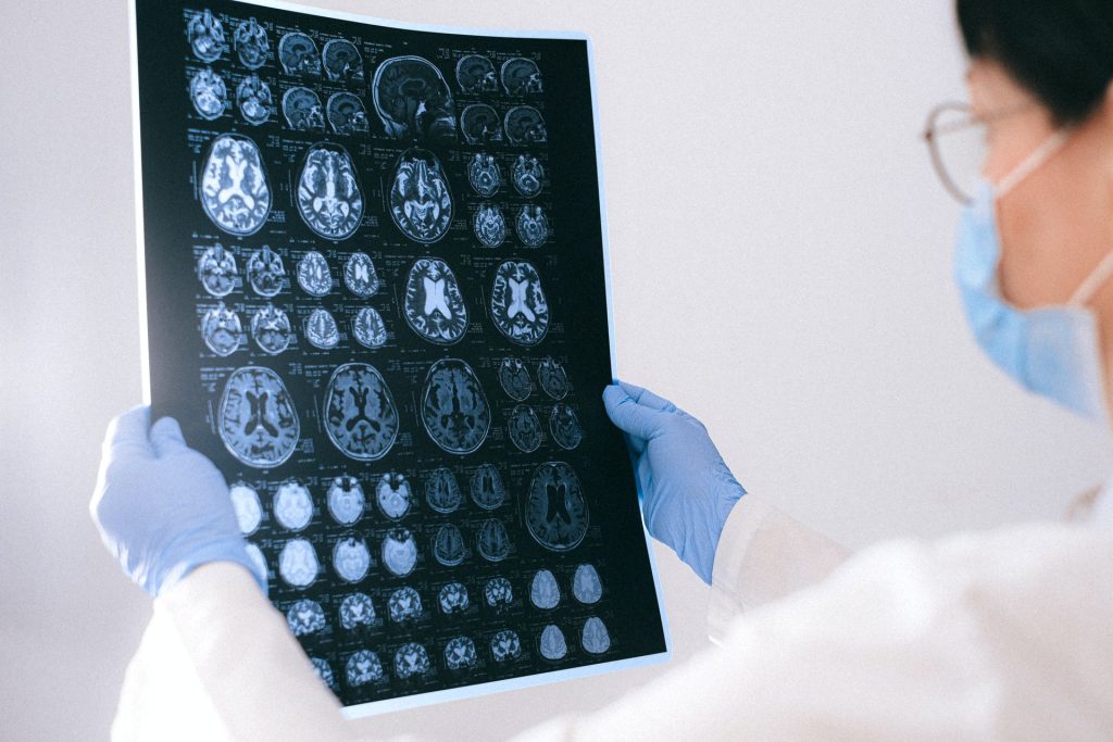
Foundations Laid for Standardised PET Examination of Diffuse Gliomas

Diffuse gliomas are malignant brain tumours that cannot be optimally examined by means of conventional MRI imaging. So-called amino acid PET (positron emission tomography) scans are better able to image the activity and spread of gliomas. An international team of researchers from the RANO Working Group have drawn up the first ever international criteria for the standardised imaging of gliomas using amino acid PET. It has published its results in the journal The Lancet Oncology.
PET uses a radioactive tracer to measure metabolic processes in the body. Amino acid PET is used in the diagnosis of diffuse gliomas, with tracers that work on a protein basis (amino acids) and accumulate in brain tumours.
The Response Assessment in Neuro-Oncology (RANO) Working Group is an international, multidisciplinary consortium founded to develop standardised new response criteria for clinical studies relating to brain tumours.
Under the joint leadership of nuclear physician Nathalie Albert from LMU and oncologist Professor Matthias Preusser from the Medical University of Vienna, the RANO group has developed new criteria for assessing the success of therapies for diffuse gliomas.
Nathalie Albert explains: “PET imaging with radioactively labelled amino acids has proven extremely valuable in neuro-oncology and permits reliable representation of the activity and extension of gliomas. Although amino acid PET has been used for years, it had not been evaluated in a structured manner before now. In contrast to MRI-based diagnostics, there have been no criteria for interpreting these PET images.” According to the researchers, the new criteria allow PET to be used in clinical studies and everyday clinical practice and create a foundation for future research and the comparison of treatments for improved therapies.
New criteria for PET examinations of brain tumours
Diffuse gliomas are malignant brain tumorus that cannot be optimally examined by means of conventional MRI imaging. So-called amino acid PET scans are better able to image the activity and spread of gliomas.
These malignant brain tumours develop out of glial cells and are generally aggressive and difficult to treat.
The RANO group has developed criteria that permit evaluation of the success of treatment using PET. Called PET RANO 1.0, these PET-based criteria open up new possibilities for the standardised assessment of diffuse gliomas.


