Tag: oral microbiome

Bacteria Subtype Linked to Growth in up to 50% of Human Colorectal Cancers

Human colon cancer cells. Credit: National Cancer Institute

Researchers at Fred Hutchinson Cancer Center have found that a specific subtype of a microbe commonly found in the mouth is able to travel to the gut and grow within colorectal cancer tumours. This microbe is also a culprit for driving cancer progression and leads to poorer patient outcomes after cancer treatment.

The findings, published in Nature, could help improve therapeutic approaches and early screening methods for colorectal cancer, which is the second most common cause of cancer deaths in adults in the U.S. according to the American Cancer Society.

Examining colorectal cancer tumours removed from 200 patients, the Fred Hutch team measured levels of Fusobacterium nucleatum, a bacterium known to infect tumours. In about 50% of the cases, they found that only a specific subtype of the bacterium was elevated in the tumour tissue compared to healthy tissue.

The researchers also found this microbe in higher numbers within stool samples of colorectal cancer patients compared with stool samples from healthy people.

“We’ve consistently seen that patients with colorectal tumours containing Fusobacterium nucleatum have poor survival and poorer prognosis compared with patients without the microbe,” explained Susan Bullman, PhD, Fred Hutch cancer microbiome researcher and co-corresponding study author. “Now we’re finding that a specific subtype of this microbe is responsible for tumour growth. It suggests therapeutics and screening that target this subgroup within the microbiota would help people who are at a higher risk for more aggressive colorectal cancer.”

In the study, Bullman and co-corresponding author Christopher D. Johnston, PhD, Fred Hutch molecular microbiologist, along with the study’s first author Martha Zepeda-Rivera, PhD, a Washington Research Foundation Fellow and Staff Scientist in the Johnston Lab, wanted to discover how the microbe moves from its typical environment of the mouth to a distant site in the lower gut and how it contributes to cancer growth.

First they found a surprise that could be important for future treatments. The predominant group of Fusobacterium nucleatum in colorectal cancer tumours, thought to be a single subspecies, is actually composed of two distinct lineages known as “clades.”

“This discovery was similar to stumbling upon the Rosetta Stone in terms of genetics,” Johnston explained. “We have bacterial strains that are so phylogenetically close that we thought of them as the same thing, but now we see an enormous difference between their relative abundance in tumours versus the oral cavity.”

By separating out the genetic differences between these clades, the researchers found that the tumour-infiltrating Fna C2 type had acquired distinct genetic traits suggesting it could travel from the mouth through the stomach, withstand stomach acid and then grow in the lower gastrointestinal tract. The analysis revealed 195 genetic differences between the clades.

Then, comparing tumour tissue with healthy tissue from patients with colorectal cancer, the researchers found that only the subtype Fna C2 is significantly enriched in colorectal tumour tissue and is responsible for colorectal cancer growth.

Further molecular analyses of two patient cohorts, including over 200 colorectal tumours, revealed the presence of this Fna C2 lineage in approximately 50% of cases.

The researchers also found in hundreds of stool samples from people with and without colorectal cancer that Fna C2 levels were consistently higher in colorectal cancer.

“We have pinpointed the exact bacterial lineage that is associated with colorectal cancer, and that knowledge is critical for developing effective preventive and treatment methods,” Johnston said.

Source: Fred Hutchinson Cancer Center

Firefighter Study Reveals how Extreme Exercise can Suppress the Immune System

Source: CC0

A study of firefighters on a punishing training course has revealed clues as to why extreme exercise temporarily weakens the immune system – a phenomenon seen in elite athletes. The findings, published in Military Medical Research, may lead to better ways to support the health of people who undergo extreme exertion, such as firefighters tackling wildfires.

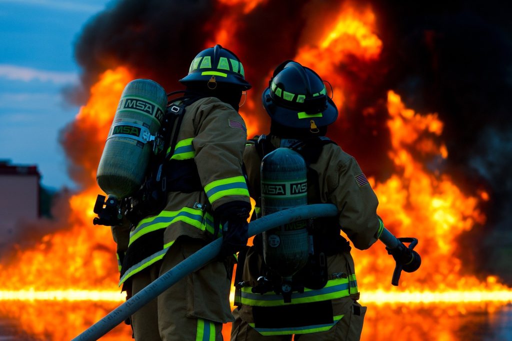
Thirteen firefighters volunteered for the study, average age 25 and male. They went through a rigorous training exercise, carrying 9 to 20kg of gear over hilly terrain during a 45-minute training exercise in the California sun. Gloves, helmets, flashlights, goggles, and more weighted them down as they sprinted through the countryside wearing fire-resistant clothing to show they were ready to serve as wildland firefighters.

After the training, they immediately gave samples of their blood, saliva, and urine for analysis. Two were excluded, one being unable to finish the course and the other arriving to late to provide a sample. The 11 participants who completed the course lost an average of 2.2% of their initial weight.

Then, the scientists from the Department of Energy’s Pacific Northwest National Laboratory (PNNL) analysed more than 4700 molecules, consisting of proteins, lipids, and metabolites, from each of the firefighters, looking to understand what happens when the body undergoes intense physical exercise. Measuring and interpreting the data from thousands of such measurements is a specialty of PNNL scientists who explore issues related to climate science and human health by analysing millions of sensitive measurements using mass spectrometry each year.

The researchers’ aim was to increase safety for first responders and others.

“Heat stress can be life threatening,” said Kristin Burnum-Johnson, a corresponding author of the study. “We wanted to take an in-depth look at what’s happening in the body and see if we’re able to detect danger from exhaustion in its earliest stages. Perhaps we can reduce the risk of strenuous exercise for first responders, athletes, and members of the military.”

As expected, the team detected hundreds of molecular changes in the firefighters. The differences before and after exercise underscored the body’s efforts at tissue damage and repair, maintenance of fluid balance, efforts to keep up with increased energy and oxygen demand, and the body’s attempts to repair and regenerate its proteins and other important substances.

But in the saliva, the team found some unexpected results. There was a change in the microbial mix of the mouth – the oral microbiome – showing that the body was increasingly on the lookout for bacterial invaders. Scientists also saw a decrease in signaling molecules important for inflammation and for fighting off viral infections.

A decrease in inflammation makes sense for people exercising vigorously; less inflammation allows people to breathe in air more quickly, meeting the body’s eager demand for more oxygen. Having fewer inflammatory signals in the respiratory system helps the body improve respiration and blood flow.

Less inflammation, more inhalation

But less inflammation leaves the body more vulnerable to viral respiratory infection, which other studies observed in elite athletes and others who exercise vigorously. Some studies have shown that a person is up to twice as likely to come down with a viral respiratory infection in the days after an especially energetic workout.

“People who are very fit might be more prone to viral respiratory infection immediately after vigorous exercise. Having less inflammatory activity to fight off an infection could be one cause,” said Ernesto Nakayasu, a corresponding author of the paper. He notes that the work provides a molecular basis for what clinicians have noticed in their patients who do strenuous workouts.

The team hopes that the findings will help explain why come people are more vulnerable to respiratory infection after a workout.

Source: DOE/Pacific Northwest National Laboratory