Tag: neutrophils

Scientists Discover that Mast Cells Gobble up Other Immune Cells

This scanning electron microscopy image captures the moment where degranulating mast cells (pseudo-colored in sepia) attract and start to incorporate living neutrophils (pseudo-colored in cyan), forming cell-in-cell structures where mast cells trap living neutrophils inside them. © Marcus Frank & Karoline Schul11z, Universitätsmedizin Rostock, Germany

When it comes to allergies, mast cells are key immune system players, releasing pro-inflammatory substances in response to allergens. Now, scientists in Germany have discovered something weird: other immune cells nested inside them like Russian dolls. But how exactly did these cells wind up there?

As reported in the journal Cell, the researchers observed mast cells observed capturing and making use of neutrophils. This surprising discovery sheds new light on how our immune system works, particularly during allergic reactions.

Mast cells, residing in tissues and critical for initiating inflammation, are filled with granules containing pro-inflammatory substances. These granules are released upon encountering potential dangers, including allergens, causing allergic reactions – which for some includes innocuous materials like pollens. But despite how common allergies are, the interaction between mast cells and other immune cells at sites of allergic responses has been largely unexplored.

The research group at the Max Planck Institute of Immunobiology and Epigenetics in Freiburg and the University of Münster used specialised microscopy to visualise the real-time dynamics of activated mast cells and other cell types during allergic reactions in living mouse tissues. The team discovered a surprising interaction: neutrophils were found inside mast cells.

“We could hardly believe our eyes: living neutrophils were sitting inside living mast cells. This phenomenon was completely unexpected and probably would not have been discovered in experiments outside a living organism and highlights the power of intravital microscopy,” says Tim Lämmermann, research leader and Director at the Institute of Medical Biochemistry at the University of Münster.

Pulling a neutrophil trick to trap neutrophils

Neutrophils are frontline immune system defenders, responding quickly and broadly to potential threats. They circulate in the blood and quickly exit blood vessels at sites of inflammation. They are well-equipped to combat pathogens by engulfing the invaders, releasing antimicrobial substances, or forming web-like traps known as ‘neutrophil extracellular traps’. Additionally, neutrophils can communicate with each other and form cell swarms to combine their individual functions for the protection of healthy tissue. While much is known about neutrophils’ role in infections and sterile injuries, their role in inflammation caused by allergic reactions is less understood.

“It quickly became clear that the double-pack immune cells were no mere coincidence. We wanted to understand how mast cells trap their colleagues and why they do it,” explains Michael Mihlan, first and co-corresponding author of the study. Once the team was able to mimic the neutrophil trapping observed in living tissue in cell culture, they we were able to identify the molecular pathways involved in this process. The researchers found that mast cells release leukotriene B4, a substance commonly used by neutrophils to initiate their own swarming behaviour.

By secreting this substance, mast cells attract neutrophils. Once the neutrophils are close enough, mast cells engulf them into a vacuole, forming a cell-in-cell structure that the researchers refer to as ‘mast cell intracellular trap’ (MIT). “It is ironic that neutrophils, which create web-like traps made of DNA and histones to capture microbes during infections, are now trapped themselves by mast cells under allergic conditions,” says Tim Lämmermann.

Recycled neutrophils to boost mast cell function

With the help of an international team, the researchers confirmed the formation of MITs in human samples and investigated the fate of the two cell types involved after trapping. They found that trapped neutrophils eventually die, and their remains get stored inside mast cells. “This is where the story takes an unexpected turn. Mast cells can recycle the material from the neutrophils to boost their own function and metabolism. In addition, mast cells can release the newly acquired neutrophil components in a delayed manner, triggering additional immune responses and helping to sustain inflammation and immune defense”, says Michael Mihlan.

“This new understanding of how mast cells and neutrophils work together adds a whole new layer to our knowledge of allergic reactions and inflammation. It shows that mast cells can use neutrophils to boost their own capabilities – an aspect that could have implications for chronic allergic conditions where inflammation occurs repeatedly,” says Tim Lämmermann. The researchers have already begun investigating this interaction in mast cell-mediated inflammatory diseases in humans, exploring whether this discovery could lead to new approaches to treating allergies and inflammatory diseases.

Source: Max Planck Institute of Immunobiology and Epigenetics

Iron is Critical for Neutrophils as Well as Red Blood Cells

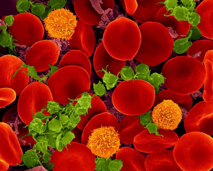
Red blood cells, platelets and T cells. Source: CC0

In a surprising discovery published in Science Advances, turning off the two proteins that regulate iron uptake results in not only anaemia but also in neutrophil levels plummeting. Iron deficiency, a known defence mechanism against infectious pathogens, can therefore a double-edged sword, as it simultaneously curbs the defensive power of this important arm of the innate immune system.

Iron is an indispensable component, needed for the blood pigment haemoglobin. The iron supply to the cells is controlled by the two proteins IRP-1 and IRP-2. If the cell lacks iron, IRP-1 and IRP-2 crank up the production of the various iron transporter proteins that take iron into the cell. IRP-1 and IRP-2 also ensure that an equally dangerous excess of iron does not occur.

IRP-1 and IRP-2 are essential for survival: mice lacking both control proteins during embryonic development die while still in the womb. But what happens when IRP-1 and IRP-2 fail in adult mice? A team led by Bruno Galy at the German Cancer Research Center (DKFZ) has now investigated this by shutting down IRP production in mice.

As the researchers had expected, the most striking change after IRPs were switched off was a pronounced decrease in red blood cells. Due to the lack of haemoglobin, these erythrocytes reached only a minimal size.

However, the researchers were surprised to see that white blood cells also decreased, mainly due to a deficiency of neutrophils, which account for up to two-thirds of white blood cells in humans.

The neutrophil decline was not caused by a mass die-off but a developmental blockade in the haematopoietic system: the precursor cells in the bone marrow no longer develop into mature neutrophils – an iron-dependent process. Other types of white blood cells, such as monocytes, were unaffected by the IRP-dependent developmental blockade.

Iron limitation is a double-edged sword

“This strong iron dependence of neutrophils was previously unknown. It possibly affects the immune defence against bacterial pathogens,” said senior author Bruno Galy. Yet iron deficiency is one of the body’s defence strategies in bacterial infections since many pathogens are dependent on iron. The body hoards the metal in certain cells to cut off access for pathogens, limiting their ability to replicate.

Galy is involved with another study also in Science Advances, which shows that iron deficiency in blood serum, as typically occurs with infections, leads to a decrease in neutrophils in mice and limits the ability of these immune cells to fight bacteria. “Iron deficiency apparently modulates the innate immune system. It suppresses the maturation of neutrophils and also throttles their defensive power,” commented Bruno Galy. “The limitation of available iron is apparently a double-edged sword: On the one hand, the body thereby prevents bacteria from spreading. On the other hand, the function of an important arm of the innate immune system suffers.”

Inflammation often leads to anaemia, as can be experienced by cancer patients. The researchers next want to investigate whether iron deficiency in chronic inflammation also impairs immune function.

Source: German Cancer Research Center (Deutsches Krebsforschungszentrum, DKFZ)

Lasers Turn Neutrophils into Medical Microbots

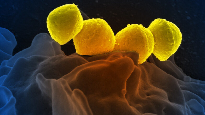
Streptococcus pyrogenese bound to human neutrophil
Streptococcus pyogenese bound to a human neutrophil. Credit: National Institute of Allergy and Infectious Diseases, National Institutes of Health

Bordering on science fiction, medicinal microrobots could help physicians better treat and prevent diseases. But a serious problem is the synthetic materials they are made of trigger immune responses. Now, for the first time, researchers report in ACS Central Science that they achieved precise control neutrophils as a natural, biocompatible microrobot by using lasers. By getting the ‘neutrobots’ to perform multiple tasks, the researchers demonstrated they could one day deliver drugs to precise locations in the body.

Microrobots being developed for medical applications would need to be administered in injections or oral capsules to get them inside the body. But these microscopic objects are often found to trigger immune reactions in small animals, resulting in the the microrobots being ejected from the body before they can carry out their tasks. By using the body’s own cells, such as neutrophils, drugs could be delivered less invasively without provoking an immune response.

Neutrophils already naturally pick up nanoparticles and dead red blood cells and can migrate through blood vessels into adjacent tissues, so they are good candidates for becoming microrobots. Previously, researchers have guided neutrophils with lasers in lab dishes, moving them around as ‘neutrobots’. However, this had not been tried in living animals. So, researchers set out to demonstrate the feasibility of light-driven neutrobots in animals using live zebrafish.

The researchers manipulated and moved neutrophils in zebrafish tails, using focused laser beams as optical tweezers. The ‘neutrobots’ could be moved up to a velocity of 1.3 µm/s, three times faster than a neutrophil’s natural speed. The optical tweezers were able to precisely and actively control the functions that neutrophils conduct as part of the immune system. For example, moving through a blood vessel wall into the surrounding tissue; carrying a plastic nanoparticle, showing potential for delivering medicine; and pushed towards red blood cell debris, a neutrophil engulfed the pieces. Surprisingly, at the same time, a different neutrophil, which wasn’t controlled by a laser, tried to naturally remove the cellular debris. Because they successfully controlled neutrobots in vivo, the researchers say this study advances the possibilities for targeted drug delivery and precise treatment of diseases.

Source: American Chemical Society

Major Mechanism for Chronic Inflammation in HIV Uncovered

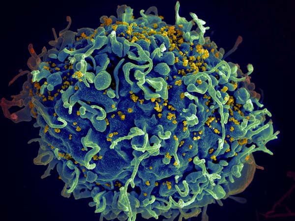
HIV invading a human cell
HIV invading a human cell: Credit NIH

In a groundbreaking study of people living with HIV, scientists found that neutrophils play a role in impaired T cell functions and counts, as well as the associated chronic inflammation that is common with the virus.

Neutrophils make up 60–80% of circulating immune cells in the blood. However, these white blood cells are extremely short-lived and cannot be frozen and thawed like other immune cells, making examining them extremely difficult, said study lead Shokrollah Elahi.

“Neutrophils live for hours to a day or two maximum,” Elahi said. “The body produces a lot of neutrophils, and they do their job and then they die and have to be regenerated in the bone marrow. But despite the fact that neutrophils are the most abundant white blood cells in the blood circulation, their role in the context of HIV has not been very well defined.”

In the study, published in the journal PLOS Biology, researchers examined fresh blood samples of 116 people living with HIV and 60 non-infected individuals. They ran comprehensive sequencing on all the genes expressed in the neutrophils from both groups to determine any differences between them.

“We found that not all HIV-infected individuals have similar types of neutrophils,” said Elahi. “As the HIV disease progresses, neutrophils become more activated and more potent, and in turn activate the body’s T cells, which likely causes some of the problems associated with HIV infection such as inflammation and rapid aging.”

Elahi said neutrophils act like an early alarm system: in response to pathogens, they release proteins to signal other immune cells to the danger. This activation can be high or low, or more or less potent, depending on the severity of the danger and the reaction of other immune cells.

One of these proteins is galectin-9, which Elahi previously linked to severe inflammation and cytokine storms in COVID patients. Elahi’s team reported that when neutrophils sense a danger such as an infection, they become stressed and release the galectin-9. As the protein begins to saturate the blood, it can interact with different immune cells. For example, the team found that galectin-9 reacted strongly with T cells and made them more susceptible to HIV infection, causing a cascading effect that leads to a hyper-immune response and inflammation.

Elahi’s prior work showed that patients with HIV and some forms of cancer had elevated levels of galectin-9 in their blood, but now he was able to identify the major source of the protein.

“We found for the very first time that the neutrophil membrane, through a complex mechanism, is covered like a blanket with galectin-9,” he said. “When neutrophils become highly activated, the secretion of galectin-9 can activate T cells through interaction with another molecule called CD44, which then promotes chronic inflammation in HIV patients.”

This ‘alarm’ reaction of shedding proteins such as galectin-9 was linked to oxidative stress, which is believed to play a role in the development of diseases including Parkinson’s, Alzheimer’s, cancer, heart failure and autism.

Based on his findings, Elahi said preventing galectin-9 shedding might be a powerful tool in reducing many of the negative effects of HIV infection. His team has already made some progress in reducing oxidative stress by using an organic antioxidant compound called phloretin and vitamin C.

“We have been looking at phloretin and vitamin C in the lab and our data are very promising,” Elahi said. “We know that both are good at reducing galectin-9 shedding, so we believe they can prevent the hyper-activation of neutrophils. We hope that our results will spark renewed investigation into the role of neutrophils in T cell activation in other acute and chronic conditions.”

Elahi noted the importance of immediate screening tests for HIV or at-risk people, saying: “If the virus is caught early and they can go on antiretroviral therapy, then it stops disease progression and reduces many of the complications associated with advanced HIV.”

Source: University of Alberta Faculty of Medicine & Dentistry