Tag: neuroscience

A Step Closer to Effective Electrical Pain Blocking

Source: Unsplash CC0

New research from the University of Connecticut has brought the drug-free technology of electrical anaesthesia for all chronic pain sufferers a step closer. 

Pain stimuli, or ‘nociceptive stimuli’ is picked up by nociceptors which send signals to the spinal cord, which passes it on to the brain where the perception of pain is manifested.

Bin Feng, associate professor in the Biomedical Engineering Department, led research which discovered how electrical stimulation of the dorsal root ganglia (DRG), sensory neural cell body clusters, can block nociceptive signal transmission to the spinal cord and prevent the brain from perceiving chronic pain signals. The findings are reported in PAIN.

Electrical devices to treat pain typically deliver electrical signals to the peripheral nervous system and spinal cord to block nociceptive signals from reaching the brain.

A major obstacle with these devices is that while some patients find them beneficial in relieving their chronic pain, others have little or no pain reduction. Despite incremental developments of neurostimulator technologies, there has not been much improvement in getting the devices to work for these patients.

“The trouble with this technology is that it can benefit a portion of patients very well, but for a larger portion of patients it has little benefit,” Prof Feng said.

One of the reasons is that such devices lag behind research into neural stimulation.

“We’re sitting on a huge pile of clinical data,” Prof Feng says. “But the science of neuromodulation remains understudied.”

Neurostimulators relieve pain according to a ‘gate control’ theory. Our bodies can detect both innocuous stimuli, like something brushing against the skin, and painful stimuli, through low- and high-threshold sensory neurons, respectively.

The spinal cord ‘gate’ can be shut by activating low-threshold sensory neurons, preventing painful nociceptive signals from high-threshold sensory neurons from crossing the spinal cord to the brain.

Neurostimulators reduce pain in patients by activating low-threshold sensory neurons with electrical pulses. This usually causes a non-painful tingling sensation in certain areas of the skin, or paresthaesia, masking the perception of pain.

Many patients receiving DRG stimulation treatment reported pain relief without the expected paraesthesia.

Seeking to understand this, Prof Feng’s lab discovered that electrical stimulation to the DRG can block transmission to the spinal cord at frequencies as low as 20 hertz. This is in contrast to previous research indicating that blocking requires kilohertz electrical stimulation.

“The cell bodies of sensory neurons form a T-junction with the peripheral and central axons in the DRG,” Feng says. “This T-junction appears to be the region that causes transmission block when DRG is stimulated.”

More remarkably, sensory nerve fibres with different characteristics are blocked by different electrical stimulation frequency ranges at the DRG, allowing the development of new neural stimulation protocols to enhance selective transmission blocking with different sensory fibre types.

“A-fibre nociceptors with large axon diameters are generally responsible for causing acute and sharp pain,” Prof Feng explained. “It is the long-lasting and dull-type pain that bothers the chronic pain patients mostIn a chronic pain condition, C-fibre nociceptors with small axon diameter and no myelin sheath play central role in the persistence of pain. Selectively blocking C-fibres while leaving A-fibres intact can be a promising strategy to target the cause of chronic pain.”

This provides evidence to place more electrodes for devices that target the DRG and surrounding neuronal tissues, letting doctors provide more precise neuromodulation.

“The next-generation neurostimulators will be more selective with fewer off-target effects,” Prof Feng said. “They should also be more intelligent by incorporating chemical and electrical sensory capabilities and ability to communicate bidirectionally to a cloud-based server.”

Prof Feng hopes that more people will be eventually able to achieve chronic pain relief with this technology. He is now working toward conducting clinical studies with his collaborators at UConn Health to test the efficacy of this method in humans.

Source: University of Connecticut

A Distinct Neural Signature for Teams ‘in the Zone’

Photo by Lagos Techie on Unsplash

Researchers have discovered that there are brain waves and regions sensitive to team flow (ie, being ‘in the zone’ together) compared to non-engaging teamwork or a solo flow.

Flow experiences are considered to be some of the most enjoyable, rewarding, and engaging experiences of all, and typically involve automatic and effortless action coupled with intense focus. The benefits of having flow experiences are still being catalogued, but include improved overall quality of life, increased self-efficacy, and a stronger sense of self.

This is the first study to objectively measure this psychological state. These neural correlates not only can be used to understand and predict the team flow experience, but could be used to monitor and predict team performance. This is an area the authors are currently investigating/
Team flow is experienced when team players get ‘in the zone’ to accomplish a task together. Successful teams experience this psychological phenomenon, ranging from sports to bands and even in the office. When teamwork reaches the team flow level, one can observe the team perform in harmony, breaking their performance limits.

In order to investigate neural processing of this team flow state, something which has been a challenge for decades, it has to be reproduced in the lab and measured.

Researchers at at Toyohashi University of Technology and California Institute of Technology found solutions to these challenges and provided the first neuroscience evidence of team flow. Using 10 teams of two playing a music video game together, the researchers measured the team members’ brain activity using EEG. In some trials, a partition separated the teammates so they couldn’t see each other while they played, allowing a solo flow state but preventing team flow.

The research team scrambled the music in other trials, thereby preventing a flow state but still enabling teamwork. Participants also answered questions after each game to assess their level of flow. The researchers also developed an objective neural method to evaluate the depth of the team flow experience. Team flow was marked by a unique signature: increased beta and gamma brain waves in the middle temporal cortex, a type of brain activity linked to information processing. In comparison to the regular teamwork state, teammates also had more synchronised brain activity during the team flow state.

Neural models from this study can inform more effective team-building strategies in areas where human performance and pleasure matters, such as sports, business and music. This will also enable improved team performance.

Enhancing performance while maintaining enjoyment will improve quality of life, which could result in reduced mental health problems.

Source: Medical Xpress

MRI and Massage Stones Help Unlock Mystery of Sensory Associations

Photo by engin akyurt on Unsplash

By using hot and cold massage stones, scientists have found that the brain’s prefrontal cortex conjures up sensations based on other sensory information, such as feeling warmth when viewing a beach.

Publishing their findings in The Journal of Neuroscience, the researchers investigated patterns of neural activity in the prefrontal cortex as well as the other regions of the brain known to be responsible for processing stimulation from all the senses and discovered significant similarities.

“Whether an individual was directly exposed to warmth, for example, or simply looking at a picture of a sunny scene, we saw the same pattern of neural activity in the prefrontal cortex,” said Dirk Bernhardt-Walther, an associate professor in the department of psychology in the Faculty of Arts & Science, and coauthor of a study published last week in the Journal of Neuroscience describing the findings. “The results suggest that the prefrontal cortex generalizes perceptual experiences that originate from different senses.”

To understand how the human brain processes the torrent of information from the environment, researchers often study the senses in isolation, with much prior work focused on the visual system. Bernhardt-Walther says that while such work is illuminating and important, it is equally important to find out how the brain integrates information from the different senses, and how it uses the information in a task-directed manner. “Understanding the basics of these capabilities provides the foundation for research of disorders of perception,” he said.

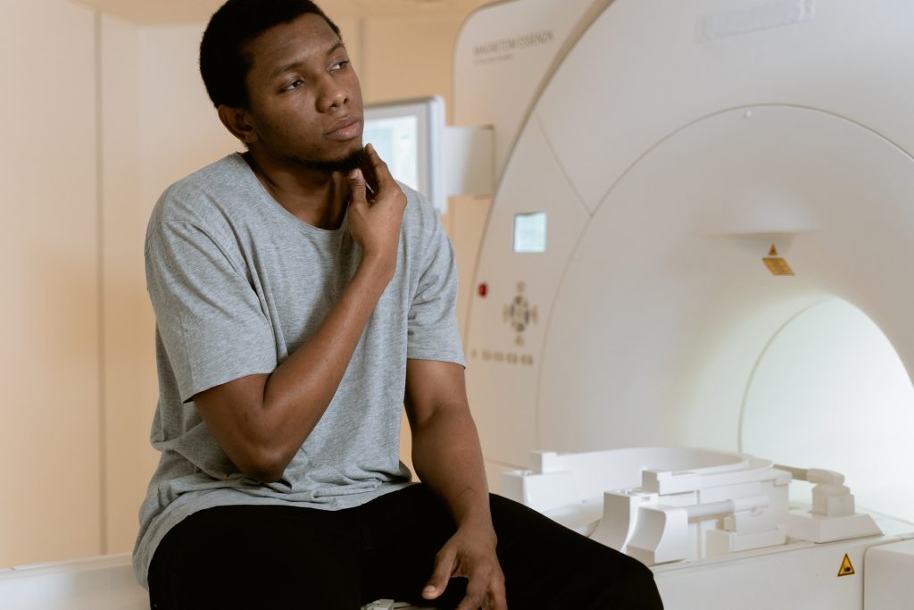
Capturing brain activity with functional magnetic resonance imaging (fMRI), the researchers conducted two experiments with the same participants, based on knowing how regions of the brain respond differently depending on the intensity of stimulation.

In the first, the participants viewed images of various scenes, such as beaches, city streets, forests and train stations, and were asked to judge if the scenes were warm or cold and noisy or quiet.

For the second experiment, participants were first handed a series of massage stones that were either heated to 45C or cooled to 9C, and later exposed to a variety of sounds such as birds, people and waves at a beach.

“When we compared the patterns of activity in the prefrontal cortex, we could determine temperature both from the stone experiment and from the experiment with pictures as the neural activity patterns for temperature were so consistent between the two experiments,” said lead author of the study Yaelan Jung, who recently completed her PhD at U of T working with Bernhardt-Walther and is now a postdoctoral researcher at Emory University.

“We could successfully determine whether a participant was holding a warm or a cold stone from patterns of brain activity in the somatosensory cortex, which is the part of the brain that receives and processes sensory information from the entire body – while brain activity in the visual cortex told us if they were looking at an image of a warm or cold scene.”

“Overall, the neural activity patterns in the prefrontal cortex produced by participants viewing the images were the same as those triggered by actual experience of temperature and noise level,” said Dr Jung.

This opens up insights into how the brain processes and represents complex real-world attributes that span multiple senses, even without directly experiencing them.

“In understanding how the human brain integrates information from different senses into higher-level concepts, we may be able to pinpoint the causes of specific inabilities to recognise particular kinds of objects or concepts,” said Bernhardt-Walther.

“Our results might help people with limitations in one sensory modality to compensate with another and reach the same or very similar conceptual representations in their prefrontal cortex, which is essential for making decisions about their environment.”

Source: University of Toronto

Carbon Fibre Electrodes Allow Unprecedented Neural Recording

Image by Robina Weemeijer on Unsplash

A tiny, implantable carbon fibre electrode has the potential to provide a long-term brain-computer interface which can record electrical signals over lengthy periods of time.

The carbon fibre electrodes were developed at the University of Michigan and demonstrated in rats. The new research shows the promise of carbon fibre electrodes in recording electrical signals from the brain without damaging brain tissue. Directly implanting carbon fiber electrodes into the brain allows the capturing of bigger and more specific signals than current technologies.

This technology could lead to advances that could give amputees and those with spinal injuries control of advanced prosthetics, stimulate the sacral nerve to restore bladder control, stimulate the cervical vagus nerve to treat epilepsy and provide deep brain stimulation as a possible treatment for Parkinson’s.  

“There are interfaces out there that can be implanted directly into the brain but, for a variety of reasons, they only last from months to a few years,” said Elissa Welle, a recent PhD graduate from the U-M Department of Biomedical Engineering. “Any time you’re opening up the skull for a procedure involving the brain, it’s a big deal.”

Brain implants are typically made from silicon due to its ability to conduct electricity and its historic use in cleanroom technology. But silicon is not very biocompatible and leads to the formulation of scar tissue over long periods. Such electodes will eventually degrade and no longer capture brain signals, requiring removal.

Carbon fibres may be the answer to getting high-quality signals with an interface that lasts years, not months. And by laser cutting and sharpening carbon fibers into tiny, subcellular electrodes in the lab with the help of a small blowtorch, U-M engineers have harnessed the potential for excellent signal capture in a form the body is more likely to accept.

“After implantation, it sits inside the brain in a way that does not interfere with the surrounding blood vessels, because it’s smaller than those blood vessels,” Welle said. “They’ll move around and adjust to an object that small, rather than get torn as they would when encountering larger implants.”

Part of the electrode’s compatibility in brain tissue is down to smaller size, but its needle-like shape may also minimise compacting of any surrounding tissue. Larger carbon-based electrodes have been shown to actually encourage neural tissue to grow instead of degrading. The team is hopeful that similar potential for their carbon fibre electrodes will be revealed by further testing.

Carbon fibre electrodes in a previous study dramatically outperformed conventional silicon electrodes with 34% of electrodes recording a neuron signal compared to 3%. Laser cutting then improved this number to 71% at 9 weeks after implantation. Flame sharpening has now enabled these high performance probes to be implanted directly into the cerebral cortex, negating the need for a temporary insertion aid, or shuttle, as well as into the rat’s cervical vagus nerve.

It is relatively easy to insert electrodes into the brain. But the researchers have also taken on the more difficult task of inserting the sharpened carbon fibre electrodes into nerves, with micrometre diameters.

Those findings show that potential for these electrodes goes beyond prosthetic manipulation, according to Cindy Chestek, a U-M associate professor of biomedical engineering, and principal investigator of the The Cortical Neural Prosthetics Lab.

“Someone who is paralysed may have no control over things like their bladder, for example,” Prof Chestek said. “We may be able to utilise these smaller electrodes to stimulate and record signals from areas that can’t be reached by larger ones, maybe the neck or spinal cord, to help give patients some level of control.”

Source: University of Michigan

Psychedelic Compound Treats Depression by Growing Neural Connections

Photo by Timothy Dykes on Unsplash

In a new study, researchers have shown that a single dose of psilocybin, a psychedelic compound with potential applications for depression, prompted long-lasting increase in connections between neurons in mice. 

For some people, psilocybin, an active compound in ‘magic mushrooms’, can produce a profound mystical experience. The psychedelic was a staple of religious ceremonies among indigenous populations of the Americas and is also a popular recreational drug. It has been the subject of some interest in treating depression. But exactly how it works in the brain and how long beneficial results might last is still unclear.
“We not only saw a 10% increase in the number of neuronal connections, but also they were on average about 10% larger, so the connections were stronger as well,” reported senior author Alex Kwan, associate professor of psychiatry and of neuroscience at Yale.

Earlier work had found promising evidence that psilocybin, as well as the anaesthetic ketamine, could decrease depression. This new study found that these compounds increase the density of dendritic spines, which are small protrusions found on nerve cells which aid in the transmission of information between neurons. The number of these neuronal connections are known to be reduced by chronic stress and depression.


Prof Kwan and first author Ling-Xiao Shao, a postdoctoral associate, imaged dendritic spines in high resolution with a laser-scanning microscope, and tracked them for multiple days in living mice. They found increases in the number of dendritic spines and in their size within 24 hours of administration of psilocybin. These changes were still evident a month later. Also, mice subjected to stress showed behavioural improvements and increased neurotransmitter activity after being given psilocybin.

It may be the novel psychological effects of psilocybin itself that spurs the growth of neuronal connections, Kwan said.

“It was a real surprise to see such enduring changes from just one dose of psilocybin,” he said.  “These new connections may be the structural changes the brain uses to store new experiences.”

Source: Yale University

Call for More Neuroscience Research in Africa

Photo by MART PRODUCTION from Pexels

A team of neuroscientists are calling for greater support of neuroscience research in Africa based on an analysis of the continent’s past two decades of research outputs.  

The findings reveal important information about the nature of funding and international collaboration comparing activity in the continent to other countries, mainly the US, UK and areas of Europe. It is hoped that the study will provide useful data to help further develop science in Africa.  

The greatest human genetic diversity is found in Africa, and Eurasian genomes have less variation than African ones; in fact, Eurasian genomes can be considered a subset of African ones. This carries important implications for understanding human diseases, including neurological disorders.

Co-lead senior author Tom Baden, Professor of Neuroscience in the School of Life Sciences and the Sussex Neuroscience research group at the University of Sussex said: “One beautiful thing about science is that there is no such thing as a truly local problem. But that also means that there should be no such thing as a local solution – research and scientific communication by their very nature must be a global endeavour.  

“And yet, currently the vast majority of research across most disciplines is carried out by a relatively small number of countries, located mostly in the global north. This is a huge waste of human potential.”  

The team, made up of experts from the University of Sussex, the Francis Crick Institute and institutions from across Africa, analysed the entirety of Africa’s outputs in neuroscience over two decades. A lot of early neuroscience research took place in Egypt, it was pointed out.

Lead author Mahmoud Bukar Maina, a Research Fellow in the School of Life Sciences and the Sussex Neuroscience research group at the University of Sussex and visiting scientist at Yobe State University, Nigeria, explained: “Even though early progress in neuroscience began in Egypt, Africa’s research in this area has not kept pace with developments in the field around the world. There are a number of reasons behind this and, for the first time, our work has provided a clear picture of why – covering both strengths and weaknesses of neuroscience research in Africa and comparing this to other continents.  

“We hope it will provide useful data to guide governments, funders and other stakeholders in helping to shape science in Africa, and combat the ‘brain drain’ from the region.”  

Co-lead senior author Lucia Prieto-Godino, a Group Leader at the Francis Crick Institute, said: “One of the reasons why this work is so important, is that the first step to solve any problem is understanding it. Here we analyse key features and the evolution of neuroscience publications across all 54 African countries, and put them in a global context. This highlights strengths and weaknesses, and informs which aspects will be key in the future to support the growth and global integration of neuroscience research in the continent.” 

The study identifies the African countries with the greatest research outputs, revealing that most research funding originates from external sources such as the USA and UK.  

The researchers argue that a sustainable African neuroscience research environment needs local funding, suggesting that greater government backing is needed as well as support from the philanthropic sector.  
Professor Baden added: “One pervasive problem highlighted in our research was the marked absence of domestic funding. In most African countries, international funding far predominates. This is doubly problematic.  

“Firstly, it takes away the crucial funding stability that African researchers would need to meaningfully embark on large-scale and long-term research projects, and secondly, it means that the international, non-African funders essentially end up deciding what research is performed across the continent. Such a system would generate profound outrage across places like Europe – how then can it be acceptable for Africa?”

A number of the researchers involved in the study are members of TReND Africa, a charity supporting scientific capacity building in Africa.  

Source: University of Sussex

Journal information: M. B. Maina et al, Two decades of neuroscience publication trends in Africa, Nature Communications (2021). DOI: 10.1038/s41467-021-23784-8 , www.nature.com/articles/s41467-021-23784-8

Molecule Found to Play a Key Role in Brain Rejuvenation

Image source: Pixabay

A new study shows that a molecule could play a key role in support cells in the brain, allowing them to repair and properly communicate.

Studies have shown that new brain cells continually formed in response to injury, physical exercise, and mental stimulation. Glial cells, and in particular oligodendrocyte progenitors, are highly responsive to external signals and injuries. They can detect changes in the nervous system and form new myelin, which forms a sheath around nerves, providing metabolic support and accurate transmission of electrical signals. However, less myelin is formed with age, and this progressive decline has been linked to the age-related cognitive and motor deficits observed in older people. Impaired myelin formation also has been reported in older individuals with neurodegenerative diseases such as Multiple Sclerosis or Alzheimer’s and identified as one of the causes of their progressive clinical deterioration.

A new study from the Neuroscience Initiative team at the Advanced Science Research Center at The Graduate Center, CUNY (CUNY ASRC) has identified a molecule called ten-eleven-translocation 1 (TET1) as a necessary component of myelin repair. shows that TET1 modifies the DNA in specific glial cells in adult brains so they can form new myelin in response to injury. The study was published in Nature Communications.

“We designed experiments to identify molecules that could affect brain rejuvenation,” said lead author Sarah Moyon, PhD, a research assistant professor with the CUNY ASRC Neuroscience Initiative. “We found that TET1 levels progressively decline in older mice, and with that, DNA can no longer be properly modified to guarantee the formation of functional myelin.”

The authors are currently exploring whether raising levels of TET1 in older mice could rejuvenate the oligodendroglial cells, restoring their regenerative functions.

Combining whole-genome sequencing bioinformatics, the authors showed that the DNA modifications induced by TET1 in young adult mice were essential to promote healthy communication among central nervous system cells and for ensuring proper function. The authors also showed that young adult mice with a genetic modification of TET1 in the myelin-forming glial cells could not produce functional myelin, and so behaved like older mice.

“This newly identified age-related decline in TET1 may account for the inability of older individuals to form new myelin,” said Patrizia Casaccia, founding director of the CUNY ASRC Neuroscience Initiative, a professor of Biology and Biochemistry at The Graduate Center, CUNY, and the study’s primary investigator. “I believe that studying the effect of aging in glial cells in normal conditions and in individuals with neurodegenerative diseases will ultimately help us design better therapeutic strategies to slow the progression of devastating diseases like multiple sclerosis and Alzheimer’s.”

The findings could also hold important implications for molecular rejuvenation of ageing brains in healthy individuals, the researchers noted. Future studies aimed at increasing TET1 levels in older mice are underway to define whether the molecule could restore new myelin formation and favour proper neuro-glial communication. The long-term goal of the team is to promote recovery of cognitive and motor functions in older people and in patients with neurodegenerative diseases.

Source: Advanced Science Research Center

Free Will not Undermined by Neuroscience

Image by Falkurian Design on Unsplash

A new article argues that recent research undermines the notion that free will is an illusion, due to the buildup of brain activity before conscious movement.

Experiments from the 1960s to 1980s measured brain signals, leading many neuroscientists to believe that our brains make decisions before we do — that human actions were initiated by electrical waves, and therefore did not reflect free, conscious thought.

However, a new article in Trends in Cognitive Science argues that recent research undermines this contention against free will.

Study co-author Adina Roskies, and Helman Family Distinguished Professor, Dartmouth College, said: “This new perspective on the data turns on its head the way well-known findings have been interpreted. The new interpretation accounts for the data while undermining all the reasons to think it challenges free will.”

The debate over free will is mostly built on 1980s research using electroencephalograms to study brain activity. The EEG-based research measured when electrical signals begin to build in the brain, relative to when a person is aware of their desire to initiate a movement. The averaged data showed a buildup before movement that became known as the ‘readiness potential‘ (RP).

That research, conducted by neurophysiologist Benjamin Libet, contended that if the RP was present before a person had a conscious thought about moving, free will therefore could not be responsible for either the buildup of electrical signals or the subsequent movement.

This part of Libet’s logic was based on a likely false premise, the researchers argue.

“Because the averaged readiness potential reliably precedes voluntary movement, people assumed that it reflected a process specifically directed at producing that movement. As it turns out, and as our model has shown, that is not necessarily the case,” explained co-author Aaron Schurger, an assistant professor of psychology at Chapman University.

The article notes new research using computational modeling that indicates that the RP’s standard interpretation should be reassessed, especially in relation to the question of free will.

The study highlights findings suggesting that the RP — the pre-movement buildup of activity — reflects the neural activity that underlies the formation of a decision to move, as opposed to the outcome of a decision to move.

“These new computational models account for the consistent finding of the readiness potential without positing anything like an RP in individual trials. The readiness potential itself is a kind of artifact or illusion, one which would be expected to appear just as it does give the experimental design, but doesn’t reflect a real brain signal that begins with the RP onset or is read out by other areas,” said Prof Roskies.

Numerous challenges exist to the idea that the RP causes humans to act: isolating RP from other electrical signals in the brain; RP presence in tasks where motor activity is not needed; and ‘noise’ in analyses trying to confirm that RP initiates movement.

False positives, where RP is observed but fails to initiate movement, and inconsistencies in the lag between brain wave buildup and movement also complicate the understanding of the connection between the electrical activity in the brain and free will. Finally, there are philosophical implications to attempting to explore free will with brain data.

Source: Medical Xpress

Journal information: Aaron Schurger et al, What Is the Readiness Potential?, Trends in Cognitive Sciences (2021). DOI: 10.1016/j.tics.2021.04.001

Researchers Discover that Humans can Readily Develop Echolocation Ability

Photo by Pawel Czerwinski on Unsplash

The ability for humans to sense their surrounding space with reflected sounds might sound like a superhero’s ability, but it is a skill that is developed by some blind people, who use clicks as a form of echolocation.

Echolocation is an ability known in dolphins, whales and bat species, which occurs when such animals emit a sound that reflects off objects in the environment, returning echoes that provide information about the surrounding space.

Existing research has shown that some blind people may use click-based echolocation to judge spaces and improve their navigation skills. Armed with this information, a team of researchers led by Dr Lore Thaler explored how people acquire this skill.

Over the course of a 10-week training programme, the team investigated how blindness and age affect learning of click-based echolocation. They also studied how learning this skill affects the daily lives of people who are blind.

Both blind and sighted people between 21 and 79 years of age participated in this study, which provided a training course of 10 weeks. Blind participants also took part in a 3-month follow up survey assessing how the training affected their daily life.

Both sighted and blind people improved considerably on all measures, and in some cases performed as well as expert echolocators did at the end of training. A surprising result was that a few sighted people even performed better than those who were blind.

However, neither age nor blindness limited participants’ rate of learning or in their ability to apply their echolocation skills to novel, untrained tasks.

Furthermore, in the follow up survey, all participants who were blind reported improved mobility, and 83% reported better independence and wellbeing.

Age or vision not a limitation

Overall, the results suggest that the ability to learn click-based echolocation is not strongly limited by age or level of vision. This has positive implications for the rehabilitation of people with vision loss or in the early stages of progressive vision loss.

Click-based echolocation is not presently taught as part of mobility training and rehabilitation for blind people. There is also the possibility that some people are reluctant to use click-based echolocation due to a perceived stigma around  the click sounds in social environments.

Despite this, the results indicate that both blind people who use echolocation and people new to echolocation are confident to use it in social situations, indicating that the perceived stigma is likely less than believed.

Source: Durham University

Journal information: Human click-based echolocation: Effects of blindness and age, and real-life implications in a 10-week training program, PLOS ONE (2021)

Precise Ultrasound Heating of Neurons Could Treat Neurological Disorders

Image source: Fakurian Design on Unsplash

A multidisciplinary team at Washington University in St. Louis has developed a new brain stimulation technique using focused ultrasound that is able to turn specific types of neurons in the brain on and off and precisely control motor activity without surgical device implantation.

Being able to turn neurons on and off can treat certain neurological disorders such as Parkinson’s disease and epilepsy. Used for over six decades, deep brain stimulation techniques have had some treatment success in neurological disorders, but those require surgical device implantation. 

The team, led by Hong Chen, assistant professor of biomedical engineering in the McKelvey School of Engineering and of radiation oncology at the School of Medicine, is the first to provide direct evidence showing noninvasive activation of specific neuron types in mammalian brains by combining an ultrasound-induced heating effect and genetics, which they have named sonothermogenetics. It is also the first work to show that the ultrasound- genetics combination can robustly control behaviour by stimulating a specific target deep in the brain.

The results of the three years of research were published online in Brain Stimulation

“Our work provided evidence that sonothermogenetics evokes behavioural responses in freely moving mice while targeting a deep brain site,” Chen said. “Sonothermogenetics has the potential to transform our approaches for neuroscience research and uncover new methods to understand and treat human brain disorders.”

Chen and colleagues delivered a viral construct containing TRPV1 ion channels to genetically-selected neurons in a mouse model. Then, they delivered small pulses of heat generated by low-intensity focused ultrasound to the selected neurons in the brain via a wearable device. The heat, only a few degrees warmer than body temperature, activated the TRPV1 ion channel, which then acted as a switch to turn the neurons on or off.

“We can move the ultrasound device worn on the head of free-moving mice around to target different locations in the whole brain,” said Yaoheng Yang, first author of the paper and a graduate student in biomedical engineering. “Because it is noninvasive, this technique has the potential to be scaled up to large animals and potentially humans in the future.”

Building on prior research from his lab, professor of biomedical engineering Jianmin Cui and his team found for the first time that ion channel activity can be influenced by ultrasound alone, possibly leading to new and noninvasive ways to control the activity of specific cells. They discovered that focused ultrasound modulated the currents flowing through the ion channels on average by up to 23%, depending on channel and stimulus intensity. Following this work, researchers found close to 10 ion channels with this capability, but all of them are mechanosensitive, not thermosensitive.

The work also builds on the concept of optogenetics, the combination of the targeted expression of light-sensitive ion channels and the precise delivery of light to stimulate neurons deep in the brain. While optogenetics has increased discovery of new neural circuits, it has limited penetration depth due to light scattering, requiring surgical implantation of optical fibres to reach deeper into the brain.

Sonothermogenetics has the promise to target any location in the mouse brain with millimetre-scale resolution without causing any damage to the brain, Chen said. She and her team are further refining the technique and validating their work.

Source: Sci Tech Daily

Journal information: Yaoheng Yang et al, Sonothermogenetics for noninvasive and cell-type specific deep brain neuromodulation, Brain Stimulation (2021). DOI: 10.1016/j.brs.2021.04.021