Tag: neurology

Exercise Protects Ageing Synapses

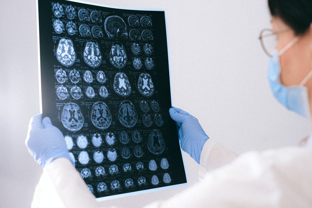
Photo by Barbara Olsen from Pexels

When elderly people stay active, their brains have more proteins that enhance the connections between neurons to maintain healthy cognition, according to a study published in Alzheimer’s & Dementia: The Journal of the Alzheimer’s Association.

This protective impact was found even in people whose brains at autopsy were riddled with amyloid and tau proteins, associated with Alzheimer’s and other neurodegenerative diseases.

“Our work is the first that uses human data to show that synaptic protein regulation is related to physical activity and may drive the beneficial cognitive outcomes we see,” said lead author Kaitlin Casaletto, PhD.

The beneficial effects of physical activity on cognition have been shown in mice but have been much harder to demonstrate in people.

Dr Casaletto collaborated with William Honer, MD, a professor of psychiatry at the University of British Columbia and senior author of the study, to leverage data from the Memory and Aging Project at Rush University in Chicago. The project tracked the late-life physical activity of elderly participants, who also agreed to donate their brains upon death.

“Maintaining the integrity of these connections between neurons may be vital to fending off dementia, since the synapse is really the site where cognition happens,” Dr Casaletto said. “Physical activity – a readily available tool – may help boost this synaptic functioning.”

The researchers found that elderly people who remained active had higher levels of proteins that facilitate the exchange of information between neurons. This result dovetailed with Prof Honer’s earlier finding that people who had more of these proteins in their brains when they died were better able to maintain their cognition late in life.

Surprisingly, the effects ranged beyond the hippocampus to include other brain regions associated with cognitive function.

“It may be that physical activity exerts a global sustaining effect, supporting and stimulating healthy function of proteins that facilitate synaptic transmission throughout the brain,” Prof Honer said.

The brains of most older adults accumulate amyloid and tau proteins that are the hallmarks of Alzheimer’s disease pathology. Many scientists believe amyloid accumulates first, then tau, causing synapses and neurons to fall apart.

Dr Casaletto previously found that synaptic integrity, whether measured in the spinal fluid of living adults or the brain tissue of autopsied adults, appeared to dampen the relationship between amyloid and tau, and between tau and neurodegeneration.

“In older adults with higher levels of the proteins associated with synaptic integrity, this cascade of neurotoxicity that leads to Alzheimer’s disease appears to be attenuated,” she said. “Taken together, these two studies show the potential importance of maintaining synaptic health to support the brain against Alzheimer’s disease.”

Source: University of California in San Francisco

New Genetic Insights into Basal Ganglia Diseases

Source: Pixabay

A new study published in Developmental Medicine & Child Neurology uncovered a number of genetic causes of basal ganglia diseases.

Basal ganglia are deep grey matter structures in the brain involved in the control of posture and voluntary movements, cognition, behaviour, and motivational states. Several conditions are known to affect basal ganglia during childhood, but many questions remain.

In a study that included 62 children with basal ganglia diseases who were followed for two years, investigators identified multiple genetic aetiologies including mitochondrial diseases (57%), Aicardi–Goutières syndrome (20%), and single-gene causes of dystonia and/or epilepsy (17%) mimicking Leigh syndrome. Radiological abnormalities included T2-hyperintense lesions (n=26) and lesions caused by calcium or manganese mineralisation (n=9).

The researchers identified three clusters: the pallidal, neostriatal, and striatal, plus the last including mtDNA defects in the oxidative phosphorylation system with prominent brain atrophy. Mitochondrial biomarkers showed poor sensitivity and specificity in children with mitochondrial disease, whereas an interferon signature was observed in all patients with Aicardi–Goutières syndrome.

Radiological imaging tests also revealed several characteristics in patients that could help lead to an earlier diagnosis of basal ganglia diseases.

Source: Wiley

About 1% of Hospitalised COVID Patients Develop Neurological Complications

49-year-old female with past medical history of mitral valve disease and tricuspid valve regurgitation who developed headache followed by cough and fever presented to the ER with right upper eyelid ptosis (drooping). Credit: Radiological Society of North America and Scott H. Faro, M.D.

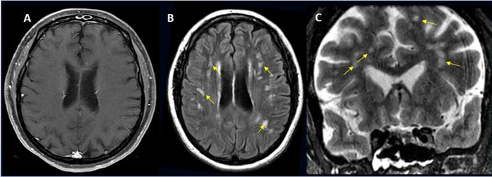
Approximately one in 100 patients hospitalised with COVID will likely develop complications of the central nervous system, according to a large international study. These can include stroke, haemorrhage, and other potentially fatal complications. The study was presented at the annual meeting of the Radiological Society of North America (RSNA).

“Much has been written about the overall pulmonary problems related to COVID, but we do not often talk about the other organs that can be affected,” said study lead author Scott H. Faro, MD, FASFNR, professor of radiology and neurology at Thomas Jefferson University. “Our study shows that central nervous system complications represent a significant cause of morbidity and mortality in this devastating pandemic.”

Dr Faro initiated the study after finding that only a small number of cases informed existing literature on central nervous system complications in hospitalised COVID patients.

To build a more complete picture, he and his colleagues analysed nearly 40 000 cases of hospitalised COVID patients, admitted between September 2019 and June 2020. Their average age was 66 years old, and two thirds were men.

Confusion and altered mental status were the most common causes of admission followed by fever. Comorbidities such as hypertension, cardiac disease and diabetes were common.

There were 442 acute neuroimaging findings most likely associated with the viral infection, with central nervous system complications in 1.2% of this large patient group.

“Of all the inpatients who had imaging such as MRI or a CT scan of the brain, the exam was positive approximately 10% of the time,” Dr Faro said. “The incidence of 1.2% means that a little more than one in 100 patients admitted to the hospital with COVID are going to have a brain problem of some sort.”

Ischaemic stroke, with an incidence of 6.2%, was the most common complication, followed by intracranial haemorrhage (3.72%) and encephalitis (0.47%).

A small percentage of unusual findings was uncovered, such as acute disseminating encephalomyelitis, an inflammation of the brain and spinal cord, and posterior reversible encephalopathy syndrome, a syndrome that mimics many of the symptoms of a stroke.

“It is important to know an accurate incidence of all the major central nervous system complications,” Dr Faro said. “There should probably be a low threshold to order brain imaging for patients with COVID.”

Source: EurekAlert!

Scientists Identify A New Recessive Neurodevelopmental Disorder

Image source: Pixabay

In the Journal of Clinical Investigation, researchers have reported a rare neurodevelopmental condition characterised by intellectual disability, ataxia with cerebellar hypoplasia and delayed puberty with hypogonadotropic hypogonadism (HH).

Patients with this unusual combination of conditions were referred to Mehul Dattani (UCL), and affected individuals were found to carry the same homozygous mutation in the PRDM13 gene, which encodes a chromatin modifying factor that contributes to regulating cell fate. Intriguingly, an unaffected heterozygous carrier of this mutation was identified by screening 42 unaffected individuals in the Maltese population, suggesting that this mutation is present at low levels in the population.

The researchers set out to model this condition and identify the underlying causes using a PRDM13-deficient mouse model. The researchers found evidence that both the cerebellar hypoplasia and reproductive phenotypes resulted from defects in the specification of specific populations of GABAergic neuronal progenitors in the developing cerebellum and hypothalamus, respectively.

The results indicate that this condition results from abnormal cell fate specification during development. Consequently, the hypoplastic cerebellum is deficient in molecular layer interneurons, which play critical roles in regulating cerebellar circuits. In the hypothalamus, fewer Kisspeptin neurons, which are important regulators of gonadotropin releasing hormone and puberty, were present in PRDM13 mutant mice.

Together, these findings identify PRDM13 as a critical regulator of neuronal cell fate in the cerebellum and hypothalamus, providing a mechanistic explanation for the co-occurrence of hypogonadism and cerebellar hypoplasia in this syndrome.

Source: King’s College London

Scientists Discover New Type of Neuron in the Retina

Source: Daniil Kuzelev on Unsplash

University of Utah scientists have discovered a new type of neuron in the retina, which will help fill in our understanding of how sensory information is relayed.

In the central nervous system a complex network of neurons communicate with each other to relay sensory and motor information. In this chain of communication, a type of neuron called interneurons serve as intermediaries . A research team led by Ning Tian, PhD, identified a previously unknown type of interneuron in the mammalian retina. Their findings were published in the journal PNAS.

This discovery is a major step forward for the field as scientists strive to build a better understanding of the central nervous system by identifying all classes of neurons and their connections.

“Based on its morphology, physiology, and genetic properties, this cell doesn’t fit into the five classes of retinal neurons first identified more than 100 years ago,” said Dr Tian. “We propose they might belong to a new retinal neuron class by themselves.”

The research team called their discovery the Campana cell after its shape, which resembles a hand bell. Campana cells relay visual signals from both types of light-sensing rod and cone photoreceptors in the retina, however their exact purpose is the subject of ongoing research. Experiments revealed that Campana cells remain activated for an unusually long time – as long as 30 seconds – in response to a 10 millisecond light flash stimulation.

“In the brain, persistent firing cells are believed to be involved in memory and learning,” said Dr Tian. “Since Campana cells have a similar behaviour, we theorise they could play a role in prompting a temporal ‘memory’ of a recent stimulation.”

Source: University of Utah

Protecting Newborns’ Brains During Rewarming Stage of Cooling Therapy

Photo by William-Fortunato on Pexels

Oxygen-deprived newborns who undergo hypothermia therapy have a higher risk of seizures and brain damage during the rewarming period, according to a new study. The finding, published online in JAMA Neurology, could lead to better ways to protect these vulnerable patients during an often overlooked yet critical period of hypothermia therapy.

“A wealth of evidence has shown that cooling babies who don’t receive enough oxygen during birth can improve their neurodevelopmental outcomes, but few studies have looked at events that occur as they are rewarmed to a normal body temperature,” said study leader Lina Chalak, MD, MSCS, Professor at UT Southwestern. “We’re showing that there’s a significantly elevated risk of seizures during the rewarming period, which typically go unnoticed and can cause long-term harm.”

Millions of newborns around the world are affected by neonatal hypoxic-ischaemic encephalopathy (HIE), brain damage initially caused by hypoxia during birth. Although the World Health Organization estimates that birth asphyxia is responsible for nearly a quarter of all neonatal deaths, those babies that survive oxygen deprivation are often left with neurological injuries, Dr Chalak explained.

To help improve outcomes, babies diagnosed with HIE are treated with hypothermia, using a cooling blanket that brings the body temperature down to as low as 33.5°C, said Dr. Chalak.

Studies initially showed that during cooling, babies with HIE commonly have symptomless seizures, which are neurological events that can further damage the brain, prompting the addition of electroencephalographic (EEG) monitoring to the hypothermia protocol. However, Dr Chalak explained, babies typically haven’t been monitored during the rewarming period, in which the temperature of the blanket is increased by 0.5°C every hour.

To better understand seizure risk during rewarming, Dr. Chalak and colleagues studied 120 babies who were enrolled in another study that compared two different cooling protocols, one longer and colder than the other. The babies in the study were also monitored with EEG to check for seizures both during the cooling and the rewarming phases of hypothermia.

When the researchers compared data from the last 12 hours of cooling and the first 12 hours of rewarming, they found that rewarming roughly tripled the odds of seizures. Additionally, babies who had seizures during rewarming, there was twice the risk of mortality or neurological disability by age 2, compared with those who didn’t have seizures during this period. This finding held true even after adjusting for differences in medical centers and the newborns’ HIE severity.

While it is not known how to prevent seizures from occurring in babies with HIE, treating seizures when they do occur can help prevent further brain damage, Dr Chalak said. Thus, monitoring during both cooling and rewarming can help protect the babies’ brains from further insults while they heal.

“This study is telling us that there’s an untapped opportunity to improve care for these babies during rewarming by making monitoring a standard part of the protocol,” said Dr Chalak.

Source: EurekAlert!

Long Stays in Space can Cause Brain Injury

Source: Wikimedia Commons

A study of five Russian cosmonauts who had stayed on the International Space Station (ISS) reveals that extended time in space causes signs of brain injury. The study is published in the scientific journal JAMA Neurology

Scientists followed five male Russian cosmonauts working on the permanently manned International Space Station (ISS), in an orbit 400km above the surface of the Earth.

Early on in spaceflight history, extended time in zero gravity was found to result in muscle atrophy and bone loss. More recently, changes in vision were discovered during long flights, a potentially serious hazard. The vision changes were ascribed to increased cerebral pressure caused by the lack of gravity no longer pulling fluid into the lower extremities. On Earth this is similar to lying with a head-down tilt, causing fluids to pool in the upper body and head.

Blood samples were taken from the cosmonauts, whose mean age was 49, 20 days before their departure to the ISS, where they had an average stay of 169 days.

After landing on Earth, follow-up blood samples were taken one day, one week, and about three weeks after landing. Concentrations of three of the biomarkers analysed – NFL, GFAP and the amyloid beta protein Aβ40 – were increased after their stay in space. The peak readings did not occur simultaneously after the men’s return to Earth, but their biomarker trends nonetheless broadly tallied over time.

“This is the first time that concrete proof of brain-cell damage has been documented in blood tests following space flights. This must be explored further and prevented if space travel is to become more common in the future,” said Henrik Zetterberg, professor of neuroscience and one of the study’s two senior coauthors.

”To get there, we must help one another to find out why the damage arises. Is it being weightless, changes in brain fluid, or stressors associated with launch and landing, or is it caused by something else? Here, loads of exciting experimental studies on humans can be done on Earth,” he continued.

Changes also seen in magnetic resonance imaging (MRI) of the brain after space travel add evidence to the notion of spaceflight causing brain injurt. Clinical tests of the men’s brain function that show deviations linked to their assignments in space further support this, but the present study was too small to investigate these associations in detail.

Prof Zetterberg and his coauthors are currently discussing follow-up studies.

“If we can sort out what causes the damage, the biomarkers we’ve developed may help us find out how best to remedy the problem,” Prof Zetterberg said.

Source: University of Gothenburg

Foetal Brain Development Mapped in Great Detail

Image source: Pixabay

Researchers at Karolinska Institute have charted a highly detailed molecular atlas of the foetal development of the brain.

The study, published in Nature, made use of single-cell technology which was performed on mice. In this way, researchers have identified almost 800 different cells that are active during foetal development – far more than previously known.

“Brain development is well described and the main cell types are known. What is new about our atlas is the high resolution and detail,” said Sten Linnarsson, head of research and professor at the Department of Medical Biochemistry and Biophysics, Karolinska Institutet.

In their work, the researchers followed the brain development of the mice from day seven, when the brain is just forming, to the end of pregnancy on day 18.

Using single-cell technology, they were able to identify the detailed composition of the brain during foetal development: what cell types exist, how many cells of each type, and how this changes at the various stages of development.

The researchers also studied gene activity in each individual cell, classifying cells according to these activity patterns.

Creating a molecular atlas

The result is a molecular atlas that accurately illustrates how all cells in the brain develop from the early embryo. The atlas shows, for example, the way early neural stem cells first increase and then decrease in number, being replaced by transitional forms in several waves that eventually mature into ready-made neurons.

The researchers also demonstrated how early stem cell lines branch much like a family tree, giving rise to several different types of mature cells. The next step is mapping out atlases of the human brain, both in adults and during foetal development.

“Atlases like this are of great importance for research into the brain, both to understand brain function and its diseases. Cells are the body’s basic building blocks and the body’s diseases are always expressed in specific cells. Genes that cause serious diseases are found in all of the body’s cells, but they cause disease only in specific cells in the brain,” said Prof Linnarsson.

Source: Karolinska Institute

Journal information: “Molecular Architecture of the Developing Mouse Brain”, Gioele La Manno, et al. Nature, online 28 July 2021, doi:10.1038/s41586-021-03775-x.

Good Outcomes for Severe Brain Injury Still Possible

Photo by Anna Shvets from Pexels

A new study adds to the growing body of evidence that decisions regarding moderate-to-severe traumatic brain injury (TBI) should not be made too soon after the injury, as a good prognosis can still emerge.

Researchers followed 484 patients with moderate-to-severe TBI and found that among the patients in a vegetative state, one quarter “regained orientation” — awareness of who, when and where they were —  within 12 months of their injury.

“Withdrawal of life-sustaining treatment based on early prediction of poor outcome accounts for most deaths in patients hospitalised with severe TBI,” said senior author Geoffrey Manley, MD, PhD,  noting that 64 of the 92 fatalities in the study occurred within two weeks of injury. Dr Manley is professor and vice chair of neurological surgery at UCSF and chief of neurosurgery at Zuckerberg San Francisco General Hospital.

“TBI is a life-changing event that can produce significant, lasting disability, and there are cases when it is very clear early on that a patient will not recover,” he said. “But results from this study show a significant proportion of our participants experienced major improvements in life functioning, with many regaining independence between two weeks and 12 months after injury.”

The patients in the study were enrolled by the brain injury research initiative TRACK-TBI, of which Dr Manley is the principal investigator. All patients were 17 and older and had presented to hospitals with level 1 trauma centers within 24 hours of injury. Their exams met criteria for either moderate TBI or severe TBI. The causes were falls, assault and primarily crashes involving a motor vehicle.

The patients, whose average ages were 35 in the severe TBI group (78 percent males) and 38 in the moderate TBI group (80 percent males), were assessed using the Glasgow Outcomes Scale Extended (GOSE), which ranges from 1 for death to 8 for “upper good recovery” and resumption of normal life. Impairment was also categorised with the Disability Rating Scale (DRS).

At two weeks post-injury, 93 percent of the severe TBI group and 79 percent of the moderate TBI group had moderate-to-severe disability, according to the DRS, and 80 percent had GOSE scores from 2 to 3, meaning they required assistance in basic everyday functioning.

But by 12 months, half of the severe TBI group and three-quarters of the moderate TBI group had GOSE scores of at least 4, indicating they could function independently at home for at least eight hours per day. Moreover, 19 percent of the severe TBI group had no disability, according to the DRS, and a further 14 percent had only mild injury, the researchers noted.

Most surprising were the findings for the 62 surviving patients who had been in a vegetative state. By the 12-month mark all patients had recovered consciousness and 1 in 4 had regained orientation. All but one survivor in this group recovered at least basic communication ability.

“These patients made the cut for favorable outcome,” said co-first author, Joseph Giacino, PhD, of Spaulding Rehabilitation Hospital, Massachusetts General Hospital and Harvard Medical School. “Their GOSE scores were 4 or higher, which meant they could be at home unsupervised for at least eight hours a day, since they were able to take care of basic needs, such as eating and toileting.”

In prior work, a significant percentage of patients with grave impairments had been shown to achieve favorable functionality after many months or years. This study coincided with the recommendation in 2018 from the American Academy of Neurology that in the first 28 days after injury, clinicians should refrain from telling families that a patient’s prognosis is beyond hope.

“While a substantial proportion of patients die or suffer lasting disability, our study adds to growing evidence that severe acute impairment does not portend uniformly poor long-term outcome,” said Manley, who is also affiliated with the UCSF Weill Institute for Neurosciences. “Even those patients in a vegetative state – an outcome viewed as dire – may improve, since this is a dynamic condition that evolves over the first year.”

Source: University of California, San Francisco

Journal information:JAMA Neurology (2021). DOI: 10.1001/jamaneurol.2021.2043

Internal Body Sensing Ability Varies with Age

Photo by Daniil Kuželev on Unsplash

A Chinese study has found that the ability to sense nervous signals such as heartbeat varies with age, peaking in young adulthood, but does not seem to be associated with autism.

Interoception is the ability to process and integrate internal signals originating from one’s body, such as heartbeats and breathing patterns. This ability is important for maintaining homeostasis. Recent findings have suggested that autism spectrum disorders are associated with a wide range of sensory integration impairments including interoceptive accuracy.

However, it is still not clear whether individuals with subclinical features of autism, which only moderately impact daily life, also exhibit similar impairments in interoceptive accuracy. It is also not clear how interoceptive ability and its association with autistic traits varies with age.

In order to address this issue, Dr Raymond Chan’s team from the Institute of Psychology of the Chinese Academy of Sciences (CAS) has developed an innovative paradigm involving eye-tracking measures to examine the multidimensional interoception and autistic traits in different age groups.

In so doing, they recruited 114 healthy university students aged 19–22 and explored the correlations among autistic traits and interoceptive accuracy using an “Eye-tracking Interoceptive Accuracy Task” (EIAT), which presents two bouncing shapes and requires participants to look at the one whiches bounces in time with their heartbeat.

Since this task requires no verbal report or button-pressing, it enables the exploration of interoceptive accuracy in preschool children and individuals with psychiatric disorders or speech impairments.

However, while autistic traits correlated significantly with the ability to describe and express emotion (alexithymia) but not with the different dimensions of interoception such as interoceptive accuracy (performance of interoceptive ability on behavioural tests), interoceptive sensibility (subjective sensitivity to internal sensations on self-report questionnaires) and interoceptive awareness (personal insight into interoceptive aptitude).

They then recruited 52 preschool children aged four to six, 50 adolescents aged 12–16 and 50 adults aged 23–54 to specifically examine the relationship of autistic traits and interoceptive accuracy across these three age groups. The researchers found that interoceptive accuracy evolves from childhood to early adulthood, and then declines with age. The highest average accuracy was seen in 12-16 year olds. The dataset showed that the developmental trajectory of interoceptive accuracy has a reverted U-shape trend peaking around early adulthood.

The findings suggest that interoceptive accuracy significantly differs between typically-developing preschool children, adolescents and adults. The study also highlights the need for future study into preschool children with suspected autism spectrum disorders.

Source: Medical Xpress