Tag: neural plasticity

Brains do Not Actually ‘Rewire’ Themselves, Scientists Argue
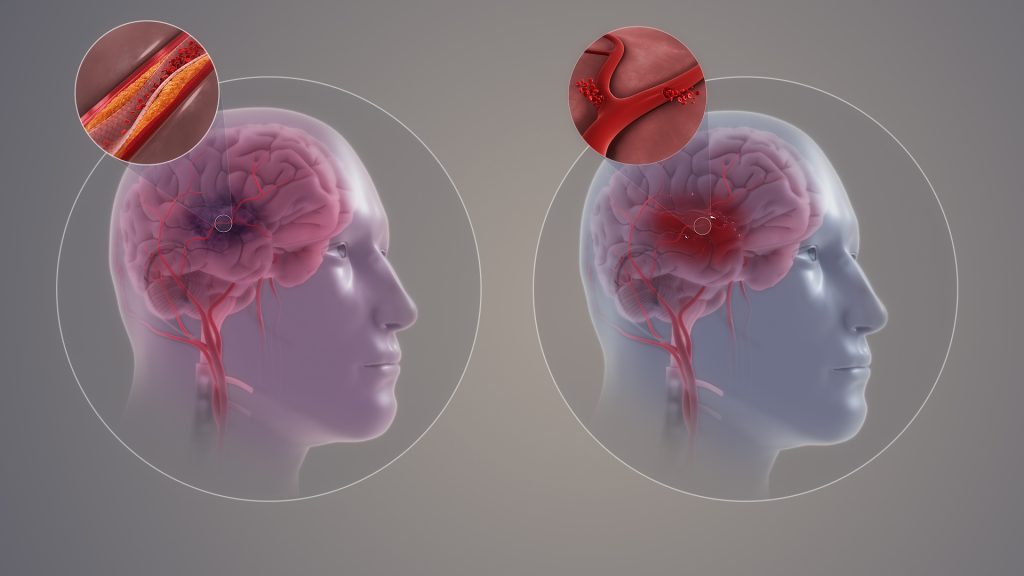
Ischaemic and haemorrhagic stroke. Credit: Scientific Animations CC4.0

Contrary to the commonly-held view, the brain does not have the ability to rewire itself to compensate for conditions such as stroke, loss of sight or an amputation, say scientists in the journal eLife.

Professors Tamar Makin of Cambridge University and John Krakauer of Johns Hopkins University argue that the notion that the brain, in response to injury or deficit, can reorganise itself and repurpose particular regions for new functions, is fundamentally flawed – despite being commonly cited in scientific textbooks. Instead, they argue that what is occurring is merely the brain being trained to utilise already existing, but latent, abilities.

One of the most common examples given is where a person loses their sight – or is born blind – and the visual cortex, previously specialised in processing vision, is rewired to process sounds, allowing the individual to use a form of ‘echolocation’ to navigate a cluttered room. Another common example is of people who have had a stroke and are initially unable to move their limbs repurposing other areas of the brain to allow them to regain control.

Krakauer, Director of the Center for the Study of Motor Learning and Brain Repair at Johns Hopkins University, said: “The idea that our brain has an amazing ability to rewire and reorganise itself is an appealing one. It gives us hope and fascination, especially when we hear extraordinary stories of blind individuals developing almost superhuman echolocation abilities, for example, or stroke survivors miraculously regaining motor abilities they thought they’d lost.

“This idea goes beyond simple adaptation, or plasticity – it implies a wholesale repurposing of brain regions. But while these stories may well be true, the explanation of what is happening is, in fact, wrong.”

In their article, Makin and Krakauer look at a ten seminal studies that purport to show the brain’s ability to reorganise. They argue, however, that while the studies do indeed show the brain’s ability to adapt to change, it is not creating new functions in previously unrelated areas – instead it’s utilising latent capacities that have been present since birth.

For example, a 1980s study by Professor Michael Merzenich at University of California, San Francisco looked at what happens when a hand loses a finger. The hand has a particular representation in the brain, with each finger appearing to map onto a specific brain region. Remove the forefinger, and the area of the brain previously allocated to this finger is reallocated to processing signals from neighbouring fingers, argued Merzenich – in other words, the brain has rewired itself in response to changes in sensory input.

Not so, says Makin, whose own research provides an alternative explanation.

In a study published in 2022, Makin used a nerve blocker to temporarily mimic the effect of amputation of the forefinger in her subjects. She showed that even before amputation, signals from neighbouring fingers mapped onto the brain region ‘responsible’ for the forefinger — in other words, while this brain region may have been primarily responsible for process signals from the forefinger, it was not exclusively so. All that happens following amputation is that existing signals from the other fingers are ‘dialled up’ in this brain region.

Makin, from the Medical Research Council (MRC) Cognition and Brain Sciences Unit at the University of Cambridge, said: “The brain’s ability to adapt to injury isn’t about commandeering new brain regions for entirely different purposes. These regions don’t start processing entirely new types of information. Information about the other fingers was available in the examined brain area even before the amputation, it’s just that in the original studies, the researchers didn’t pay much notice to it because it was weaker than for the finger about to be amputated.”

Another compelling counterexample to the reorganisation argument is seen in a study of congenitally deaf cats, whose auditory cortex appears to be repurposed to process vision. But when they are fitted with a cochlear implant, this brain region immediately begins processing sound once again, suggesting that the brain had not, in fact, rewired.

Examining other studies, Makin and Krakauer found no compelling evidence that the visual cortex of individuals that were born blind or the uninjured cortex of stroke survivors ever developed a novel functional ability that did not otherwise exist.

Makin and Krakauer do not dismiss stories such as blind people navigating using hearing, or individuals who have experienced a stroke regain their motor functions. They argue instead that rather than completely repurposing regions for new tasks, the brain is enhancing or modifying its pre-existing architecture — and it is doing this through repetition and learning.

Understanding the true nature and limits of brain plasticity is crucial, both for setting realistic expectations for patients and for guiding clinical practitioners in their rehabilitative approaches, they argue.

Makin added: “This learning process is a testament to the brain’s remarkable – but constrained – capacity for plasticity. There are no shortcuts or fast tracks in this journey. The idea of quickly unlocking hidden brain potentials or tapping into vast unused reserves is more wishful thinking than reality. It’s a slow, incremental journey, demanding persistent effort and practice. Recognising this helps us appreciate the hard work behind every story of recovery and adapt our strategies accordingly.

“So many times, the brain’s ability to rewire has been described as ‘miraculous’ – but we’re scientists, we don’t believe in magic. These amazing behaviours that we see are rooted in hard work, repetition and training, not the magical reassignment of the brain’s resources.”

The original text of this story is licensed under a Creative Commons Licence.

Source: University of Cambridge

We may now Know the Reason why SSRIs Take so Long to Kick in

Source: CC0

Selective serotonin reuptake inhibitors (SSRIs) normally take a few weeks before any improvements manifest, but the reasons why it takes so long have remained unclear since their first introduction 50 years ago. Now, new research provides the first human evidence that this is due to physical changes in the brain, which leads to greater brain plasticity developing over the first few weeks of SSRI intake. This may also begin to explain one of the mechanisms of how antidepressants work.

This work is presented at the ECNP conference in Barcelona, and also has been accepted in a peer-reviewed journal.

Clinician have long been puzzled as to why SSRIs take a relatively long time before having an effect. Researchers in Copenhagen, Innsbruck, and University of Cambridge have undertaken a randomised, double-blind placebo-controlled study in a group of healthy volunteers which shows a gradual difference in how many nerve cell connections (synapses) the brain cells have between those taking the antidepressants and a control group, depending on how long the treatment lasts.

In the study, 17 volunteers were given a 20mg daily dose of the SSRI escitalopram, with 15 volunteers given a placebo. Between three and five weeks after starting the trial, their brains were scanned with a PET (Positron Emission Tomography) scanner, which showed the amount of synaptic vesicle glycoprotein 2A in the brain: this is an indicator of the presence of synapses, so the more of the protein is found in an area, the more synapses are present in that area (ie, greater synaptic density). These scans showed significant between-group differences in how the synapse density evolved over time.

Researcher Professor Gitte Knudsen (of Copenhagen University Hospital) said:

“We found that with those taking the SSRI, over time there was a gradual increase in synapses in the neocortex and the hippocampus of the brain, compared to those taking placebo. We did not see any effect in those taking placebo.”

The neocortex, which takes up around half of the brain’s volume, deals with higher functions, such as sensory perception, emotion, and cognition. The hippocampus, which is found deep in the brain, handles functions of memory and learning.

Professor Knudsen continued, “This points towards two main conclusions. Firstly, it indicates that SSRIs increase synaptic density in the brain areas critically involved in depression. This would go some way to indicating that the synaptic density in the brain may be involved in how these antidepressants function, which would give us a target for developing novel drugs against depression. The second point is that our data suggest that synapses build up over a period of weeks, which would explain why the effects of these drugs take time to kick in.

Commenting, Professor David Nutt (Imperial College, London) said “The delay in therapeutic action of antidepressants has been a puzzle to psychiatrists ever since they were first discerned over 50 years ago. So these new data in humans that uses cutting edge brain imaging to demonstrate an increase in brain connections developing over the period that the depression lifts are very exciting.  Also they provide more evidence enhancing serotonin function in the brain can have enduring health benefits.”

This is an independent comment, Professor Nutt was not involved in this work..

Source: EurekAlert!

Novel Drug Shown to Repair Damage after Stroke

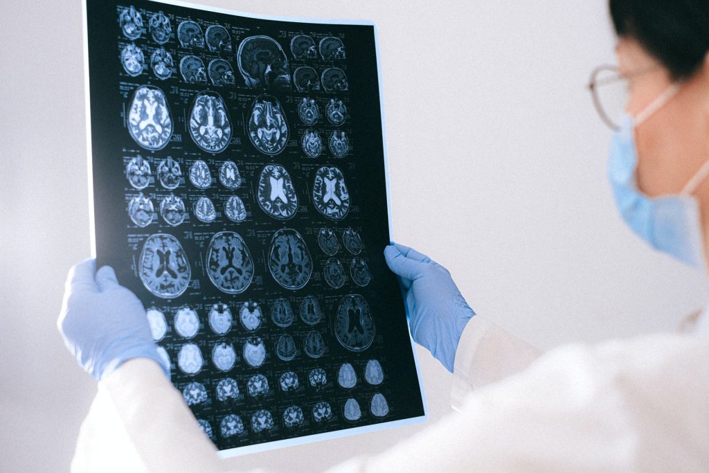
MRI images of the brain
Photo by Anna Shvets on Pexels

A pioneering new study from the University of Cincinnati shows promise that a new drug may help repair damage caused by strokes. The preclinical study appears in the journal Cell Reports.

Currently, there are no FDA approved drugs to repair the damage caused by a stroke. The study found that the new drug, NVG-291-R, enables nervous system repair and significant functional recovery in an animal model of severe ischaemic stroke. Deleting the gene for the drug’s molecular target also shows similar effect on neural stem cells. The drug has also proven to be safe and well-tolerated in volunteers with multiple sclerosis.

“We are very excited about the data showing significant improvement in motor function, sensory function, spatial learning and memory,” said Agnes (Yu) Luo, PhD, associate professor UC and the study’s senior author.

Prof Luo said the drug would be a “substantial breakthrough” if the early results translate into clinical settings. Further study and validation of results from independent groups will be needed to determine if the drug is similarly effective to repair the damage of ischaemic strokes in human patients. Additional studies will be needed to research if NVG-291-R effectively repairs damage caused by haemorrhagic strokes in both animal models and human patients.

“Most therapies being researched today primarily focus on reducing the early damage from stroke,” Assoc Prof Luo said. “However, our group has focused on neurorepair as an alternative and now has shown that treatment with NVG-291-R not only results in neuroprotection to reduce neuronal death but also robust neuroreparative effects.”

The drug proved to be effective even when treatment began as late as seven days after the stroke’s onset.

“The only current FDA-approved drug for treatment of stroke does not repair damage and must be administered within 4.5 hours of stroke onset.” Luo said. “Most therapies being researched need to be applied within 24–48 hours of a stroke’s onset. A product that works to repair damage from stroke even a week after symptom onset would change the paradigm for stroke treatment.”

Jerry Silver, PhD, co-author of the study and professor of neurosciences at CWRU’s School of Medicine, said the study showed the drug repaired damage in at least two ways: creating new neuronal connections and enhancing migration of new neurons derived from neuronal stem cells to the damage site.

“NVG-291-R’s ability to enhance plasticity was demonstrated by using staining techniques that clearly showed an increase in axonal sprouting to the damaged part of the brain,” Prof Silver said. “This enhanced plasticity is an excellent validation of the same powerful mechanisms that we and other researchers were able to demonstrate using NVG-291-R in spinal cord injury.”

Source: University of Cincinnati

Neural Plastic Changes can Help in Cervical Spinal Cord Injuries

MRI images of the brain
Photo by Anna Shvets on Pexels

By studying damage involving the connection between the brain’s hemispheres, researchers are finding new ways to leverage neural plasticity to promote functional recovery after a spinal cord injury.

In a study published in JCI Insight, the team of researchers used models in the lab to investigate a unilateral spinal cord injury similar to Brown-Sequard Syndrome, a rare neurological condition where damage to the spinal cord in a person results in weakness or paralysis on one side of the body and a loss of sensation on the opposite side.

Assistant Professor Wei Wu at Indiana University School of Medicine, said that the spinal cord injury model damaged the connection between the left hemisphere of the brain and the right side of the body, leading to significant loss of function in the right forelimb.

“The skilled function of upper limbs is very important for the quality of life in the patients with cervical spinal cord injury, but such functional recovery is very difficult to achieve in the severe injury,” said Asst Prof Wu, first author of the paper. “We found that the intact corticospinal system in the opposite side of the brain and spinal cord can be modulated to at least partially take over the control of the forelimb that is damaged by the spinal cord injury, resulting in a forelimb functional improvement.”

Since each hemisphere controls the opposite side of the body, researchers discovered a spontaneous shift of the neural circuits after injury from the left hemisphere to the right. Although there are connections between the right hemisphere of the brain and the right side of the body through some relayed pathways after injury, Asst Prof Wu said that’s not sufficient to support the motor recovery.

Using optogenetics to stimulate the right hemisphere of the brain, the researchers modulated the motor cortex. Additional neural circuits were shifted from the left side to the right side of the brain to dramatically increase and improve forelimb function.

“New circuits in the whisker, jaw forelimb and neck areas in the right hemisphere of the brain are recruited to control the right forelimb,” Asst Prof Wu said. “Interestingly, the beneficial neural plastic changes emerge both in the brain and the distal spinal cord after the optogenetic neuromodulation was applied on the motor cortex.”

Asst Prof Wu said results of the study showed significant improvement to the forelimb; however, there are still many challenges ahead, since complete digital recovery was not achieved.

The research team will continue explore this transhemispheric neural reorganisation to further improve the functional recovery after the spinal cord injury, Asst Prof Wu said. He hopes that these findings will be applied to treatments for spinal cord injuries.

Source: Indiana University School of Medicine