Tag: nanoparticles

Crafting a ‘Key’ to Cross the Blood-brain Boundary

Source: Pixabay CC0

Researchers led by Michael Mitchell of the University of Pennsylvania are close to gaining access through the blood-brain barrier, a long-standing boundary in biology, by granting molecules a special ‘key’ to gain access.

Their findings, published in the journal Nano Letters, present a model that uses lipid nanoparticles (LNPs) to deliver mRNA, offering new hope for treating conditions like Alzheimer’s disease and seizures.

“Our model performed better at crossing the blood-brain barrier than others and helped us identify organ-specific particles that we later validated in future models,” says Mitchell, associate professor of bioengineering at Penn’s School of Engineering and Applied Science, and senior author on the study.

“It’s an exciting proof of concept that will no doubt inform novel approaches to treating conditions like traumatic brain injury, stroke, and Alzheimer’s.”

Search for the key

To develop the model, Emily Han, a PhD candidate and NSF Graduate Research Fellow in the Mitchell Lab and first author of the paper, explains that it started with a search for the right in vitro screening platform, saying, “I was combing through the literature, most of the platforms I found were limited to a regular 96-well plate, a two-dimensional array that can’t represent both the upper and lower parts of the blood-brain barrier, which correspond to the blood and brain, respectively.”

Han then explored high-throughput transwell systems with both compartments but found they didn’t account for mRNA transfection of the cells, revealing a gap in the development process.

This led her to create a platform capable of measuring mRNA transport from the blood compartment to the brain, as well as transfection of various brain cell types including endothelial cells and neurons.

“I spent months figuring out the optimal conditions for this new in vitro system, including which cell growth conditions and fluorescent reporters to use,” Han explains.

“Once robust, we screened our library of LNPs and tested them on animal models. Seeing the brains express protein as a result of the mRNA we delivered was thrilling and confirmed we were on the right track.”

The team’s platform is poised to significantly advance treatments for neurological disorders.

It’s currently tailored for testing a range of LNPs with brain-targeted peptides, antibodies, and various lipid compositions.

However, it could also deliver other therapeutic agents like siRNA, DNA, proteins, or small molecule drugs directly to the brain after intravenous administration.

What’s more, this approach isn’t limited to the blood-brain barrier as it shows promise for exploring treatments for pregnancy-related diseases by targeting the blood-placental barrier, and for retinal diseases focusing on the blood-retinal barrier.

Next Steps

The team is eager to use this platform to screen new designs and test their effectiveness in different animal models.

They are particularly interested in working with collaborators with advanced animal models of neurological disorders.

“We’re collaborating with researchers at Penn to establish brain disease models,” Han says.

“We’re examining how these LNPs impact mice with various brain conditions, ranging from glioblastoma to traumatic brain injuries. We hope to make inroads towards repairing the blood-brain barrier or target neurons damaged post-injury.”

Source: University of Pennsylvania

Nanoparticles from Coffee Grounds could Stall Neurodegenerative Disease Development

Photo by Mike Kenneally on Unsplash

Researchers may potentially have found a preventive solution for neurodegenerative disorders in the most unlikely of sources: used coffee grounds. The researchers found caffeic-acid based Carbon Quantum Dots (CACQDs) have the potential to protect brain cells from the damage caused by several neurodegenerative diseases – if the condition is triggered by factors such as obesity, age and exposure to pesticides and other toxic environmental chemicals.

Carbon Quantum Dots are essentially simple nanoparticles made of carbon that have found a growing number of applications, including bioimaging thanks to its fluorescent properties and as photochemical catalysts. Their active surfaces can be doped with different elements for desired effects, are biocompatible and can be produced simply from a range of organic substances such as lemon juice and used tea leaves.

The University of Texas at El Paso team behind the study was led by Jyotish Kumar, a doctoral student in the Department of Chemistry and Biochemistry, and overseen by Mahesh Narayan, PhD, a professor and Fellow of the Royal Society of Chemistry in the same department. Their work is described in the journal Environmental Research.

“Caffeic-acid based Carbon Quantum Dots have the potential to be transformative in the treatment of neurodegenerative disorders,” Kumar said.

“This is because none of the current treatments resolve the diseases; they only help manage the symptoms. Our aim is to find a cure by addressing the atomic and molecular underpinnings that drive these conditions.”

Neurodegenerative diseases, when they are in their early stages and are caused by lifestyle or environmental factors, share several traits.

These include elevated levels of free radicals in the brain, and the aggregation of fragments of amyloid-forming proteins that can lead to plaques or fibrils in the brain.

Kumar and his colleagues found that CACQDs were neuroprotective across test tube experiments, cell lines and other models of Parkinson’s disease when the disorder was caused by a pesticide called paraquat.

The CACQDs, the team observed, were able to remove free radicals or prevent them from causing damage and inhibited the aggregation of amyloid protein fragments without causing any significant side effects.

The team hypothesises that in humans, in the very early stage of a condition such as Alzheimer’s or Parkinson’s, a treatment based on CACQDs can be effective in preventing full-on disease.

“It is critical to address these disorders before they reach the clinical stage,” Narayan said.

“At that point, it is likely too late. Any current treatments that can address advanced symptoms of neurodegenerative disease are simply beyond the means of most people. Our aim is to come up with a solution that can prevent most cases of these conditions at a cost that is manageable for as many patients as possible.”

Caffeic acid belongs to a family of compounds called polyphenols, which are plant-based compounds known for their antioxidant, or free radical-scavenging properties. Caffeic acid is unique because it can penetrate the blood-brain barrier and is thus able to exert its effects upon the cells inside the brain, Narayan said.

In the simple one-step ‘green chemistry’ method, the team ‘cooked’ caffeic acid at 230°C for two hours to reorient the caffeic acid’s carbon structure and form CACQDs. The CACQDs were then extracted according to a molecular weight cutoff of 1kDa.

The sheer abundance of coffee grounds is what makes the process both economical and sustainable, Narayan said.

Source: University of Texas at El Paso

Gold Nanoparticles Ease IBD Inflammatory Symptoms

Gold bars
Photo by Jingming Pan on Unsplash

In a Chinese study published in Fundamental Research, researchers explored a treatment for inflammatory bowel disease (IBD) using ultrasmall gold nanoparticles. Previous studies showed that these nanoclusters effectively eliminate a variety of reactive oxygen species (ROS), elevated levels of which are commonly found in the gastrointestinal tract of IBD patients.

IBD includes ulcerative colitis and Crohn’s disease, both of which tend to be debilitating, lifelong conditions that can prove fatal in severe cases. Currently, there is no cure for IBD. The main clinical treatments are drugs such as aminosalicylic acid preparations and corticosteroids, but they are often accompanied by gastrointestinal problems, anaemia, and various intestinal complications. Finding alternative, more effective options is a priority for researchers.

The team found that administering gold (Au25) nanoclusters orally to mice suffering from colitis eliminated ROS, increased antioxidant enzymes, and inhibited pro-inflammatory cytokines, without any obvious side effects. According to paper author Fei Wang of China’s The Seventh Affiliated Hospital of Sun Yat-Sen University, a reduction in the inflammation in the gastrointestinal tracts of the mice was observed within 24 hours. She added: “And the fact that these nanoclusters can be administered orally, means there is no need for invasive procedures.”

Additionally, the team found that the nanoclusters have a number of benefits when compared with natural enzymes used in traditional IBD treatments, including lower cost, better stability, mass synthesis and easier storage. Wang explained: “The storage of Au25 nanoclusters was not affected by pH, temperature or solution medium, and their good physiological stability and acid resistance meant they were easily able to access the inflamed colon. They also have good biocompatibility and chemical stability and can remove a variety of ROS.”

Wang concluded: “Au25 nanoclusters offer a promising strategy in the research field of nanomedicine therapy for IBD. We believe this study demonstrates their value as a scientific basis and experimental basis for the clinical treatment of IBD.”

Source: EurekAlert!

Nanoparticle Could Boost Polymyxin B for Gram-negative Sepsis

Patient's hand with IV drip
Photo by Anna Shvets on Pexels

To treat Gram-negative sepsis, Purdue University researchers are developing an injectable nanoparticle that can safely deliver Polymyxin B at high enough levels to inactivate endotoxins. Their research is published in Science Advances.

With an estimated annual mortality of between 30 and 50 deaths per 100 000 population,this condition ranks in the top 10 causes of death. One in three patients who die in a hospital has sepsis. Sepsis is a systemic illness caused by microbial invasion of normally sterile parts of the body, occurring when the body’s immune response to an infection or injury goes unchecked. The condition makes blood vessels leaky, leading to inflammation and blood clots, leading to impaired blood flow and possible death.

Professor Yoon Yeo leads a Purdue University team developing biocompatible nanoparticles that treat sepsis systemically through intravenous injection.

Prof Yeo said Polymyxin B, a traditional antibiotic, can inactivate endotoxins that cause a specific type of sepsis, but it may be too toxic for systemic application. For sepsis therapy, it mostly has been tested in extracorporeal blood cleaning, which is cumbersome and time consuming.

“Our nanoparticle formulations reduce dose-limiting toxicity of Polymyxin B without losing its ability to inactivate endotoxins,” Prof Yeo said.

In mouse models of sepsis, 100% treated with the Purdue nanoparticle were protected from excessive inflammation and survived.

“This technology holds promise as a safe, convenient option for patients and physicians,” Prof Yeo said.

Source: Purdue University

Injectable Nanoparticles That Could Slow Internal Bleeding

Photo by Camilo Jimenez on Unsplash

Researchers at MIT have found the ideal size for injectable nanoparticles that could slow traumatic internal bleeding, buying more time for a patient to reach a hospital for further treatment.

In a rat study, the researchers showed that polymer nanoparticles particles in an intermediate size range, (about 150nm in diameter) were the most effective at stopping bleeding. These particles also were much less likely to travel to the lungs or other off-target sites, which larger particles often do. The results were published in ACS Nano.

“With nano systems, there is always some accumulation in the liver and the spleen, but we’d like more of the active system to accumulate at the wound than at these filtration sites in the body,” said senior author Paula Hammond, Professor at MIT.

Nanoparticles that can stop bleeding, also called haemostatic nanoparticles, can be made in a variety of ways. One of the most commonly used strategies is to create nanoparticles made of a biocompatible polymer conjugated with a protein or peptide that attracts platelets, the blood cells that initiate blood clotting.

In this study, the researchers used a polymer known as PEG-PLGA, conjugated with a peptide called GRGDS, to make their particles. Most of the previous studies of polymeric particles to stop bleeding have focused on particles ranging in size from 300–500nm. However, few, if any studies have systematically analysed how size affects the function of the nanoparticles.

“We were really trying to look at how the size of the nanoparticle affects its interactions with the wound, which is an area that hasn’t been explored with the polymer nanoparticles used as haemostats before,” Hong says.

Studies in animals have shown that larger nanoparticles can help to stop bleeding, but those particles also tend to accumulate in the lungs, which can cause unwanted clotting there. In the new study, the MIT team analysed a range of nanoparticles, including small (< 100nm), intermediate (140–220nm), and large (500–650nm).

They first analysed the nanoparticles in the lab to see how how they interacted with platelets in various conditions, to see how well platelets bound to them. They found that, flowing through a tube, the smallest particles bound best to platelets, while the largest particles stuck best to surfaces coated with platelets. However, in terms of the ratio particles to platelets, the intermediate-sized particles were the lowest.

“If you attract a bunch of nanoparticles and they end up blocking platelet binding because they clump onto each other, that is not very useful. We want platelets to come in,” said lead author, Celestine Hong, an MIT graduate student. “When we did that experiment, we found that the intermediate particle size was the one that ended up with the greatest platelet content.”

The researchers injected the different size classes of nanoparticles into mice to see how long they would circulate for, and where they would end up in the body. As with previous studies, the largest nanoparticles tended accumulated in the lungs or other off-target sites.

The researchers then used a rat model of internal injury to study which particles would be most effective at stopping bleeding. They found that the intermediate-sized particles appeared to work the best, and that those particles also showed the greatest accumulation rate at the wound site.

“This study suggests that the bigger nanoparticles are not necessarily the system that we want to focus on, and I think that was not clear from the previous work. Being able to turn our attention to this medium-size range can open up some new doors,” Prof Hammond said.

The researchers now hope to test these intermediate-sized particles in larger animal models, to get more information on their safety and the most effective doses. They hope that eventually, such particles could be used as a first line of treatment to stop bleeding from traumatic injuries long enough for a patient to reach the hospital.

Source: Massachusetts Institute of Technology

‘A-Maize-ing’ Nanoparticles Target Cancer Cells Directly

Computer=generated depiction of nanoparticles

Researchers have recently developed novel nanoparticles derived from maize that can target cancer cells directly, via an immune mechanism. The results of this study, published in Scientific Reports, are encouraging, and the technique has demonstrated efficacy in treating tumour-bearing laboratory mice with no adverse effects.

Nanoparticles, or particles whose size varies between 1 and 100nm, have shown tremendous potential in many areas of science and technology, including therapeutics. However, conventional, synthetic nanoparticles are complicated and expensive to produce and alternatives such as extracellular vesicles (EVs) have mass production challenges.
Another recently emerging option is that of plant-derived nanoparticles (NPs), which can be easily produced in high levels at relatively lower costs. Like EVs, these nanoparticle-based systems also contain bioactive molecules, including polyphenols (which are known antioxidants) and microRNA, and they can serve as vehicles for targeted drug delivery.

Recently, researchers from the Tokyo University of Science (TUS) developed anti-cancer bionanoparticles, using corn (maize) as the raw material.
Lead researcher Professor Makiya Nishikawa explained: “By controlling the physicochemical properties of nanoparticles, we can control their pharmacokinetics in the body; so, we wanted to explore the nanoparticulation of edible plants. Maize, or corn, is produced in large quantities worldwide in its native form as well as in its genetically modified forms. That is why we selected it for our study.” 

The team centrifuged a super-sweet corn juice and then filtered it through a syringe filter with a 0.45μm pore size, then ultracentrifuged to obtain NPs derived from corn. The corn-derived NPs (cNPs) were approximately 80nm in diameter with a tiny net negative charge of -17mV.

The research team then set up experiments to see whether these cNPs were being taken up by various types of cells. In a series of promising results, the cNPs were taken up by multiple types of cells, including the clinically relevant colon26 tumor cells (cancer cells derived from mice), RAW264.7 macrophage-like cells, and normal NIH3T3 cells. RAW264.7 cells are commonly used as in vitro screens for immunomodulators.

The results were astounding: of the three types of cells, cNPs only significantly inhibited the growth of colon26 cells, indicating their selectivity for carcinogenic cell lines. Moreover, cNPs were able to successfully induce the release of tumour necrosis factor-α (TNF-α) from RAW264.7 cells. TNFα is primarily secreted by macrophages, natural killer cells, and lymphocytes, which help mount an anticancer response. “The strong TNFα response was encouraging and indicated the role of cNPs in treating various types of cancer,” explains Dr. Daisuke Sasaki, first author of the study and an instructor and researcher at TUS.

A luciferase-based assay revealed that the potent combination of cNPs and RAW264.7 cells significantly suppressed the proliferation of colon26 cells. Finally, the research team studied the effect of cNPs on laboratory mice bearing subcutaneous tumours. Once again, the results were astonishing: daily injections of cNPs into colon26 tumours significantly suppressed tumour growth, without causing serious side effects, or weight loss.

“By optimising nanoparticle properties and by combining them with anticancer drugs, we hope to devise safe and efficacious drugs for various cancers,” observed an optimistic Prof Nishikawa.

Source: Tokyo University of Science

Nanoparticle and Antibiotic Polytherapy Defeats AMR Bacteria

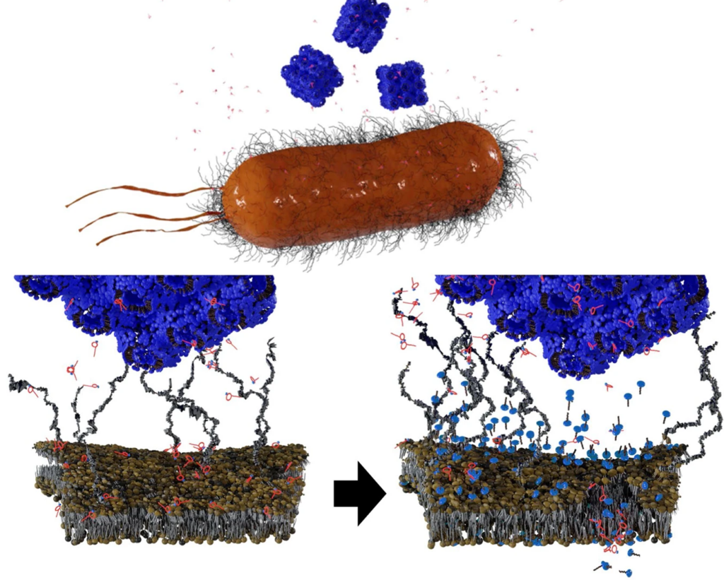
Polytherapy with PMB and cubosomes result in interactions with the bacterial OM in two consecutive ways: PMB initially interacts with the outer leaflet of OM via electrostatic interactions, leading to destabilised areas. Cubosomes then contact with the bilayer, causing further membrane perturbations via a lipid-exchange process. Credit: Monash University/Lai et al.

Researchers from Monash University have discovered a potential new method to circumvent antibiotic resistance, by means of a nanoparticle and antibiotic polytherapy. This approach could also reduce antibiotic intake.

The World Health Organisation (WHO) has declared antimicrobial resistance (AMR) to be among the top 10 global public health threats. A recent report found that in 2019, 1.27 million deaths were directly attributable to AMR infections – more than deaths from either HIV or TB.

AMR occurs when pathogens evolve to no longer respond to medicines, consequently infections become increasingly difficult or impossible to treat.

The study, which appears in Nature Communications, has found that the use of nanoparticles in combination with other antibiotics, is an effective strategy to improve bacterial killing.

For Gram-negative bacteria, polymyxins have been used as drugs of last resort as they disrupt the bacterial outer membrane (OM), causing it to become more permeable, causing cell contents to leak out and kill the bacteria.

The strategy involves administering polymyxin B (PMB) alongside cube-shaped nanoparticles called cubosomes. The PMB disrupted the OM first, but not enough to kill the cell. When the accompanying cubosome bound to the OM, disrupting it further, successfully killing the cell. Interestingly, loading PMB into the cubosomes as a carrier had little effect; in fact, the cubosome strengthened the OM.

“This is a stunning finding in how we deliver medicine and how the medicine we take impacts us in the future,” said lead researcher Dr Hsin-Hui Shen. 

This approach also means that lower dosages of antibiotics could be used. “Instead of looking for new antibiotics to counteract superbugs, we can use the nanotechnology approach to reduce the dose of antibiotic intake, effectively killing multidrug-resistant organisms.”

It has been 30 years since the discovery of the last new antibiotic, and in coming years, the growing crisis of antibiotics resistance will result in increased mortality from basic infections because they have developed antimicrobial resistance.

Without effective antimicrobials, the WHO warns that the success of modern medicine in treating infections, including during major surgery and cancer chemotherapy, would be at increased risk.

While nanoparticles had been used for a long time before as antimicrobial carriers,  “but the use of nanoparticles in polytherapy treatments with antibiotics in order to overcome antimicrobial resistance has been overlooked,” explained Dr Shen. “The use of nanoparticles-antibiotics combination therapy could reduce the dose intake in the human body and overcome the multidrug resistance.”

Research will now progress to the testing phase.

Source: Monash University

New Coating Makes the Nanomedicine Go Down

Upon injection into the blood, nanomedicines (blue spheres) are immediately attacked by proteins of the immune system called complement proteins (orange). Complement proteins cause rapid destruction of the nanomedicine, and also induce an anaphylaxis-like reaction. By attaching complement-degrading proteins (yellow ninjas made of protein) to the surface of nanomedicines, Penn researchers have largely solved this problem, potentially allowing more diseases to be safely treated by nanomedicine. Credit: University of Pennsylvania

In nanomedicine, immune reactions against the nanoparticles that contain the medicine or vaccine, reducing its effectiveness. Researchers have now come up with a new method to prevent the body from treating nanomedicines like foreign invaders, by covering those nanoparticles with a coating to suppress the immune response.

As soon as they are injected into the bloodstream, unmodified nanoparticles are swarmed by complement proteins, triggering an inflammatory response and preventing the nanoparticles from reaching their treatment targets. Penn Medicine researchers, whose findings are published in Advanced Materials, have devised a coating for nanoparticles that suppresses complement activation.

Nanoparticles are tiny capsules, typically made from proteins or fat-related molecules, that contain certain types of treatment or vaccine. The best-known examples of nanoparticle-delivered medicines are mRNA COVID vaccines.

“It turned out to be one of those technologies that just works right away and better than anticipated,” said study co-senior author Jacob Brenner, MD, PhD.

RNA- or DNA-based therapies generally need delivery systems to get them through the bloodstream into target organs. Harmless viruses often have been used as carriers or “vectors” of these therapies, but nanoparticles are increasingly considered safer alternatives. Nanoparticles also can be tagged with antibodies or other molecules that make them hone in precisely on targeted tissues.

The complement attack problem has been a serious impediment to nanomedicine. Circulating complement proteins treat nanoparticles as if they were bacteria, immediately coating nanoparticle surfaces and summoning macrophages to engulf them. Researchers have attempted to reduce the problem by pre-coating nanoparticles with camouflaging molecules, such as forming a watery, protective shell around nanoparticles using polyethylene glycol (PEG).

But nanoparticles camouflaged with substances like PEG still draw at least some complement attack. In general, nanoparticle-based medicines that move through the bloodstream (mRNA COVID vaccines are injected into muscle, not the bloodstream) have had a very low efficiency in getting to their target organs, usually under 1%.

In the study, the researchers came up with a new approach to protect nanoparticles, based on natural complement-inhibitor proteins that circulate in the blood, attaching to human cells to help protect them from complement attack.

In vitro tests using standard PEG-protected nanoparticles with one of these complement inhibitors, called Factor I, provided dramatically better protection from complement attack. In mice, the same strategy prolonged the half-life of standard nanoparticles in the bloodstream, allowing a much larger fraction of them to reach their targets.

“Many bacteria also coat themselves with these factors to protect against complement attack, so we decided to borrow that strategy for nanoparticles,” said co-senior author Jacob Myerson, PhD, a senior research scientist in the Department of Systems Pharmacology and Translational Therapeutics at Penn.

In a set of experiments in mouse models of severe inflammatory illness, the researchers also showed that attaching Factor I to nanoparticles prevents the hyper-allergic reaction that otherwise could be fatal.

Further testing will be needed before nanomedicines incorporating Factor I can be used in people, but in principle, the researchers said, attaching the complement-suppressing protein could make nanoparticles safer and more efficient as therapeutic delivery vehicles so that they could be used even in severely ill patients.

The researchers now plan other protective strategies for medical devices, such as catheters, stents and dialysis tubing, which are similarly susceptible to complement attack. They also plan to investigate other protective proteins.

“We’re recognising now that there’s a whole world of proteins that we can put on the surface of nanoparticles to defend them from immune attack,” Dr Brenner said.

Source: University of Pennsylvania School of Medicine

‘Nanotraps’ Capture COVID Virus and Prevent Infection

Researchers have developed an entirely new treatment for COVID: ‘Nanotraps’ that capture the viruses inside the body, allowing the immune systems to destroy them

The “Nanotraps” mimick the human cells the virus normally attaches to, and bind it to their surface, keeping the virus from reaching other cells and target it for destruction by the immune system. It is possible that Nanotraps could be used on SARS-CoV-2 variants, and could be administered as a nasal spray.

“Since the pandemic began, our research team has been developing this new way to treat COVID-19,” said Assistant Professor Jun Huang, whose lab led the research. “We have done rigorous testing to prove that these Nanotraps work, and we are excited about their potential.”

Postdoc Min Chen and graduate student Jill Rosenberg targeted the spike mechanism that SARS-CoV-2 uses to lock onto ACE2 proteins on human cells.

To create a trap that would bind to the virus in the same way, they designed nanoparticles with a high density of ACE2 proteins on their surface. Other nanoparticles were designed with neutralising antibodies on their surfaces.

ACE2 proteins and neutralising antibodies have both been used in COVID treatments, but by mounting them onto nanoparticles, a much more effective and robust means for trapping the virus was created.

The nanoparticles are smaller than cells, 500 nanometres in diameter, allowing them to reach deep inside tissue and trap the virus.

No evidence of toxicity was seen in tests with mice, and they then tested the Nanotraps against a non-replicating virus called a pseudovirus in human lung cells in tissue culture plates and saw that they completely prevented viral entry into the cells.

 When the nanoparticle binds to the virus (about 10 minutes after injection), it chemically signalled macrophages to engulf and destroy the nanoparticle and the attached virus. Macrophages normally engulf nanoparticles, so this merely sped up the process.

Testing the Nanotraps on a pair of donated lungs kept alive with a ventilator, they found that they completely prevented infection.

They also collaborated with researchers at Argonne National Laboratory to test the Nanotraps with a live virus (rather than a pseudovirus) in an in vitro system. They found a 10 times better performance than with neutralising antibodies or ACE2 inhibitor.

The researchers plan further tests, including live virus and its variants.

“That’s what is so powerful about this Nanotrap,” Rosenberg said. “It’s easily modulated. We can switch out different antibodies or proteins or target different immune cells, based on what we need with new variants.”

Storage is simple, as the Nanotraps can be kept in a standard freezer, and administration is simple, using a nasal spray. The researchers said it is also possible to serve as a vaccine by optimisation of the Nanotrap formulation.

Source: Phys.Org

Journal information: Min Chen et al, Nanotraps for the containment and clearance of SARS-CoV-2, Matter (2021). DOI: 10.1016/j.matt.2021.04.005