Extra Year of Education does Not Protect the Brain

Thanks to a ‘natural experiment’ involving 30 000 people, researchers at Radboud university medical centre were able to very precisely determine the effect of an extra year of education to the brain in the long term. To their surprise, they found no effect on brain structure and no protective benefit of additional education against brain ageing. Their findings appear in eLife.
It is well-known that education has many positive effects. People who spend more time in school are generally healthier, smarter, and have better jobs and higher incomes than those with less education. However, whether prolonged education actually causes changes in brain structure over the long term and protects against brain ageing, was still unknown.
It is challenging to study this, because alongside education, many other factors influence brain structure, such as the conditions under which someone grows up, DNA traits, and environmental pollution. Nonetheless, researchers Rogier Kievit (PI of the Lifespan Cognitive Dynamics lab) and Nicholas Judd from Radboudumc and the Donders Institute found a unique opportunity to very precisely examine the effects of an extra year of education.
Ageing
In 1972, a change in the law in the UK raised the number of mandatory school years from 15 to 16, while all other circumstances remained constant. This created an interesting ‘natural experiment’, an event not under the control of researchers which divides people into an exposed and unexposed group. Data from approximately 30 000 people who attended school around that time, including MRI scans taken much later (46 years after), is available. This dataset is the world’s largest collection of brain imaging data.
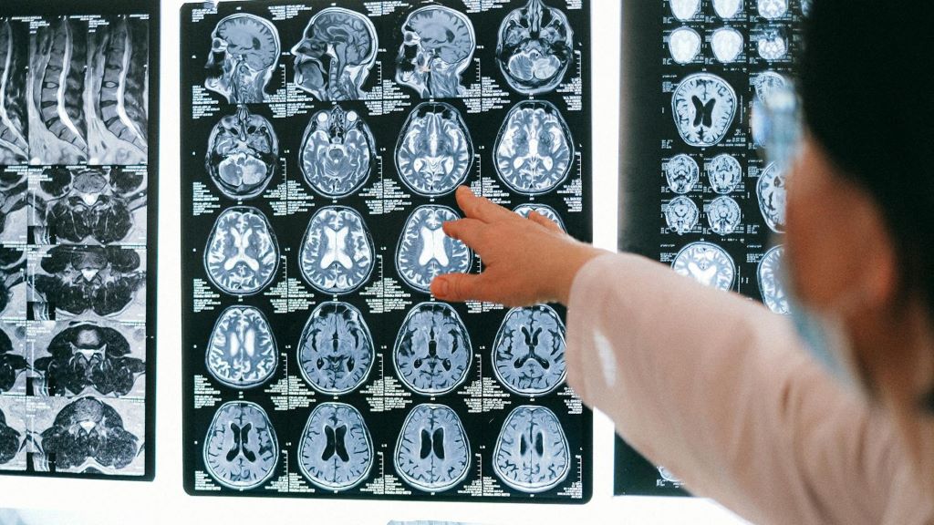
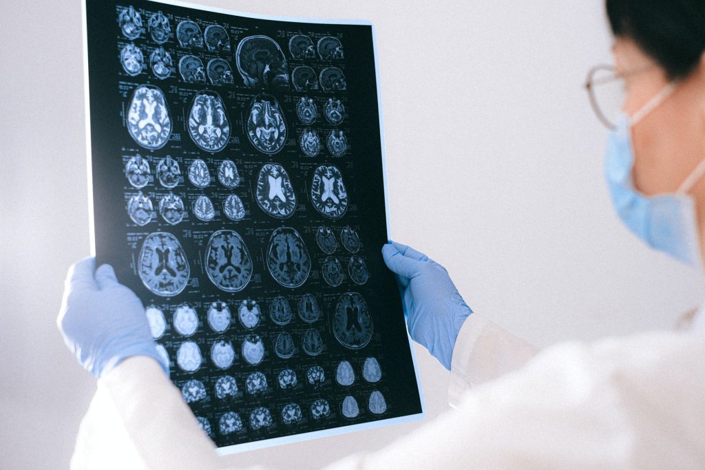
The researchers examined the MRI scans for the structure of various brain regions, but they found no differences between those who attended school longer and those who did not. ‘This surprised us’, says Judd. ‘We know that education is beneficial, and we had expected education to provide protection against brain aging. Aging shows up in all of our MRI measures, for instance we see a decline in total volume, surface area, cortical thickness, and worse water diffusion in the brain. However, the extra year of education appears to have no effect here.’
Brain structure
It’s possible that the brain looked different immediately after the extra year of education, but that wasn’t measured. “Maybe education temporarily increases brain size, but it returns to normal later. After all, it has to fit in your head,” explains Kievit. “It could be like sports: if you train hard for a year at sixteen, you’ll see a positive effect on your muscles, but fifty years later, that effect is gone.” It’s also possible that extra education only produces microscopic changes in the brain, which are not visible with MRI.
Both in this study and in other, smaller studies, links have been found between more education and brain benefits. For example, people who receive more education have stronger cognitive abilities, better health, and a higher likelihood of employment. However, this is not visible in brain structure via MRI. Kievit notes: “Our study shows that one should be cautious about assigning causation when only a correlation is observed. Although we also see correlations between education and the brain, we see no evidence of this in brain structure.”


