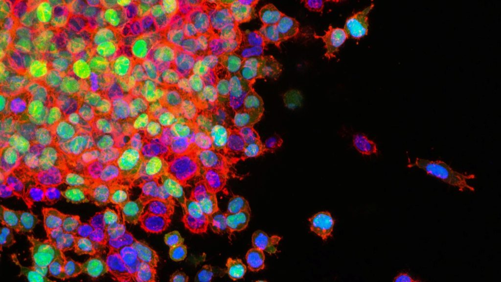
In a breakthrough for understanding metastases, researchers have found that, as metastatic cancers spread to different parts of the body, they adapt their metabolism to the tissue in which they grow. The findings, which help further break down the puzzle of metastasis, are published in PNAS.
Metabolism in the body is an important target for cancer treatments, where the focus is on stopping the progress of cancer cells.
“Obviously, the local environment affects the cancer cells more than previously known. The metastatic tumours should show the same metabolic properties no matter where in the body they are located, but we discovered that the cancer cells largely adapted their metabolism to the new tissue in order to continue to develop and grow. This is important knowledge, which shows that we cannot consider the metastases as their original tumours,” says Fariba Roshanzamir, PhD in Systems and Synthetic Biology at Chalmers and the study’s lead author.
Cutting off cancer metabolism
Fariba Roshanzamir works in Professor Jens Nielsen’s research group at Chalmers and has, together with Swedish and international colleagues, been able to establish the groundbreaking results. The study focused primarily on triple-negative breast cancer but the conclusions can, according to the researchers, be applied to all types of metastatic cancer. This opens new doors to develop more effective treatments.
“If we manage to shut down the metabolism in a tumour, it will stop working and this study provides important keys to better understand what to target. Selecting metabolic inhibitors that specifically target the metastases in the organs to which the tumour has spread, rather than treating them as their original tumours, is of great importance to be able to find good strategies for treatments in the future,” she says.
Higher rates of brain metastases in patients with inflammatory breast cancer, a rare subtype of breast cancer, have been observed in studies, but detailed information is lacking. Now, a new study published in CANCER indicates that patients inflammatory breast cancer face a higher risk that their cancer will metastasise to the brain.
To provide insights into the incidence and risk factors for brain metastases in this patient population, Laura E.G. Warren, MD, and colleagues analysed data on 372 patients with stage III inflammatory breast cancer and 159 with stage IV inflammatory breast cancer.
Over a median follow-up of five years, the incidence of brain metastases at one, two, and five years was 5%, 9%, and 18% among patients who presented with stage III disease, and 17%, 30%, and 42% among those with stage IV disease. Patients with triple-negative breast cancer faced a particularly high risk, and when they did experience brain metastases, their survival time was shorter than those with hormone receptor–positive or HER2-positive breast cancer who experienced brain metastases. Higher risks of brain metastases were also seen in patients whose cancer had metastasised to other parts of the body besides the brain, especially when this occurred at a young age.
“The relatively high incidence of brain metastases seen in the study population highlights the need for future research on the potential role for surveillance brain imaging for high-risk patients. There is an open, phase II, single arm study at Dana-Farber Cancer Institute examining this question,” said Dr. Warren. “It also emphasises the need to obtain brain imaging in patients with inflammatory breast cancer presenting with neurologic symptoms given the high incidence of brain metastases in this population.” Most patients in this study who were diagnosed with brain metastases had neurologic symptoms, but because some patients may have undetected, asymptomatic brain metastases, the true incidence in patients with inflammatory breast cancer is likely even higher than what Dr Warren and her colleagues observed.
An accompanying editorial notes that when considering whether to implement routine brain imaging tests in patients with inflammatory breast cancer, it will be important to determine whether earlier detection of brain metastases leads to improvements in both survival and quality of life.
Cambridge University scientists have discovered that cancer cells ‘hijack’ a process used by healthy cells to spread around the body, challenging how cancer metastasis is currently understood. Publishing in Nature Genetics, the team found that blocking the activity of a sodium channel protein in cells in mice with cancer triggers metastasis.
To their surprise, the investigators also discovered that this process extends beyond just cancer: when they removed NALCN from cancer-free mice, this caused their healthy cells to leave their original tissue and travel around the body where they joined other organs.
They found, for example, that healthy pancreas cells migrated to the kidney where they became healthy kidney cells. This suggests that metastasis isn’t an abnormal process limited to cancer as previously thought, but is in fact a normal process used by healthy cells that has been exploited by cancers to generate metastases.
Group Leader of the study, Professor Richard Gilbertson, said: “These findings are among the most important to have come out of my lab for three decades. Not only have we identified one of the elusive drivers of metastasis, but we have also turned a commonly held understanding of this on its head, showing how cancer hijacks processes in healthy cells for its own gains. If validated through further research, this could have far-reaching implications for how we prevent cancer from spreading and allow us to manipulate this process to repair damaged organs.”
Metastasis has been challenging to treat because of the difficulty in identifying key drivers of this process that could be targeted by drugs. Now that they have identified NALCN’s role in metastasis, the team are looking into various ways to restore its function, including using existing drugs on the market.
Lead researcher on the study Dr Eric Rahrmann, said: “We are incredibly excited to have identified a single protein that regulates not only how cancer spreads through the body, independent of tumour growth, but also normal tissue cell shedding and repair. We are developing a clearer picture on the processes that govern how cancer cells spread. We can now consider whether there are likely existing drugs which could be repurposed to prevent this mechanism from triggering cancer spreading in patients.”
NALCN stands for sodium (Na+) leak channel, non-selective. Sodium leak channels are expressed predominately in the central nervous system but are also found throughout the rest of the body, controlling the amount of sodium that goes in and out of the cell. Controlling this process also alters the balance of electricity across the cell membrane. It is still unclear why these channels seem to be implicated so directly in cancer metastasis.
Microscopy image of mouse sciatic nerves showing axons (red) wrapped by Schwann cells (green) with their nuclei depicted in blue.
Credit: A. Alvarez-Prats and T. Balla, Eunice Kennedy Shriver National Institute of Child Health and Human Development/NIH
In addition to forming the myelin sheath along peripheral nerves and supporting neighbouring neurons, Schwann cells have also been found to play an important immune modulating function, starting and shutting off inflammation. This function not only helps nerve repair, but may also turn nerve tumours benign. These new findings were reported in the journal Glia.
The research has revealed that Schwann cells produce signalling molecules that can activate other immune cells. In particular, however, they are able to stop inflammatory reactions in order to prevent excessive tissue damage and allow the nerve to regenerate.
“This is essential, because inflammation releases free radicals against which nerve fibers cannot protect themselves. Therefore, the inflammation must be cleared quickly, which is precisely what Schwann cells do,” explained study designer Dr. Sabine Taschner-Mandl, who designed the study and heads a research group at St. Anna CCRI.
Do Schwann cells protect against malignancy?
These findings also have implications for protection against malignancy After nerve injury, Schwann cells engage a ‘repair’ mode that is also found in benign infantile nerve tumours. There, it causes the tumour cells to mature and thus reach a stage where they lose their aggressive properties and no longer divide unchecked
“Based on the current results, we now suspect that the immune cell functions of Schwann cells also become effective in childhood nerve tumours. This is because in cancer, there is always a kind of inflammation bubbling away that never comes to a halt. In benign nerve tumours, ganglioneuromas, the accompanying chronic inflammation could be stopped by Schwann cells similar to nerve healing, because unlike malignancies, ganglioneuromas have many Schwann cells in their microenvironment. We also see that a lot of immune cells migrate into these tumours, for which the Schwann cells could also be responsible,” said Dr Taschner-Mandl.
Healthy Inflammation: First Activate, Then Shut Down
In particular, the current study shows that Schwann cells can influence T cells, which are key in cancer defence. Schwann cells – both those in nerve regeneration and those in benign tumours – carry MHC-I and MHC-II molecules on their surface that are important for T-cell regulation. Via these molecules, Schwann cells present recognition features of material they have previously taken up from their environment.
“We mimicked an inflammatory response in the laboratory and detected a whole range of additional stimulatory and inhibitory surface molecules that are also necessary for T cell activation,” explained Jakob Berner, MSc, co-first author of the study and interim PhD student at St. Anna CCRI. “Our experiments show that Schwann cells are able to take up large amounts of material via phagocytosis.”
As the first immune response to a nerve cut, Schwann cells secrete substances that attract T cells, macrophages and other immune cells. Now it turned out that not only a reaction between the classical immune cells takes place, but also between Schwann cells and T cells.
While Schwann cells initially fuel the inflammatory response by releasing interferon-gamma, they can later shut it down by up-regulating the T-cell inhibitory PD-L1 molecule.
“First activate, then shut down – that’s the normal process of an inflammatory response. If this were also the case in cancer, then it could curb cancer growth,” Dr Taschner-Mandl theorised. Researchers are now investigating whether and how these findings could be applied to cancer treatment.
Giving standard chemotherapy drugs in a specific sequence for certain types of metastatic breast cancer can cut costs while preserving quality of life, according to a study in theJournal of Clinical Oncology.
The study, led by researchers from UNC Lineberger Comprehensive Cancer Center and UNC Gillings School of Global Public Health, developed three different computer models to predict how a hypothetical set of 10 000 patients with specific types of metastatic breast cancer would respond to different sequences and types of chemotherapy. For this study, the patient’s cancer was either endocrine resistant or was triple-negative breast cancer.
Many chemotherapy choices are available to treat metastatic breast cancer. While oncologists may prefer certain drugs to use early in treatment, the best order in which to give the drugs is unclear. The researchers consulted oncologists and experts in the field to choose which chemotherapy drugs were preferred choices to include in the study.
Mimicking clinical practice, and based upon existing data, the researchers then assumed that if a person started treatment with one drug, they would change to a second-choice treatment after their cancer stopped responding to the first drug, or if the side effects weren’t tolerable. The purpose of the study was to test whether putting the drugs in one sequence compared to another could keep the patient on treatment for similar times while decreasing their side effect and/or cost burden.
“The cost of cancer drugs in the US has rapidly increased, even for generics. As a society, we urgently need more strategies to reduce cancer drug costs without compromising outcomes, and our analysis provides quantifiable evidence to help providers choose lower priced, but equally effective sequences of drugs,” said Stephanie B. Wheeler, PhD, MPH, professor of health policy & management at UNC Gillings and associate director of community outreach and engagement at UNC Lineberger and corresponding author of the article. “More spending on cancer care does not necessarily confer greater health benefits.”
The costs calculated in this study were inclusive of medical and nonmedical costs borne by patients, including lost productivity. In this simulation, after two years, nearly all women would have completed the first three sets of treatment, but the cancer would cause the death of about one-third of the women. Productivity days lost due to sickness were similar across chemotherapy sequences, so most of the cost difference was due to drug savings. In the simulation, patients were placed in three groups, depending on what treatments they had already received for earlier episodes of breast cancer.
Outcomes in the three groups were:
For people who had not previously received the common chemotherapy drug categories, including a taxane (e.g., paclitaxel) or an anthracycline (e.g., capecitabine), treatment with paclitaxel then capecitabine followed by doxorubicin corresponded to the highest expected gains in quality of life and lowest costs.
For people who had previously received a taxane and an anthracycline drug, treatment with carboplatin, followed by capecitabine, followed by eribulin, corresponded to the highest expected gains in quality of life and lowest costs.
For people who had previously received a taxane but not an anthracycline, treatment sequences beginning with capecitabine or doxorubicin, followed by eribulin, were most cost-effective.
“The drugs we studied are already recommended and reimbursed for the treatment of metastatic breast cancer, but the optimal sequencing of them has been unclear, which has led to considerable variation in physician preference and practice. Our study suggests that treatment sequencing approaches that minimise costs early may improve the value of care,” Wheeler said. “The implications of this study are fairly straightforward for medical oncologists and those developing value-based clinical pathways to implement in practice now.”
Associate professor Katherine E. Reeder-Hayes, one of the study’s authors, said the treatment choices for metastatic breast cancer are constantly changing, and new options for targeted therapy have emerged even since this study was conducted. “Many oncologists and patients find that there aren’t any more targeted therapies that fit the cancer’s molecular profiles, so they are left with the choice of a number of chemotherapy drugs that may feel pretty similar or have an unclear balance of pros and cons.
“In that scenario, I hope our study will help expand the framework that we use to make these decisions from one where we just think about the biologic action of the drug to one where we also consider the bigger picture of what the treatment experience is like for the patient, including their financial burden, investment of time, and side effects,” Reeder-Hayes added. “The most potent drug isn’t always the next best choice depending on what the patient values and wants to accomplish with their treatment.”
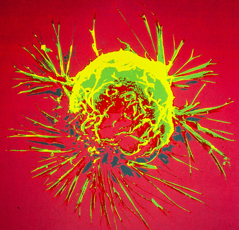
Lung cancer cells metastasising. Source: National Cancer Institute on Unsplash
Researchers have eliminated cells derived from untreatable metastatic cancer by disrupting the cellular components that are responsible for converting oxygen into chemical energy. The results are published in the Journal of the American Chemical Society.
Treatment of cancer is a a long battle because surviving cancer cells often evolve into aggressive, untreatable forms. Hence, treatment plans often involve multiple drug combinations and/or radiation therapy in order to prevent cancer relapse. To combat the variety of cancer cell types, modern drugs have been developed to target specific biochemical processes that are unique within each cell type.
However, cancer cells are highly adaptive and able to develop mechanisms to avoid the effects of the treatment. “We want to prevent such adaptation by invading the main pillar of cellular life – how cells breathe – that means take up oxygen – and thus produce chemical energy for growth,” explained David Ng, group leader at the Max Planck Institute for Polymer Research (MPI-P).
The research team produced a synthetic drug that travels into cells where it reacts to conditions found inside and triggers a chemical process. This allows the drug’s molecules to bind together and form tiny hairs that are a thousand times thinner than human hair. “These hairs are fluorescent, so you can look at them directly with a microscope as they form,” said first author Zhixuan Zhou, an Alexander von Humboldt fellow.
The scientists monitored the oxygen consumption in different cell types and found that these tiny hairs prevent all of them from converting oxygen into the energy deliver molecule ATP. The process worked even for those cells derived from untreatable metastatic cancer, with the cells dying off within four hours. After some more years of research, the scientists hope that they can develop a new method to treat up-to-now untreatable cancer.
Weil, Ng and colleagues have shown an exciting outcome under controlled laboratory culture and will continue to unravel deeper insights on the basis of how these tiny hairs prevent the conversion of oxygen to chemical energy. With further development, these objects could in the future possibly also be manipulated to control other cellular processes to address other important diseases.
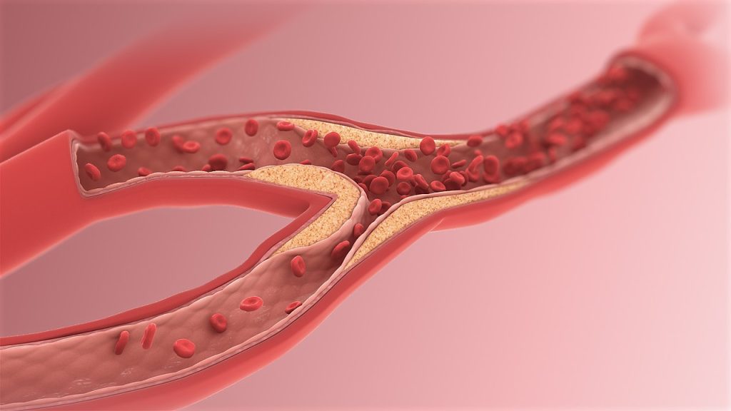
Researchers have identified a key signalling molecule for cancer metastasis. one which is already known for its involvement in atherosclerosis, suggesting a possible treatment approach for both diseases simultaneously. The discovery was published in the International Journal of Cancer.
In order to become malignant, metastasising cancer, tumour cells undergo a series of transformations involving interactions with the immune system. Growing evidence exists that in tumour progression to metastasis, inflammation of blood vessel-lining endothelial cells is a key process.
A team of researchers led by Professor Kyoko Hida at Hokkaido University have discovered that, in malignant tumours, endothelial cells accumulate low-density lipoprotein (LDL) and neutrophils. Neutrophils are immune suppressor cells which are known to contribute to tumour progression.
Previous work by the team had revealed that blood vessels in malignant tumorus expressed a high level of proteoglycans, and it is known that cancerous tissue is inflamed – similar to what is seen in atherosclerosis.
The research team showed that metastasising tumors, in contrast to non-metastasising ones, accumulate proteoglycan molecules; these, in turn, attach to and accumulate LDL to the walls of blood vessels, where it becomes oxidised. There are also high levels of its receptor, LOX-1, in the blood vessel-lining endothelial cells of metastasising tumours. This, they found, causes these cells to produce inflammation signals that attract neutrophils. Using a mouse model, they proved that the suppression of LOX-1 can significantly reduce tumour malignancy, and also that LOX-1 overexpression caused an increase in signalling molecules attracting neutrophils.
This sequence of interactions observed in malignant tumours is not novel: it occurs in atherosclerosis. “Atherosclerosis and cancer appear to be completely different diseases, but they share several common pathophysiological features in the blood vessels,” said Prof Hida.
Though some questions remain, especially on the mechanism of how neutrophils contribute to cancer malignancy, this study is the first to explicitly prove the mechanistic commonalities between cardiovascular disease and cancer progression and trace the mechanism involving LDL accumulation and LOX-1 expression in in vivo tumour tissue.
“Our present study focused on the importance of LOX-1 in endothelial cells as a common factor between cancer and atherosclerosis,” Prof Hida explained. “The presence of neutrophils in tumours is a telltale sign of tumor progression.”
The study also points to a promising approach for treating and preventing malignant cancer (and cardiovascular disease) by targeting neutrophil recruitment to endothelial cells. Prof Hida concluded: “The number of patients with cancer who die not of cancer, but of cardiovascular events, is increasing. Targeting the LOX-1/oxidised LDL axis might be a promising strategy for the treatment of the two diseases concomitantly.”
Researchers have discovered that, counterintuitively, certain cells move faster in thicker fluid – such as mucus as opposed to blood – because their ruffled edges sense the viscosity of their environment and adapt to increase their speed.
The researchers’ combined results in cancer and fibroblast cells suggest that the viscosity of a cell’s surrounding environment is an important contributor to disease. The findings, published in Nature Physics, may help explain tumour progression, scarring in mucus-filled lungs affected by cystic fibrosis, and the wound-healing process.
“This link between cell viscosity and attachment has never been demonstrated before,” noted Sergey Plotnikov, assistant professor at the University of Toronto and a co-corresponding author of the study. “We found that the thicker the surrounding environment, the stronger the cells adhere to the substrate and the faster they move – much like walking on an icy surface with shoes that have spikes, versus shoes with no grip at all.”
Understanding why cells behave in this surprising way is important because cancer tumours create a viscous environment, which means spreading cells can move into tumours faster than non-cancerous tissues. Since the researchers observed that cancer cells speed up in a thickened environment, they concluded that the development of ruffled edges in cancer cells may contribute to cancer spreading to other areas of the body.
Targeting the spreading response in fibroblasts, on the other hand, may reduce tissue damage in the mucus-filled lungs affected by cystic fibrosis. Because ruffled fibroblasts move quickly, they are the first type of cells to move through the mucus to the wound, contributing to scarring rather than healing. These results also imply that cell movement might be controlled by changing the viscosity of the lung’s mucus.
“By showing how cells respond to what’s around them, and by describing the physical properties of this area, we can learn what affects their behaviour and eventually how to influence it,” says Ernest Iu, PhD student at the University of Toronto and study co-author.
Plotnikov added, “For example, perhaps if you put a liquid as thick as honey into a wound, the cells will move deeper and faster into it, thereby healing it more effectively.”
Asst Prof Plotnikov and Iu used advanced microscopy techniques to measure the traction that cells exert to move, and changes in structural molecules inside the cells. They compared cancer and fibroblast cells, which have ruffled edges, to cells with smooth edges. They determined that ruffled cell edges sense the thickened environment, triggering a response that allows the cell to pull through the resistance – the ruffles flatten down, spread out and latch on to the surrounding surface.
The experiment originated at Johns Hopkins, where assistant professor Yun Chen, lead author of the study, and Matthew Pittman, PhD student and first author, were first examining the movement of cancer cells. Pittman created a viscous, mucus-like polymer solution, deposited it on different cell types, and saw that cancer cells moved faster than non-cancerous cells when migrating through the thick liquid. To further probe this behaviour, Asst Prof Chen collaborated with U of T’s Plotnikov, who specialises in the push and pull of cell movement.
Plotnikov was amazed at the change in speed going into thick, mucus-like liquid. “Normally, we’re looking at slow, subtle changes under the microscope, but we could see the cells moving twice as fast in real time, and spreading to double their original size,” he explained.
Typically, cell movement depends on myosin proteins, which help muscles contract. Asst Prof Plotnikov and Iu reasoned that stopping myosin would prevent cells from spreading, however were surprised when evidence showed the cells still sped up despite this action. They instead found that columns of the actin protein inside the cell, which contributes to muscle contraction, became more stable in response to the thick liquid, further pushing out the edge of the cell.
The teams are now investigating how to slow the movement of ruffled cells through thickened environments, which may open the door to new treatments for people affected by cancer and cystic fibrosis.
A new study appearing in The Lancet Oncology suggests that a targeted radiation therapy is as effective as standard care for patients with lung cancer brain metastasis.
The findings suggests that patients could benefit from this targeted approach as it is known to have have fewer negative cognitive consequences.
In non-small-cell (NSLC) lung cancer, about 57% of patients present with metastatic disease, and 20% present with brain metastases. Brain metastasis is currently treated with whole brain radiation therapy, which targets the entire brain. While this approach treats even microscopic tumours, it results in memory problems and decreases cognitive function. The alternative, stereotactic radiosurgery, spares healthy brain tissue by precisely targeting the tumour, has been shown to have less severe cognitive consequences but has not yet been studied in patients with small cell lung cancer that has metastasised to the brain.
“For many years, it made sense to treat these patients with whole brain radiation because their survival was quite poor,” said Karolina Gaebe, a research student in Dr Sunit Das’s lab, who led the study.
“For them, long-term consequences of the treatment were not as crucial as reducing the impact of disease in the short-term. But now, as treatments for their lung cancer have improved, these patients are surviving much longer.”
The researchers set out to learn more after noticing patients with longer survival times were also living with severe cognitive impairments due to the treatments for their brain metastases. They wanted to understand whether a more targeted brain radiation regimen might be as beneficial for these patients, as has been demonstrated for most other cancer types.
As a first step, they undertook this meta-analysis, reviewing current literature to examine survival and brain outcomes following stereotactic radiosurgery for patients with small cell lung cancer that had spread to the brain. The team analysed data from 31 studies and included 18 130 patients, the largest cohort of small cell lung cancer patients with brain metastases to be studied so far.
The next steps are to conduct a large clinical trial to investigate cognitive outcome differences between stereotactic radiosurgery and whole brain radiation therapy for such patients.
“Because this is a meta-analysis, we can’t use this as absolute evidence that all patients should be treated in this way,” Dr Das said. “But essentially, this means that we need to challenge our standing worldwide paradigms for treating patients with this disease and revisit the idea that these patients should receive whole brain radiation therapy.”
Scanning Electron Micrograph of a breast cancer cell. Credit: NIH
Researchers have found that bacteria lurking inside tumours promote cancer metastasis. They do so by enhancing the strength of host cells against mechanical stress in the bloodstream, promoting cell survival during tumour progression, researchers report in the journal Cell.
“Our study reveals that the cancer cell’s behaviour is also controlled by the microbes hiding inside tumours, the majority of which were originally thought to be sterile,” said senior author Shang Cai of the Westlake Laboratory of Life Sciences and Biomedicine. “This microbial involvement is distinct from the genetic, epigenetic, and metabolic components that most cancer drugs target.”
“However, our study does not mean that using antibiotics during cancer treatment will benefit patients,” he cautioned. “Therefore, it is still an important scientific question of how to manage the intratumor bacteria to improve cancer treatment in the future.”
It is known that microbes play a critical role in affecting cancer susceptibility and tumour progression, particularly in colorectal cancers. New evidence suggests however that, in a broad range of cancer types, they also form integral components of the tumour tissue itself, such as pancreatic cancer, lung cancer, and breast cancer. Microbial features are linked to cancer risk, prognosis, and treatment responses, yet the biological functions of tumour-resident microbes in tumour progression remain unclear.
Whether these microbes are actually drivers of tumour progression has been an intriguing question. “Tumour cells hijacked by microbes could be more common than previously thought, which underscores the broad clinical value of understanding the exact role of the tumour-resident microbial community in cancer progression,” Cai explained.
To find answers, Cai’s team utilised a mouse model of breast cancer with significant amounts of bacteria inside cells, similar to human breast cancer. The bacteria were found to be capable of travelling through the circulatory system with the cancer cells, playing critical roles in tumour metastasis. These passenger bacteria have the capacity to modulate the cellular actin network, promoting cell survival against mechanical stress in circulation.
“We were surprised initially at the fact that such a low abundance of bacteria could exert such a crucial role in cancer metastasis. What is even more astonishing is that only one shot of bacteria injection into the breast tumour can cause a tumour that originally rarely metastasises to start to metastasise,” Cai said. “Intracellular microbiota could be a potential target for preventing metastasis in broad cancer types at an early stage, which is much better than to have to treat it later on.”
While intratumour bacteria was found to have a clear role in promoting cancer cell metastatic colonisation, the authors did not exclude the possibility that the gut microbiome and immune system may act together with intratumour bacteria to determine cancer progression. Future in-depth analyses of how bacteria invade tumour cells, how intracellular bacteria are integrated into the host cell system, and how bacteria-containing tumor cells interact with the immune system will help inform how to properly deploy antibiotics in cancer treatment.