Tag: memory loss

How does Oxygen Depletion Disrupt Memory Formation in the Brain?

Scientists identify a positive molecular feedback loop which could explain stroke-induced memory loss.
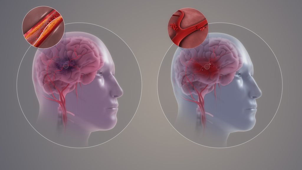
Ischaemic and haemorrhagic stroke. Credit: Scientific Animations CC4.0

In learning, neurons communicate with each other, and the connections between them getting stronger with repetition. This is known as long-term potentiation or LTP.  

Another type of LTP occurs when the brain is deprived of oxygen temporarily – anoxia-induced long-term potentiation or aLTP. aLTP blocks the former process, thereby impairing learning and memory. Therefore, some scientists think that aLTP might be involved in memory problems seen in conditions like stroke. 

Researchers at the Okinawa Institute of Science and Technology (OIST) and their collaborators have studied the aLTP process in detail. They found that maintaining aLTP requires the amino acid glutamate, which triggers nitric oxide (NO) production in both neurons and brain blood vessels. This process forms a positive glutamate-NO-glutamate feedback loop. Their study, published in iScience, indicates that the continuous presence of aLTP could potentially hinder the brain’s memory strengthening processes and explain the memory loss observed in certain patients after experiencing a stroke.  

The brain’s response to low oxygen 

When there is a lack of oxygen in the brain, the neurotransmitter glutamate is released from neurons in large amounts. This increased glutamate causes the production of NO. NO produced in neurons and brain blood vessels boosts glutamate release from neurons during aLTP. This glutamate-NO-glutamate loop continues even after the brain gets enough oxygen. 

“We wanted to know how oxygen depletion affects the brain and how these changes occur,” stated Dr Han-Ying Wang, a researcher in the former Cellular and Molecular Synaptic Function Unit at OIST and lead author of the study,. “It’s been known that nitric oxide is involved in releasing glutamate in the brain when there is a shortage of oxygen, but the mechanism was unclear.”  

During a stroke, when the brain is deprived of oxygen, amnesia – the loss of recent memories – can be one of the symptoms. Investigating the effects of oxygen deficiency on the brain is important because of the potential medicinal benefits. “If we can work out what’s going wrong in those neurons when they have no oxygen, it may point in the direction of how to treat stroke patients,” Dr Patrick Stoney, a scientist in OIST’s Sensory and Behavioral Neuroscience Unit, explained. 

Brain tissues from mice were placed in a saline solution, mimicking the natural environment in the living brain. Normally, this solution is oxygenated to meet the high oxygen demands of brain tissue. However, replacing the oxygen with nitrogen allowed the researchers to deprive the cells of oxygen for precise lengths of time.  

The tissues were then examined under a microscope and electrodes were placed on them to record electrical activity of the individual cells. The cells were stimulated in a way that mimics how they would be stimulated in living mice. 

Stopping memory and learning activity 

The aLTP process is activated when the brain is deprived of oxygen
The aLTP process is activated when the brain is temporarily deprived of oxygen and glutamate levels increase. If aLTP is maintained for an extended period, this hijacks the normal functioning of the memory strengthening process (LTP), resulting in memory loss. Blocking nitric oxide (NO) synthesis or the molecular pathways that boost glutamate release eventually stops aLTP. Credit: Wang et al., 2024 

The scientists found that maintaining aLTP requires NO production in both neurons and in blood vessels in the brain. Collaborating scientists from OIST’s Optical Neuroimaging Unit showed that in addition to neurons and blood vessels, aLTP requires the activity of astrocytes, another type of brain cell. Astrocytes connect and support communication between neurons and blood vessels. 

“Long-term maintenance of aLTP requires continuous synthesis of nitric oxide. NO synthesis is self-sustaining, supported by the NO-glutamate loop, but blocking molecular steps for NO-synthesis or those that trigger glutamate release eventually disrupt the loop and stop aLTP,” Prof. Tomoyuki Takahashi, leader of the former Cellular and Molecular Synaptic Function Unit at OIST, explained.  

Notably, the cellular processes that support aLTP are shared by those involved in memory strengthening and learning (LTP). When aLTP is present, it hijacks molecular activities required for LTP and removing aLTP can rescue these memory enhancing mechanisms. This suggests that long-lasting aLTP may obstruct memory formation, possibly explaining why some patients have memory loss after a short stroke. 

Prof Takahashi emphasised that the formation of a positive feedback loop formed between glutamate and NO when the brain is temporarily deprived of oxygen is an important finding. It explains long-lasting aLTP and may offer a solution for memory loss caused by a lack of oxygen.  

Source: Okinawa Institute of Science and Technology

Amnesia from Head Injury Reversed in Early Mouse Study

Photo by Olga Guryanova on Unsplash

A mouse-based study to investigate memory loss in people who experience repeated head impacts, such as athletes, suggests the condition could potentially be reversed. The research in mice finds that amnesia and poor memory following head injury is due to inadequate reactivation of neurons involved in forming memories.

The study, conducted by researchers at Georgetown University Medical Center in collaboration with Trinity College Dublin, Ireland, is reported in the Journal of Neuroscience.

Importantly for diagnostic and treatment purposes, the researchers found that the memory loss attributed to head injury was not a permanent pathological event driven by a neurodegenerative disease.

Indeed, the researchers could reverse the amnesia to allow the mice to recall the lost memory, potentially allowing cognitive impairment caused by head impact to be clinically reversed.

The Georgetown investigators had previously found that the brain adapts to repeated head impacts by changing the way the synapses in the brain operate, which can cause trouble in memory storage and retrieval.

In their new study, investigators were able to trigger mice to remember memories that had been forgotten due to head impacts.

“Our research gives us hope that we can design treatments to return the head-impact brain to its normal condition and recover cognitive function in humans that have poor memory caused by repeated head impacts,” says the study’s senior investigator, Mark Burns, PhD, a professor and Vice-Chair in Georgetown’s Department of Neuroscience and director of the Laboratory for Brain Injury and Dementia.

In the new study, the scientists gave two groups of mice a new memory by training them in a test they had never seen before. One group was exposed to a high frequency of mild head impacts for one week (similar to contact sport exposure in people) and one group were controls that didn’t receive the impacts. The impacted mice were unable to recall the new memory a week later.

“Most research in this area has been in human brains with chronic traumatic encephalopathy (CTE), which is a degenerative brain disease found in people with a history of repetitive head impact,” said Burns.

“By contrast, our goal was to understand how the brain changes in response to the low-level head impacts that many young football players regularly experience.”

Researchers have found that, on average, college football players receive 21 head impacts per week with defensive ends receiving 41 head impacts per week.

The number of head impacts to mice in this study were designed to mimic a week of exposure for a college football player, and each single head impact by itself was extraordinarily mild.

Using genetically modified mice allowed the researchers to see the neurons involved in learning new memories, and they found that these memory neurons (the “memory engram”) were equally present in both the control mice and the experimental mice.

To understand the physiology underlying these memory changes, study first author Daniel P. Chapman, PhD, said, “We are good at associating memories with places, and that’s because being in a place, or seeing a photo of a place, causes a reactivation of our memory engrams. This is why we examined the engram neurons to look for the specific signature of an activated neuron. When the mice see the room where they first learned the memory, the control mice are able to activate their memory engram, but the head impact mice were not. This is what was causing the amnesia.”

The researchers were able to reverse the amnesia to allow the mice to remember the lost memory using lasers to activate the engram cells.

“We used an invasive technique to reverse memory loss in our mice, and unfortunately this is not translatable to humans,” Burns adds.

“We are currently studying a number of non-invasive techniques to try to communicate to the brain that it is no longer in danger, and to open a window of plasticity that can reset the brain to its former state.”

Source: Georgetown University Medical Center

  1. Daniel P. Chapman, Sarah D. Power, Stefano Vicini, Tomás J. Ryan, Mark P. Burns. Amnesia after repeated head impact is caused by impaired synaptic plasticity in the memory engramThe Journal of Neuroscience, 2024; e1560232024 DOI: 10.1523/JNEUROSCI.1560-23.2024

Not all Memories Lost to Sleep Deprivation are Gone Forever

Sleeping man
Photo by Mert Kahveci on Unsplash

Sleep deprivation is bad for memorisation, something which still doesn’t deter many med students from late night cramming. Researchers however have discovered that memories learned during sleep deprivation is not necessarily lost, it is just difficult to recall. Publishing in the journal Current Biology, the researchers have found a way to make this ‘hidden knowledge’ accessible again days after studying whilst sleep-deprived using optogenetic approaches and the asthma drug roflumilast.

University of Groningen neuroscientist Robbert Havekes and his team have extensively studied how sleep deprivation affects memory processes. “We previously focused on finding ways to support memory processes during a sleep deprivation episode,” says Havekes. However, in his latest study, his team examined whether amnesia as a result of sleep deprivation was a direct result of information loss, or merely caused by difficulties retrieving information. “Sleep deprivation undermines memory processes, but every student knows that an answer that eluded them during the exam might pop up hours afterwards. In that case, the information was, in fact, stored in the brain, but just difficult to retrieve.”

Priming the hippocampus

To find out, the researchers selectively introduced optogenetic proteins into neurons that are activated during a learning experience, enabling recall of a specific experience by shining a light on the cells. “In our sleep deprivation studies, we applied this approach to neurons in the hippocampus, the area in the brain where spatial information and factual knowledge are stored,” says Havekes.

First, the genetically engineered mice were given a spatial learning task in which they had to learn the location of individual objects, a process heavily reliant on neurons in the hippocampus. The mice then had to perform this same task days later, but this time with one object moved to a new location. The mice that were deprived of sleep for a few hours before the first session failed to detect this spatial change, which suggests that they cannot recall the original object locations. “However, when we reintroduced them to the task after reactivating the hippocampal neurons that initially stored this information with light, they did successfully remember the original locations,” says Havekes. “This shows that the information was stored in the hippocampus during sleep deprivation, but couldn’t be retrieved without the stimulation.”

Memory problems

The molecular pathway set off during the reactivation is also targeted by the drug roflumilast, which is used by patients with asthma or COPD. Havekes says: “When we gave mice that were trained while being sleep deprived roflumilast just before the second test, they remembered, exactly as happened with the direct stimulation of the neurons.” Since roflumilast is approved for use in humans and can enter the brain, this may lead to testing to see if it can recover ‘lost’ memories for humans..

It might be possible to stimulate the memory accessibility in people with age-induced memory problems or early-stage Alzheimer’s disease with roflumilast,” says Havekes. “And maybe we could reactivate specific memories to make them permanently retrievable again, as we successfully did in mice.” If a subject’s neurons are stimulated with the drug while they try and ‘relive’ a memory, or revise information for an exam, this information might be reconsolidated more firmly in the brain. “For now, this is all speculation of course, but time will tell.”

Source: University of Groningen.

New Drug Targets for Memory Loss

Image by Falkurian Design on Unsplash

Researchers have identified specific drug targets within memory-encoding neural circuits, opening up possibilities for new treatments of a range of brain disorders.

Memory loss is a main feature of a number of neurological and psychiatric disorders including Alzheimer’s disease and schizophrenia. Presently, there are few, very limited memory loss treatments and the search for safe and effective drug therapies has, until now, borne little fruit.

The research was done in collaboration with colleagues at the international biopharmaceutical company Sosei Heptares. The findings, published in Nature Communications, identify specific receptors for the neurotransmitter acetylcholine that re-route information flowing through memory circuits in the hippocampus. Acetylcholine is released in the brain during learning and is critical for the acquisition of new memories. Until now, the only effective treatment for the symptoms of cognitive or memory impairment seen in diseases such as Alzheimer’s is using drugs that broadly boost acetylcholine. However, this leads to multiple adverse side effects. The discovery of specific receptor targets that have the potential to provide the positive effects whilst avoiding the negative ones is promising.

Lead author Professor Jack Mellor from the University of Bristol’s Center for Synaptic Plasticity, said: “These findings are about the fundamental processes that occur in the brain during the encoding of memory and how they may be regulated by brain state or drugs targeting specific receptor proteins. In the long-term, the discovery of these specific targets opens up avenues and opportunities for the development of new treatments for the symptoms of Alzheimer’s disease and other conditions with prominent cognitive impairments. The academic-industry partnership is important for these discoveries and we hope to continue working together on these projects.”

Dr Miles Congreve, Chief Scientific Officer at Sosei Heptares, added: “These important studies have helped us to design and select new, exquisitely targeted therapeutic agents that mimic the effects of acetylcholine at specific muscarinic receptors, without triggering the unwanted side effects of earlier and less-well targeted treatments. This approach has the exciting potential to improve memory and cognitive function in patients with Alzheimer’s and other neurological diseases.”

“It is fascinating how the brain prioritises different bits of information, working out what is important to encode in memory and what can be discarded. We know there must be mechanisms to pull out the things that are important to us but we know very little about how these processes work. Our future program of work aims to reveal how the brain does this using acetylcholine in tandem with other neurotransmitters such as dopamine, serotonin and noradrenaline,” said Professor Mellor.

Source: University of Bristol