Tag: medical technology

The Promise of Plant-based Vaccines

Photo by Bill Oxford on Unsplash

Recent advances in the development and testing of plant-made vaccines has rekindled interest in plant-produced pharmaceuticals, including edible drugs, for human use. Technology and manufacturing advances could boost the uptake of such therapeutics, wrote Hugues Fausther-Bovendo and Gary Kobinger in an article published in Science

Currently, therapeutic proteins such as antibodies, hormones, cytokines, and proteins in vaccines are mostly produced in bacteria or eukaryotic systems, including chicken eggs and mammalian or insect cell cultures. In 1986, scientists proposed the use of plants for the production of these proteins in what is termed ‘molecular farming’. Such a production process can be less costly and produce fewer contaminants. 

Thus far, just one therapeutic protein derived from plants for human use has been approved (in 2012, for Gaucher disease). More recently in 2019, a plant-produced influenza virus vaccine completed phase III clinical trials with promising results, and phase III trials for a plant-made vaccine COVID vaccine started in early 2021. Plant-produced proteins have a number of advantages for vaccine development, according to Fausther-Bovendo and Kobinger, in particular the strong immune response the plant components of virus-like particles in vaccines can generate, which may reduce the need for adjuvants. 

Also interesting to consider are oral, plant-made therapeutics, said Fausther-Bovendo and Kobinger. Possibly needing minimal processing, they could avoid expensive, lengthy manufacturing. 

Edible vaccines – still predominantly in the preclinical stage of development – are also currently under development, the authors note. Compared to the proof-of-concept edible vaccines first tested decades ago, which generated weak immune responses, newly developed edible plant-made vaccines are now capable of provoking stronger immune responses, thanks to improved technology. 

Because doses for therapeutics are much higher than for vaccines, investment in manufacturing infrastructure must increase to achieve large-scale manufacturing of plant therapeutic products, Fausther-Bovendo and Kobinger said.

Source: EurekAlert!

Tiny Generators Tap Body Motion for Medical Applications

Researchers have created biocompatible generators which harvest body motion to produce electrical impulses for medical applications such as wound healing.

Piezoelectric materials such as ceramics and crystals can generate an electrical charge when mechanically stressed, and are used in many devices such as ultrasound transducers, vibration sensors, and cell phones. In medicine, electrostimulation using piezoelectric devices has been shown to be beneficial for accelerating wound and bone fracture healing, maintaining muscle tone in stroke victims, and chronic pain reduction. However, lack of biocompatibility has stalled progress in the field.

Now bioengineers at the University of Wisconsin’s Department of Materials Science and Engineering, led by Professor Xudong Wang, have developed implantable piezoelectric therapeutic devices. These thin, flexible devices make use of the piezoelectric properties of non-rigid, nontoxic biological materials such as silk, collagen, and amino acids.
The team came up with a method for self-assembly of small patch-like constructs that use the amino acid lysine as the piezoelectric generator. The self-assembly process incorporates a biocompatible polymer shell that surrounds the lysine as the polymer/lysine solution evaporates. Chemical interactions between the inner layer of lysine and the polymer coating orient the lysine into the crystal structure necessary for it to produce electric current when flexed.

“This work is an outstanding example of using the chemical properties of the materials to create a self-assembling product,” explained David Rampulla, director of the Division of Discovery Science and Technology at the National Institute of Biomedical Imaging and Bioengineering. “The process used is rapid and inexpensive, making production of such wafers for therapeutic applications feasible. That the wafers are biodegradable opens the possibility for creating electrotherapies that could be used to accelerate healing of an injured bone or muscle, for example, and then degrade and disappear from the body.”

In one of a number of tests, wafers were placed in the leg and chest of rats, movements of which compressed the piezoelectric wafers enough to create an electrical output. Blood tests performed after the transplanted wafer dissolved showed normal levels of blood cells and other metabolites, indicating no harmful effects from the dissolved device.

Prof Wang emphasises the simplicity of the elegant work. “We believe the technology opens a vast array of possibilities including real-time sensing, accelerated healing of wounds and other types of injuries, and electrical stimulation to treat pain and other neurological disorders. Importantly, our rapid self-assembling technology dramatically reduces the cost of such devices, which has the potential to greatly expand the use of this very promising form of medical intervention.”

The results were reported in the journal Science.

Source: Medical Xpress

A New Snake Venom ‘Super Glue’ For Wound Closures

Photo by David Clode on Unsplash

A novel snake venom ‘super glue’ has been developed, that can stop life-threatening bleeding in under a minute.

Over the past 20 years, bioengineer Kibret Mequanint, a professor at the University, has developed an array of biomaterials-based medical devices and therapeutic technologies – some of which are either now licensed to medical companies or are in the advanced stage of preclinical testing.

This latest work focuses on a blood clotting enzyme called reptilase or batroxobin, which is found in the venom of lancehead snakes (Bothrops atrox), which are amongst the most venomous snakes in South America.

Prof Mequanint and the international research team designed a body tissue adhesive that takes advantage of the clotting property of this enzyme, incorporating it into a modified gelatin that can be packaged into a small, handy tube for easy application.

“During trauma, injury and emergency bleeding, this ‘super glue’ can be applied by simply squeezing the tube and shining a visible light, such as a laser pointer, over it for a few seconds. Even a smartphone flashlight will do the job,” said Prof Mequanint.

Compared to the industry gold standard for clinical and field surgeons, clinical fibrin glue, the new tissue sealant has 10 times the adhesive strength to resist detachment or washout from bleeding. The blood clotting time is also much shorter, halving the 90 seconds for fibrin glue to 45 seconds for this new adhesive.

This novel biotechnology could reduce blood loss and save more lives. Tests were performed in models of major bleeding, such as deep skin cuts, ruptured aortae, and severely injured livers.

“We envision that this tissue ‘super glue’ will be used in saving lives on the battlefield, or other accidental traumas like car crashes,” said Prof Mequanint. “The applicator easily fits in first aid kits too.”

Besides its trauma application, the new snake venom ‘super glue’ can be used in surgical wound closures.

The study was published in the journal Science Advances..

“The next phase of study which is underway is to translate the tissue ‘super glue’ discovery to the clinic,” said Prof Mequanint.

Source: University of Western Ontario

Journal information: Guo, Y., et al. (2021) Snake extract–laden hemostatic bioadhesive gel cross-linked by visible light. Science Advances.doi.org/10.1126/sciadv.abf9635.

Manufacturer Shuts Down its Robot Mid-surgery

Photo by Piron Guillaume on Unsplash

One of a series of lawsuits against the company that makes the da Vinci surgical robot alleges that the company shut down its robot mid-surgery, forcing the surgeons to switch to an open surgery.

Several hospitals have launched a legal battle against the company Intuitive Surgical, the manufacturer of the da Vinci surgical robot. They allege that the company’s monopoly position forces hospitals to buy its maintenance services and spare parts at inflated prices even though cheaper alternatives are available.

One hospital alleges that, after it said that it was considering a service contract with a third party, Intuitive Surgical remotely shut down its surgical robot “in the middle of a procedure”, forcing the surgeon “to convert the procedure to open surgery with the patient on the operating table”.

Separately, malfunctions of the instrument arms have been reported, requiring additional, sometimes larger, incisions in patients in order to complete the surgical procedure manually. Use of the robotic technology also requires longer operating and anesthesia times as well as several complications occurring from the use of the da Vinci Surgical System itself.

Intuitive Surgical sells its da Vinci surgical robot to hospitals for anywhere from $500,000 to $2.5 million each. However, a majority of Intuitive Surgical’s $4 billion of annual revenue comes from the parts and services that are required to keep the robots running. Its executives are among the most highly paid in the healthcare industry.
Franciscan Health, Valley Medical Center and Kaleida Health filed class-action lawsuits. These hospitals that claim Intuitive Surgical has a monopoly on minimally invasive surgical robots, giving the company a “near-stranglehold” on the parts and services market for the robots.

One lawsuit alleges hospitals cannot have their da Vinci robots serviced by third parties because Intuitive Surgical forces hospitals to sign “multi-year, exclusive servicing agreements” at rates that are much higher than other vendors’. Hospitals also allege they are coerced into buying new, expensive instruments and attachments for their robots (called EndoWrists) after 10 uses, even if the parts are in good working condition. A limited extension of these uses has been launched by the company. The lawsuit alleges that Intuitive Surgical engineers have threatened hospitals with turning the machines into “paperweights” should hospitals seek outside vendors for parts or repairs.

While Intuitive Surgical has faced antitrust lawsuits from third-party repair and service companies since 2019, these hospital class-action lawsuits are new.

In an email, an Intuitive Surgical spokesperson told MedPage Today that the medical robotics company “does not have the ability to remotely shut down a da Vinci system during a surgical procedure underway at hospital.”

“There is risk associated with deviating from the validated processes cleared by regulatory authorities,” the spokesperson stated. “Continued use beyond an instrument’s determined useful life may reduce safety, precision, and dexterity. Further, third parties may use incompatible or unvalidated parts or processes in servicing or repairing the systems, which could cause damage and put patient safety at risk.”

Source: Axios

Liquid Metal Sensors Recreate a Sense of Touch

Photo by ThisisEngineering RAEng on Unsplash

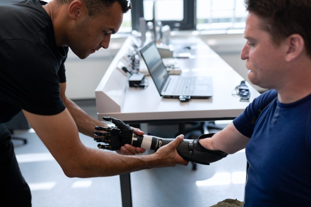
To recreate a sense of ‘touch’, researchers have incorporated stretchable tactile sensors using liquid metal on the fingertips of a prosthetic hand. 

When manipulating an object, humans are heavily reliant on sensation in their fingertips, each of which has over 3000 pressure-sensitive touch receptors. While there are many high-tech, dexterous prosthetics available today, they all lack the sensation of ‘touch‘, resulting in objects inadvertently being dropped or crushed by a prosthetic hand.
To make a prosthetic hand interface that feels more natural and intuitive, researchers from Florida Atlantic University’s College of Engineering and Computer Science and collaborators incorporated stretchable tactile sensors using liquid metal on a prosthetic hand’s fingertips. Encapsulated within silicone-based elastomers, this technology provides key advantages over traditional sensors, including high conductivity, compliance, flexibility and stretchability.

For the study, published in the journal Sensors, researchers used individual fingertips on the prosthesis to distinguish between different speeds of a sliding motion along different textured surfaces. The four different textures had one variation: the distance between the ridges. To detect the textures and speeds, researchers trained four machine learning algorithms. For each of the ten surfaces, 20 trials were performed to test the ability of the machine learning algorithms to distinguish between the different textured surfaces.

Results showed that integrating tactile information from the fingertip sensors simultaneously distinguished between complex, multi-textured surfaces – demonstrating a new form of hierarchical intelligence. The algorithms could accurately distinguish between the fingertip speeds. This new technology could improve prosthetic hand control and provide haptic feedback for amputees to restore a sense of touch.

“Significant research has been done on tactile sensors for artificial hands, but there is still a need for advances in lightweight, low-cost, robust multimodal tactile sensors,” said senior author Erik Engeberg, PhD, an associate professor in the Department of Ocean and Mechanical Engineering. “The tactile information from all the individual fingertips in our study provided the foundation for a higher hand-level of perception enabling the distinction between ten complex, multi-textured surfaces that would not have been possible using purely local information from an individual fingertip. We believe that these tactile details could be useful in the future to afford a more realistic experience for prosthetic hand users through an advanced haptic display, which could enrich the amputee-prosthesis interface and prevent amputees from abandoning their prosthetic hand.”

Researchers compared four different machine learning algorithms for their successful classification capabilities. The time-frequency features of the liquid metal sensors were extracted to train and test the machine learning algorithms. Of these, a neural network algorithm generally performed the best at the speed and texture detection with a single finger and had a 99.2 percent accuracy to distinguish between ten different multi-textured surfaces using four liquid metal sensors from four fingers simultaneously.

“The loss of an upper limb can be a daunting challenge for an individual who is trying to seamlessly engage in regular activities,” said Stella Batalama, Ph.D., dean, College of Engineering and Computer Science. “Although advances in prosthetic limbs have been beneficial and allow amputees to better perform their daily duties, they do not provide them with sensory information such as touch. They also don’t enable them to control the prosthetic limb naturally with their minds. With this latest technology from our research team, we are one step closer to providing people all over the world with a more natural prosthetic device that can ‘feel’ and respond to its environment.”

Source: Florida Atlantic University

Journal information: Abd, M.A., et al. (2021) Hierarchical Tactile Sensation Integration from Prosthetic Fingertips Enables Multi-Texture Surface Recognition. Sensors. doi.org/10.3390/s21134324.

MRI and Ultrasound Combo Opens Blood-brain Barrier

In a mouse model study of MRI-guided focused ultrasound-induced blood-brain barrier (BBB) opening at MRI field strengths ranging from ­approximately 0 T (outside the magnetic field) to 4.7 T, the static magnetic field dampened the detected microbubble cavitation signal and decreased the BBB opening volume. Credit: Washington University School of Medicine in St. Louis

Using a combination of ultrasound, MRI field strength and microbubbles can open the blood-brain barrier (BBB) and allow therapeutic drugs to reach the diseased brain location with MRI guidance. 

Using the physical phenomenon of cavitation, it is a promising technique that has been shown safe in patients with various brain diseases, such as Alzheimer’s diseases, Parkinson’s disease, ALS, and glioblastoma.
While MRI has been commonly used for treatment guidance and assessment in preclinical research and clinical studies, until now, researchers did not know the impact that MRI scanner’s magnetic field had on the BBB opening size and drug delivery efficiency.

Hong Chen, associate professor of biomedical engineering at Washington University in St. Louis, and her lab have found for the first time that the magnetic field of the MRI scanner decreased the BBB opening volume by 3.3-fold to 11.7-fold, depending on the strength of the magnetic field, in a mouse model. The findings were in Radiology.

Prof Chen conducted the study on four groups of mice. After they were injected microbubbles, three groups received focused-ultrasound sonication at different strengths of the magnetic field: 1.5 T (teslas), 3 T and 4.7 T, and one group was never exposed to the field. 

The researchers found that the microbubble cavitation activity, or the growing, shrinking and collapse of the microbubbles, decreased by 2.1 decibels at 1.5 T; 2.9 decibels at 3 T; and 3 decibels at 4.7 T, compared with those that had received the dose outside of the magnetic field. Additionally, the magnetic field decreased the BBB opening volume by 3.3-fold at 1.5 T; 4.4-fold at 3 T; and 11.7-fold at 4.7 T. No tissue damage from the procedure was seen.

Following focused-ultrasound sonication, the team injected a model drug, Evans blue dye, to investigate whether the magnetic field affected drug delivery across the BBB. The images showed that the fluorescence intensity of the Evans blue was lower in mice that received the treatment in one of the three strengths of magnetic fields compared with mice treated outside the magnetic field. The Evans blue trans-BBB delivery was decreased by 1.4-fold at1.5 T, 1.6-fold at 3.0 T and 1.9-fold at 4.7 T when compared with those treated outside of the magnetic field.

“The dampening effect of the magnetic field on the microbubble is likely caused by the loss of bubble kinetic energy due to the Lorentz force acting on the moving charged lipid molecules on the microbubble shell and dipolar water molecules surrounding the microbubbles,” said Yaoheng (Mack) Yang, a doctoral student in Prof Chen’s lab and the lead author of the study.

“Findings from this study suggest that the impact of the magnetic field needs to be considered in the clinical applications of focused ultrasound in brain drug delivery,” Prof Chen said.

In addition to brain drug delivery, cavitation is also used in several other therapeutic techniques, such as histotripsy, the use of cavitation to mechanically destroy regions of tissue, and sonothrombolysis, a therapy used after acute ischaemic stroke. The magnetic field’s damping effect on cavitation is expected to affect the treatment outcomes of other cavitation-mediated techniques when MRI-guided focused-ultrasound systems are used.

Source: Washington University in St. Louis

Journal information: Yang, Y., et al. (2021) Static Magnetic Fields Dampen Focused Ultrasound–mediated Blood-Brain Barrier Opening. Radiology. doi.org/10.1148/radiol.2021204441

Carbon Fibre Electrodes Allow Unprecedented Neural Recording

Image by Robina Weemeijer on Unsplash

A tiny, implantable carbon fibre electrode has the potential to provide a long-term brain-computer interface which can record electrical signals over lengthy periods of time.

The carbon fibre electrodes were developed at the University of Michigan and demonstrated in rats. The new research shows the promise of carbon fibre electrodes in recording electrical signals from the brain without damaging brain tissue. Directly implanting carbon fiber electrodes into the brain allows the capturing of bigger and more specific signals than current technologies.

This technology could lead to advances that could give amputees and those with spinal injuries control of advanced prosthetics, stimulate the sacral nerve to restore bladder control, stimulate the cervical vagus nerve to treat epilepsy and provide deep brain stimulation as a possible treatment for Parkinson’s.  

“There are interfaces out there that can be implanted directly into the brain but, for a variety of reasons, they only last from months to a few years,” said Elissa Welle, a recent PhD graduate from the U-M Department of Biomedical Engineering. “Any time you’re opening up the skull for a procedure involving the brain, it’s a big deal.”

Brain implants are typically made from silicon due to its ability to conduct electricity and its historic use in cleanroom technology. But silicon is not very biocompatible and leads to the formulation of scar tissue over long periods. Such electodes will eventually degrade and no longer capture brain signals, requiring removal.

Carbon fibres may be the answer to getting high-quality signals with an interface that lasts years, not months. And by laser cutting and sharpening carbon fibers into tiny, subcellular electrodes in the lab with the help of a small blowtorch, U-M engineers have harnessed the potential for excellent signal capture in a form the body is more likely to accept.

“After implantation, it sits inside the brain in a way that does not interfere with the surrounding blood vessels, because it’s smaller than those blood vessels,” Welle said. “They’ll move around and adjust to an object that small, rather than get torn as they would when encountering larger implants.”

Part of the electrode’s compatibility in brain tissue is down to smaller size, but its needle-like shape may also minimise compacting of any surrounding tissue. Larger carbon-based electrodes have been shown to actually encourage neural tissue to grow instead of degrading. The team is hopeful that similar potential for their carbon fibre electrodes will be revealed by further testing.

Carbon fibre electrodes in a previous study dramatically outperformed conventional silicon electrodes with 34% of electrodes recording a neuron signal compared to 3%. Laser cutting then improved this number to 71% at 9 weeks after implantation. Flame sharpening has now enabled these high performance probes to be implanted directly into the cerebral cortex, negating the need for a temporary insertion aid, or shuttle, as well as into the rat’s cervical vagus nerve.

It is relatively easy to insert electrodes into the brain. But the researchers have also taken on the more difficult task of inserting the sharpened carbon fibre electrodes into nerves, with micrometre diameters.

Those findings show that potential for these electrodes goes beyond prosthetic manipulation, according to Cindy Chestek, a U-M associate professor of biomedical engineering, and principal investigator of the The Cortical Neural Prosthetics Lab.

“Someone who is paralysed may have no control over things like their bladder, for example,” Prof Chestek said. “We may be able to utilise these smaller electrodes to stimulate and record signals from areas that can’t be reached by larger ones, maybe the neck or spinal cord, to help give patients some level of control.”

Source: University of Michigan

A Step to Towards Electrically Restoring Oral Sensation and Function

Photo by Maria Lysenko on Unsplash

In an effort towards restoring oral functionality lost to nerve or brain damage, researchers at Texas A&M University have determined the minimum electrical stimulation needed to provide sensation in various parts of the mouth.

Sensorimotor feedback loops involve the brain interpreting incoming signals from sensory nerves and then ordering motor nerves to execute a particular movement. Sensorimotor loops play a vital role in voluntary functions, like walking or holding an object, and involuntary movements, like sneezing or blinking.

Within the mouth, both sensory and motor nerves are richly supplied. In particular, sensorimotor nerves in the soft palate and tongue coordinate several intraoral movements related to swallowing, speech and respiration. Damage to either the sensory or motor nerve fibres due to neurotrauma or disease can therefore compromise these essential functions and worsening the quality of life for afflicted individuals.

Electrical nerve stimulation might help jumpstart the nerves into action, much like how a pacemaker can electrically stimulate nerves in the heart, causing the heart muscle to contract. Unlike a pacemaker however, the parameters of the electrical currents needed for proper stimulation of different parts of the mouth have not been investigated.

“Electrical stimulation can modulate nerve currents or action potentials, which are the mode of communication to and from the brain,” said Hangue Park, assistant professor in the Department of Electrical and Computer Engineering. “And so, electrical stimulation should be carefully applied, because if not, then it might cause undesirable effects, or it might not stimulate anything at all.”

To investigate the minimum stimulation currents needed, Park and his team place tiny metal electrodes in a standard dental retainer. These electrodes were positioned in subjects’ mouths to stimulate either their soft palate or the side and tip of the tongue, which are dense in sensory nerves. The researchers slowly changed the amplitude of the stimulation current, keeping the frequency fixed. Subjects reported when they began feeling a sensation and when the sensation was uncomfortable, and the same experiment was repeated with a higher frequency of current.

After compiling their data, the team determined the average perception and discomfort thresholds for the tongue and soft palate. In addition, they produced an equivalent circuit of the intraoral cavity to duplicate the electrical properties of that area. This circuit, the researchers said, can help to further study the effects of electrical stimulation offline without requiring human subjects.

The researchers noted that their next steps would be to electrically stimulate the intraoral region and investigate how these simulations change chewing, swallowing and other behaviours.

“Sensorimotor systems can be extremely vulnerable to damage due to neural defects, aging and neurodegenerative diseases,” Park said. “In this study, we have begun to lay the groundwork for electrically stimulating parts of the mouth that control involuntary and voluntary movements. Our work is a seminal study and it is important so that we can, in the near future, help people that face enormous challenges doing everyday tasks that we take for granted.”

Source: Texas A&M University

Journal information: Park, B., et al. (2021) Electrical Characterization of the Tongue and the Soft Palate using Lumped-Element Model for Intraoral Neuromodulation. IEEE Transactions on Biomedical Engineering. doi.org/10.1109/TBME.2021.3070867.

Breakthrough AI Development for Premature Baby Care

Photo by Hush Naidoo on Unsplash

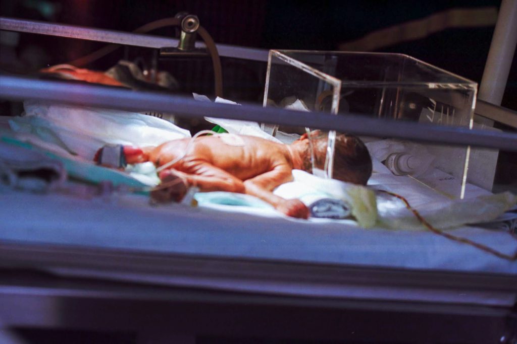
Researchers believe they have made a breakthrough in the science of keeping premature babies alive.

As part of her PhD work, James Cook University engineering lecturer Stephanie Baker led a pilot study that used a hybrid neural network to accurately predict how much risk individual premature babies face. This study was published in the journal Computers in Biology and Medicine.

Complications resulting from premature birth are the leading cause of death in children under five and over 50% of neonatal deaths occur in preterm infants, she said. In 2005, 12.9 million births, or 9.6% of all births worldwide, were preterm.

“Preterm birth rates are increasing almost everywhere. In neonatal intensive care units, assessment of mortality risk assists in making difficult decisions regarding which treatments should be used and if and when treatments are working effectively,” said Ms Baker.

To better guide their care, preterm babies are often given a score that indicates the risk they face.

“But there are several limitations of this system. Generating the score requires complex manual measurements, extensive laboratory results, and the listing of maternal characteristics and existing conditions,” noted Ms Baker.

She said the alternative was to measure variables that do not change (eg, birthweight) that prevents recalculation of the infant’s risk on an ongoing basis and does not show their response to treatment.

“An ideal scheme would be one that uses fundamental demographics and routinely measured vital signs to provide continuous assessment. This would allow for assessment of changing risk without placing unreasonable additional burden on healthcare staff,” said Ms Baker.

She said the JCU team’s research had culminated in the Neonatal Artificial Intelligence Mortality Score (NAIMS), a hybrid neural network that relies on simple demographics and trends in heart and respiratory rate to determine mortality risk.

“Using data generated over a 12 hour period, NAIMS showed strong performance in predicting an infant’s risk of mortality within 3, 7, or 14 days.

“This is the first work we’re aware of that uses only easy-to-record demographics and respiratory rate and heart rate data to produce an accurate prediction of immediate mortality risk,” said Ms Baker.

According to Ms Baker, the technique was fast with no invasive procedures or knowledge of medical histories needed.

“Due to the simplicity and high performance of our proposed scheme, NAIMS could easily be continuously and automatically recalculated, enabling analysis of a baby’s responsiveness to treatment and other health trends,” said Ms Baker.

She said NAIMS had proved accurate when tested against hospital mortality records of preterm babies and had the added advantage over existing schemes of being able to perform a risk assessment based on any 12 hour period of data gathered during the patient’s stay.

Ms Baker said the next step in the process was partnering with local hospitals to gather more data and undertake further testing.

“Additionally, we aim to conduct research into the prediction of other outcomes in neo-natal intensive care, such as the onset of sepsis and patient length of stay,” said Ms Baker.

Source: James Cook University

Journal information: Baker, S., et al. (2021) Hybridized neural networks for non-invasive and continuous mortality risk assessment in neonates. Computers in Biology and Medicine. doi.org/10.1016/j.compbiomed.2021.104521.

New Machine Learning Tools Could Save Teeth

Photo by Kevin Bation on Unsplash

Machine learning tools could help identify those at greatest risk for tooth loss and refer them for further dental assessment for early interventions to avert or delay the conditions.

The study by researchers at the Harvard School of Dental Medicine compared five different algorithms using various combinations of variables to screen for risk. The results showed those that factored medical characteristics and socioeconomic variables, including race, education, arthritis, and diabetes, outperformed algorithms that relied on dental clinical indicators alone.

“Our analysis showed that while all machine-learning models can be useful predictors of risk, those that incorporate socioeconomic variables can be especially powerful screening tools to identify those at heightened risk for tooth loss,” said study lead investigator Hawazin Elani, assistant professor of oral health policy and epidemiology at HSDM.

The approach could be used to screen people globally and in a variety of health care settings even by non-dental professionals, she added. This approach could be applied around the world, even allowing non-dental professionals to screen patients.

Tooth loss can affect quality of life, well-being, nutrition, and social interactions. It is also associated with dementia. If the earliest signs of dental disease are identified, then the process can be delayed or averted with prompt treatment. However, many people with dental disease may not see a dentist until the process is too far gone. This is where screening tools could help identify those at highest risk and refer them for further assessment, the team said.

For the study, the researchers used data on nearly 12 000 adults from the National Health and Nutrition Examination Survey to design and test five machine-learning algorithms and assess their predictions for both complete and incremental tooth loss among adults based on socioeconomic, health, and medical characteristics.

A key point is that algorithms were designed to assess risk without a dental exam, though anyone at risk for tooth loss would still need one. The study’s findings point to the importance of socioeconomic factors.

“Our findings suggest that the machine-learning algorithm models incorporating socioeconomic characteristics were better at predicting tooth loss than those relying on routine clinical dental indicators alone,” Elani said. “This work highlights the importance of social determinants of health. Knowing the patient’s education level, employment status, and income is just as relevant for predicting tooth loss as assessing their clinical dental status.”

Low socioeconomic status populations have long been known to have greater rates of tooth loss, likely due to lack of regular access to dental care, among other reasons, the team said.

“As oral health professionals, we know how critical early identification and prompt care are in preventing tooth loss, and these new findings point to an important new tool in achieving that,” said Jane Barrow, associate dean for global and community health and executive director of the Initiative to Integrate Oral Health and Medicine at HSDM. “Dr. Elani and her research team shed new light on how we can most effectively target our prevention efforts and improve quality of life for our patients.”

Source: Harvard Medical School

Journal information: Hawazin W. Elani et al, Predictors of tooth loss: A machine learning approach, PLOS ONE (2021). DOI: 10.1371/journal.pone.0252873