Tag: medical research

Uncovering Albumin’s Role in Fertility and Inflammation
Source: CC0

Researchers have discovered that albumin (Alb), one of the most abundant proteins in the body, activates a proton channel (hHv1), also widespread in the body, giving sperm the ability to penetrate and fertilise an egg, and also allowing white blood cells to produce inflammatory mediators to fight infection.

The study explored the physiological connection between Alb and human voltage-gated proton channels (hHv1), which are both essential to cell biology in health and diseases. Researchers also demonstrated the mechanism by which Alb binds directly to hHv1 to activate the channel. This research explains how sperm are triggered to fertilise, and neutrophils are stimulated to release mediators in the innate immune response, describing a new role for Alb in physiology that will operate in the many tissues expressing hHv1.

“We found that the interaction of Alb and hHv1 activates sperm when they leave semen and enter the female reproductive tract because Alb is low in semen and high in the reproductive tract. We now understand why albumin supplementation improves IVF,” explained first author Ruiming Zhao, PhD, from the Department of Physiology & Biophysics at UCI School of Medicine. “We also found the same Alb/hHv1 interaction allows the white blood cells called neutrophils to produce and secrete the inflammatory mediators that kill bacteria and fight infection. However, it’s important to note that the inflammatory response itself can lead to disease.”

Alb’s stimulating role in the physiology of sperm and neutrophils via hHv1 pointed to its having other enhancing or deleterious roles in the other tissues, including the central nervous system, heart and lungs, and influencing cancers of the breast and gastrointestinal tract.

“It is exciting to discover that a common protein has the power to activate the proton channel.  This finding suggests new strategies to block or enhance fertility, and to augment or suppress the innate immune response and inflammation,” said senior author Steve A. N. Goldstein, MD, PhD, vice chancellor of Health Affairs at UCI.

hHv1 is involved in many biological processes in addition to the capacitation of sperm and the innate immune responses included in the study.  The channels have notable roles in proliferation of cancer cells, tissue damage during ischaemic stroke, and hypertensive kidney injury. Because Alb’s presence and involvement varies, the potentiation of hHv1 by Alb can be either beneficial or detrimental in different diseases or conditions.

“We have modeled the structural basis for binding of Alb to the channel that leads to activation and changes in cellular function, and we are now conducting in vivo studies of viral and bacterial infections.  Our next steps include studies of the effects of inhibitors of the Alb-hHv1 interaction on infection, inflammation and fertility,” said Goldstein.  

Source: University of California, Irvine

Journal information: Ruiming Zhao et al, Direct activation of the proton channel by albumin leads to human sperm capacitation and sustained release of inflammatory mediators by neutrophils, Nature Communications (2021). DOI: 10.1038/s41467-021-24145-1

Bias Against Both Sexes Found in Clinical Trials

Photo by Tim Mossholder on Unsplash

Though evenly split overall, research shows that women and men in disease trials are not represented according to the population affected.

Clinical trial sample populations should be proportionate to the population affected by the disease, as some diseases are more prevalent or manifest differently in one sex versus the other. Neglecting one sex in clinical trials can skew medical evidence toward therapies for the neglected population.
The study cross-analysed over 20 000 US clinical trials between 2000-2020, and found that women are underrepresented in clinical trials in cardiology, oncology, neurology, immunology and haematology. Meanwhile, men are underrepresented in clinical trials in musculoskeletal disease and trauma, psychiatry and preventive medicine. 

This study, published JAMA Network Open, is the first to examine sex bias in all US human clinical trials relative to disease burden (which is the prevalence of disease based on factors such as sex and ethnicity).

“Sex bias in clinical trials can negatively impact both men and women by creating gendered data gaps that then drive clinical practice,” said first study author Dr Jecca Steinberg, a medical resident in the department of obstetrics and gynecology at Northwestern University Feinberg School of Medicine. “Neglecting one sex in clinical trials — the gold standard scientific exploration and discovery — excludes them from health innovation and skews medical evidence toward therapies with worse efficacy in that sex.”

Underrepresentating either sex in clinical trials can lead to less optimal health outcomes; differences exist for women and men in medical test results, disease progression, treatment response, drug metabolism and surgical outcomes, Dr Steinberg said.

These differences stem from variations in body size, composition, and hormones. Women’s smaller body sizes and higher fat contents typically result in varied drug responses. One study showed that aspirin has differential effects on the sexes with regard to primary protection against strokes and heart attacks. Aspirin only lowered women’s risk of stroke but had no effect on the risk of myocardial infarction or death.

“Identifying areas of research in which sex bias disadvantages males is important to improving population health,” Dr Steinberg said. “Our novel finding that men are underrepresented in trials related to mental health and trauma assumes greater urgency in American society where suicide, violence and substance use increasingly contribute to growing morbidity and premature male mortality in the US.”

Clinical trials for preventative medicine are more likely to have greater enrollment of women, the study found, adding credence to the notion that women, more than men, seek out preventive services and access to health care.  

Women-specific underrepresentation remains
“One of the top reasons for Food and Drug Administration drug recall is adverse effects in women,” Dr Steinberg said. “Millions and millions of dollars go into these trials, so to relatively neglect women in the trial population is a waste.

“A greater allocation of resources for female-focused trials could be critical to improving care for women and discerning the heterogenous manifestations of diseases within the female population.”

For example, women with heart disease often have different reactions to medications and experience different symptoms from men, such as feeling abdominal pain rather than their left arm. If a clinical trial implements its intervention based on symptoms predominantly exhibited by the male population, it could miss testing interventions in women with cardiac arrest. 

The reduced representation of women relative to disease burden specifically in oncology and cardiology clinical trials is especially troubling, said Steinberf, because cardiologic and oncologic diseases are among the leading causes of death among women in the US.

Participating in clinical trials is also one of the only ways to access cutting edge therapies, especially oncology, so the relative deficiency of one sex contributes to disparities in health outcomes, Steinberg said.  

“One of our hopes from this study is that scientists and physicians will read about our findings and be inspired to say, ‘Why is that happening in my field?’ and then address it,” Dr Steinberg said.

Source: Northwestern University

Call for More Neuroscience Research in Africa

Photo by MART PRODUCTION from Pexels

A team of neuroscientists are calling for greater support of neuroscience research in Africa based on an analysis of the continent’s past two decades of research outputs.  

The findings reveal important information about the nature of funding and international collaboration comparing activity in the continent to other countries, mainly the US, UK and areas of Europe. It is hoped that the study will provide useful data to help further develop science in Africa.  

The greatest human genetic diversity is found in Africa, and Eurasian genomes have less variation than African ones; in fact, Eurasian genomes can be considered a subset of African ones. This carries important implications for understanding human diseases, including neurological disorders.

Co-lead senior author Tom Baden, Professor of Neuroscience in the School of Life Sciences and the Sussex Neuroscience research group at the University of Sussex said: “One beautiful thing about science is that there is no such thing as a truly local problem. But that also means that there should be no such thing as a local solution – research and scientific communication by their very nature must be a global endeavour.  

“And yet, currently the vast majority of research across most disciplines is carried out by a relatively small number of countries, located mostly in the global north. This is a huge waste of human potential.”  

The team, made up of experts from the University of Sussex, the Francis Crick Institute and institutions from across Africa, analysed the entirety of Africa’s outputs in neuroscience over two decades. A lot of early neuroscience research took place in Egypt, it was pointed out.

Lead author Mahmoud Bukar Maina, a Research Fellow in the School of Life Sciences and the Sussex Neuroscience research group at the University of Sussex and visiting scientist at Yobe State University, Nigeria, explained: “Even though early progress in neuroscience began in Egypt, Africa’s research in this area has not kept pace with developments in the field around the world. There are a number of reasons behind this and, for the first time, our work has provided a clear picture of why – covering both strengths and weaknesses of neuroscience research in Africa and comparing this to other continents.  

“We hope it will provide useful data to guide governments, funders and other stakeholders in helping to shape science in Africa, and combat the ‘brain drain’ from the region.”  

Co-lead senior author Lucia Prieto-Godino, a Group Leader at the Francis Crick Institute, said: “One of the reasons why this work is so important, is that the first step to solve any problem is understanding it. Here we analyse key features and the evolution of neuroscience publications across all 54 African countries, and put them in a global context. This highlights strengths and weaknesses, and informs which aspects will be key in the future to support the growth and global integration of neuroscience research in the continent.” 

The study identifies the African countries with the greatest research outputs, revealing that most research funding originates from external sources such as the USA and UK.  

The researchers argue that a sustainable African neuroscience research environment needs local funding, suggesting that greater government backing is needed as well as support from the philanthropic sector.  
Professor Baden added: “One pervasive problem highlighted in our research was the marked absence of domestic funding. In most African countries, international funding far predominates. This is doubly problematic.  

“Firstly, it takes away the crucial funding stability that African researchers would need to meaningfully embark on large-scale and long-term research projects, and secondly, it means that the international, non-African funders essentially end up deciding what research is performed across the continent. Such a system would generate profound outrage across places like Europe – how then can it be acceptable for Africa?”

A number of the researchers involved in the study are members of TReND Africa, a charity supporting scientific capacity building in Africa.  

Source: University of Sussex

Journal information: M. B. Maina et al, Two decades of neuroscience publication trends in Africa, Nature Communications (2021). DOI: 10.1038/s41467-021-23784-8 , www.nature.com/articles/s41467-021-23784-8

Researchers Discover that Humans can Readily Develop Echolocation Ability

Photo by Pawel Czerwinski on Unsplash

The ability for humans to sense their surrounding space with reflected sounds might sound like a superhero’s ability, but it is a skill that is developed by some blind people, who use clicks as a form of echolocation.

Echolocation is an ability known in dolphins, whales and bat species, which occurs when such animals emit a sound that reflects off objects in the environment, returning echoes that provide information about the surrounding space.

Existing research has shown that some blind people may use click-based echolocation to judge spaces and improve their navigation skills. Armed with this information, a team of researchers led by Dr Lore Thaler explored how people acquire this skill.

Over the course of a 10-week training programme, the team investigated how blindness and age affect learning of click-based echolocation. They also studied how learning this skill affects the daily lives of people who are blind.

Both blind and sighted people between 21 and 79 years of age participated in this study, which provided a training course of 10 weeks. Blind participants also took part in a 3-month follow up survey assessing how the training affected their daily life.

Both sighted and blind people improved considerably on all measures, and in some cases performed as well as expert echolocators did at the end of training. A surprising result was that a few sighted people even performed better than those who were blind.

However, neither age nor blindness limited participants’ rate of learning or in their ability to apply their echolocation skills to novel, untrained tasks.

Furthermore, in the follow up survey, all participants who were blind reported improved mobility, and 83% reported better independence and wellbeing.

Age or vision not a limitation

Overall, the results suggest that the ability to learn click-based echolocation is not strongly limited by age or level of vision. This has positive implications for the rehabilitation of people with vision loss or in the early stages of progressive vision loss.

Click-based echolocation is not presently taught as part of mobility training and rehabilitation for blind people. There is also the possibility that some people are reluctant to use click-based echolocation due to a perceived stigma around  the click sounds in social environments.

Despite this, the results indicate that both blind people who use echolocation and people new to echolocation are confident to use it in social situations, indicating that the perceived stigma is likely less than believed.

Source: Durham University

Journal information: Human click-based echolocation: Effects of blindness and age, and real-life implications in a 10-week training program, PLOS ONE (2021)

Neural Connectivity can Predict Epilepsy Outcomes

Image source: Unsplash

Researchers have found that neuron connectivity patterns within brain regions can better indicate disease progression and treatment outcomes for people with brain disorders such as epilepsy.

Many brain diseases lead to cell death and the removal of connections within the brain. A team led by Dr Marcus Kaiser from the School of Medicine at the University of Nottingham looked at epilepsy patients undergoing surgery. Their findings were published in Human Brain Mapping.

They found that changes in the local network within brain regions can predict disease progression, and also whether surgery will be successful or not.

The team found that looking at connectivity within regions of the brain, showed superior results compared to only observing fibre tract connectivity between brain regions, which is the current method. Dividing the surface of the brain into 50 000 network nodes of comparable size, each brain region could be studied as a local network with 100-500 nodes. There were distinct changes seen in these local networks in patients suffering from epileptic seizures.

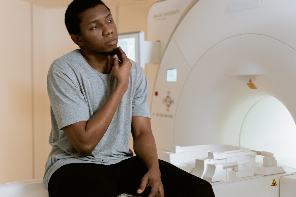
Employing diffusion tensor imaging, a special measurement protocol for MRI scanners, the team of scientists showed that fibres within and between brain regions are removed for patients.

However, they found that connectivity within regions better predicted whether surgical removal of brain tissue was successful in preventing future seizures.

Dr Kaiser, Professor of Neuroinformatics at the University of Nottingham, explained: “When someone has an epileptic seizure, it ‘spreads’ through the brain. We found that local network changes occurred for regions along the main spreading pathways for seizures. Importantly, regions far away from the starting point of the seizure, for example in the opposite brain hemisphere, were involved.

“This indicates that the increased brain activity during seizures leads to changes in a wide range of brain regions. Furthermore, the longer patients suffered, the more regions showed local changes and the more severe were these changes.”

The researchers from the involved universities, along with the company Biomax, evaluated the scans of 33 temporal lobe epilepsy patients and 36 control subjects.

Project partners used the NeuroXM™ knowledge management platform to develop a knowledge model for high-resolution connectivity with more than 50 000 cortical nodes and several millions of connections and corresponding automated processing pipelines accessible through Biomax’s neuroimaging product NICARA™.

Project manager Dr Markus Butz-Ostendorf from Biomax said: “Our software can be easily employed at hospitals and can also be combined with other kinds of data from genetics or from other imaging approaches such as PET, CT, or EEG.”

Professor Yanjiang Wang, who is one of the corresponding authors, and Ms Xue Chen, both from China University of Petroleum (East China), commented: “Local connectivity was not only better in overall predictions but particularly successful in identifying patients where surgery did not lead to any improvement, identifying 95% of such cases compared to 90% when used connectivity between regions”.

Source: University of Nottingham

Researchers Close in on Genetic Cure for Congenital Deafness

Researchers are a step closer in the quest to use gene therapy to enable people born deaf to hear, having uncovered a new role for a key protein.

The study, published in Molecular Biology of the Cell, focused on a large gene responsible for an inner-ear protein called otoferlin. Otoferlin mutations are linked to severe congenital hearing loss, a common type of deafness in which patients can hear almost nothing.

“For a long time otoferlin seemed to be a one-trick pony of a protein,” explained Colin Johnson, associate professor of biochemistry and biophysics in the Oregon State UniversityCollege of Science. “A lot of genes will find various things to do, but the otoferlin gene had appeared only to have one purpose and that was to encode sound in the sensory hair cells in the inner ear. Small mutations in otoferlin render people profoundly deaf.”

Because the otoferlin gene is too big as it normally is to package into a delivery vehicle for molecular therapy, Prof Johnson’s team explored the use of a shortened version.

Research led by graduate student Aayushi Manchanda showed the shortened version needed to have part of the gene known as the transmembrane domain, for a surprising reason: without it, the sensory cells matured slowly.

“That was surprising since otoferlin was known to help encode hearing information but had not been thought to be involved in sensory cell development,” Johnson said.

For years, scientists in Prof Johnson’s lab have been working with the otoferlin molecule and in 2017 they identified a shortened form of the gene that can function in the encoding of sound.

To find out if the transmembrane domain of otoferlin needed to be part of the shortened version of the gene, Manchanda shortened the transmembrane domain in zebrafish.

Zebrafish are a small freshwater species that is very popular as a research organism. They grow rapidly, from a cell to a swimming fish in about five days, and share a remarkable similarity to humans at the molecular, genetic and cellular levels due to the conservation of mammalian genes early in their evolution. Embryonic zebrafish are transparent and easily maintained, and are amenable to genetic manipulation.

“The transmembrane domain tethers otoferlin to the cell membrane and intracellular vesicles but it was not clear if this was essential and had to be included in a shortened form of otoferlin,” Manchanda said. “We found that the loss of the transmembrane domain results in the sensory hair cells producing less otoferlin as well as deficits in hair cell activity. The mutation also caused a delay in the maturation of the sensory cells, which was a surprise. Overall the results argue that the transmembrane domain must be included in any gene therapy construct.”

At the molecular level, Manchanda found that a lack of transmembrane domain led to otoferlin not properly linking the neurotransmitter-filled synaptic vesicles to the cell membrane, resulting in less neurotransmitter being released.

“Our study suggests otoferlin’s ability to tether the vesicles to the cell membrane is a key mechanistic step for neurotransmitter release during the encoding of sound,” Manchanda said.

Source: EurekaAlert!

Validity of Screen Time Studies Questioned

Photo by Tracy le Blanc from Pexels


In the largest study of its kind to date, a systematic review and meta-analysis of people’s perceptions of their screen time compares to their actual usage, estimates of usage were found to be accurate only in about five per cent of studies.

Multiple studies have linked increasing amounts of ‘screen time’, looking at and using devices such as computers, tablets and smartphones, to a wide range of negative health outcomes such as depression and inactivity.

The international team say their results cast doubt on the validity of research on the impact of screen time on mental health, and its influences on government policy, as the vast majority rely on participants’ self-reported estimates on the amount of time spent on digital devices, rather than logs of actual usage, or tracked time. This research was published in Nature Human Behaviour

For lead researcher Dr Doug Parry at Stellenbosch University, the studies highlight how much our current perceptions of technology are built up on long-lasting, unchallenged assumptions.

“For decades, researchers have relied on estimates of how we use various technologies to study how people use digital media and the potential outcomes this behaviour can lead to. Our findings suggest that much of this work may be on unstable footing.”

“The screen time discrepancies highlight that we simply do not know enough yet about the actual effects (both positive and negative) of our media use. Researchers, journalists, members of the public, and crucially policy makers need to question the quality of evidence when they consider research on media uses and effects. We can no longer simply take claims of harmful effects at face value.”

The researchers also investigated whether questionnaires and scales addressing ‘problematic’ media use, such as excessive or so-called ‘addictive’ media use, were suitable substitutes for logged usage. There was even less of an association with usage logs to these measures.

Exhaustive literature survey

The research identified every existing study that compares logged or tracked media use measures with equivalent self-reports. Screening more than 12 000 articles for inclusion, they found 47 studies that included both types of measures. From here they were able to identify and extract 106 comparisons, based on 50 000 individuals, to address the question of how closely self-report estimates relate to logs of actual usage.

“These highly flawed studies are over-inflating the relationships between digital media use and typically negative outcomes, such as mental health symptoms and cognitive impairments, which of course explains the pervading view that smartphones among other technologies are bad for us,” commented Dr Brit Davidson from the University of Bath’s School of Management.

“Media and technology use takes the blame for everything from increases in teenage depression and suicide to higher incidence of Attention Deficit Hyperactivity Disorder (ADHD) and violence. If we want to properly investigate harms, we must first tackle assumptions about screen time and disentangle how people are actually using their phones or other technologies of interest.

“Importantly, these questionable studies are also being used to influence policy. The UK and Canada both have forms of screen time guidelines based on poorly conducted research, which is clearly worrying and hard to reverse.”

The researchers hope that the findings will prompt a change in how technology use is measured, as well as how society regards technology use, leading to a better understanding of our relationship with technology.

Source: News-Medical.Net

Journal information: Parry, D. A., et al. (2021) A systematic review and meta-analysis of discrepancies between logged and self-reported digital media use. Nature Human Behaviour. doi.org/10.1038/s41562-021-01117-5.

Two-way Signalling Discovered in Certain Neurons

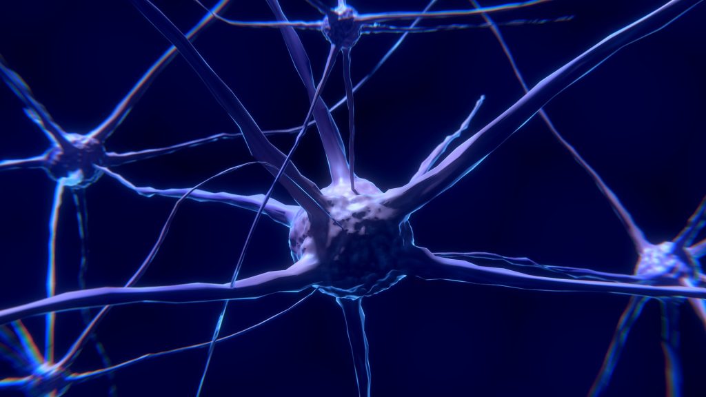
It was long thought that information travelled in a one-way direction, but a new study has revealed that information also travels in the opposite direction at a key synapse in the hippocampus, the brain region responsible for learning and memory. 

Now, Peter Jonas and his group at the Institute of Science and Technology Austria (IST Austria) have demonstrated that information can also travel in the opposite direction at a key synapse in the hippocampus. At the ‘mossy fibre synapse’, the post-synaptic CA3 neuron influences the firing of the post-synaptic ‘mossy fibre neuron’. Their work was published in Nature Communications.

“We have shown, for the first time, that a retrograde information flow is physiologically relevant for pre-synaptic plasticity,” said Yuji Okamoto, a postdoc in the group of Peter Jonas at IST Austria and co-first author of the paper published in Nature Communications.

In the neuronal network, the mossy fibre synapse play a key role in information storage. Synaptic transmission is plastic, meaning that a variable amount of neurotransmitter is released into the synapse. To understand the mechanism of plasticity at work in this synapse, Okamoto precisely stimulated the pre-synaptic terminal of the mossy fibre synapse in rats and at the same time recorded electrical properties at the post-synaptic neuron. “We need to know the synapse’s exact properties—with the numerical values, eg, for its conductance—to create an exact model of this synapse. With his exact measurements, Yuji managed to obtain these numbers,” added Peter Jonas, co-corresponding author with postdoc David Vandael.

Smart teacher balances student’s workload

The researchers found that, unexpectedly, the post-synaptic neuron has an influence on plasticity in the pre-synaptic neuron. Previously the assumption was that the mossy fibre was a ‘teacher synapse’, inducing firing in the post-synaptic neuron. “Instead, we find that this synapse acts like a ‘smart teacher’, who adapts the lessons when students are overloaded with information. Similarly, the pre-synaptic mossy fibre detects when the post-synaptic neuron can’t take more information: When activity increases in the post-synaptic neuron, the pre-synaptic neuron reduces the extent of plasticity,” explained Jonas.

This finding raises the question of how the post-synaptic neuron sends information about its activity status to the pre-synaptic neuron. Pharmacological evidence suggests a role for glutamate, one of the key neurotransmitters used by neurons to send signals to other cells. Glutamate is also the transmitter released from pre-synaptic mossy fibre terminals. When calcium levels increase in the post-synaptic neuron—a sign that the neuron is active—the post-synaptic neuron may release vesicles with glutamate into the synapse. The glutamate travels back to the pre-synaptic neuron, against the usual flow of neuronal information.

“This retrograde modulation of plasticity likely helps to improve information storage in the downstream hippocampal network,” said Jonas, adding: “Once again, exact measurements have shown that reality is more complex than a simplified model would suggest.”

Source: Institute of Science and Technology Austria

Journal information: David Vandael et al. Transsynaptic modulation of presynaptic short-term plasticity in hippocampal mossy fiber synapses, Nature Communications (2021). DOI: 10.1038/s41467-021-23153-5

New Antimalarial Compound Traps Parasites in Cells

Photo by Егор Камелев on Unsplash

To combat the growing resistance of malaria to current treatments, researchers at the Francis Crick Institute and the Latvian Institute of Organic Synthesis have designed a new antimalarial compound which interrupts the malaria parasite life cycle by trapping them in their host cells.

While drugs and mosquito control have reduced levels of malaria over recent decades, with malaria being effectively wiped out in North America by the 1950s, the parasite still kills over 400 000 people every year, 90% of whom live in sub-Saharan Africa. It has now developed resistance to many existing antimalarial drugs, meaning new treatments that work in different ways are urgently needed.

If we can effectively trap malaria in the cell by blocking the parasite’s exit route, we could stop the disease in its tracks and halt its devastating cycle of invading cells.
Mike Blackman

The researchers developed an array of compounds designed to prevent the parasites bursting out of blood cells, a vital replication step. One compound in particular was found to be very effective in human cell tests.

“Malaria parasites invade red blood cells where they replicate many times, before bursting out into the bloodstream to repeat the process. It’s this cycle and build-up of infected red blood cells which causes the symptoms and sometimes fatal effects of the disease,” says Mike Blackman, lead author and group leader of the Malaria Biochemistry Laboratory at the Crick.  

“If we can effectively trap malaria in the cell by blocking the parasite’s exit route, we could stop the disease in its tracks and halt its devastating cycle of invading cells.”

Blocking the parasite’s emergence

The compound works by blocking an enzyme called SUB1, needed for them to burst out of cells. Current antimalarials kill the parasite within the cell, so the researchers hope this alternative drug action will overcome the resistance the parasite has acquired.

The compound can penetrate both the cell wall and the compartment within where the parasites reside.

The researchers are further refining the compound making it smaller and more potent. Further tests are needed before it can be trialled in humans.

Study author Chrislaine Withers-Martinez and researcher in the Malaria Biochemistry Laboratory, said: “Many existing antimalarial drugs are plant derived and while they’re incredibly effective, we don’t know the precise mechanisms behind how they work. Our decades of research have helped us identify and understand pathways crucial to the malaria life cycle allowing us to rationally design new drug compounds based on the structure and mechanism of critical enzymes like SUB1.

“This approach, which has already been highly successful at finding new treatments for diseases including HIV and Hepatitis C, could be key to sustained and effective malaria control for many years to come.” 

Source: Francis Crick Institute

How Legionnaire’s Disease Digs In

A bunker from World War II, emulating how Legionella makes a protective shelter. Image by herb1979 from Pixabay

Scientists have discovered how the bacteria that causes Legionnaires’ disease digs in and makes a tiny shelter inside the cells of humans and other hosts. 

The findings, published in Science, could offer insights into how other bacteria are able to survive inside cells, knowledge that could lead to new treatments for a wide variety of infections.

Discovered in 1976, Legionella, an aerobic gram-negative bacillus is responsible for Legionnaires’ disease, a condition of severe pneumonia. Spread through aerosolised water particles, it is a common cause of hospital and community-acquired pneumonia.

“Many infectious bacteria, from listeria to chlamydia to salmonella, use systems that allow them to dwell within their host’s cells,” explained study leader Vincent Tagliabracci, Ph.D., assistant professor of molecular biology at UTSW and member of the Harold C Simmons Comprehensive Cancer Center. “Better understanding the tools they use to make this happen is teaching us some interesting biochemistry and could eventually lead to new targets for therapy.”

Dr Tagliabracci’s lab studies atypical kinases, unusual forms of enzymes that put phosphates onto proteins or lipids, changing their function. Legionella is a particularly rich source of these noncanonical kinases. According to the Centers for Disease Control and Prevention, nearly 10 000 cases of Legionnaires’ disease were reported in the US in 2018, though the true incidence is believed to be higher.      

After identifying a new Legionella atypical kinase named MavQ, Dr Tagliabracci and colleagues used a live-cell imaging technique in concert with a relatively new molecular tagging method to see where MavQ is found in infected human cells. However, rather than residing in a specific location, the researchers were surprised to see that the protein moved back and forth between the endoplasmic reticulum – a network of membranes important for protein and lipid synthesis – and bubble- or tube-shaped structures within the cell.

Further research suggests that MavQ, along with a partner molecule called SidP, remodels the endoplasmic reticulum so that Legionella can strip off sections of the membrane to help create and sustain the vacuole, a structure that the parasitic bacteria uses to shelter inside cells, protecting it from immune attack.

Dr Tagliabracci said that he suspects other bacterial pathogens may use similar mechanisms to co-opt existing host cell structures to create their own protective shelters. 

 Source: University of Texas

Journal information: Ting-Sung Hsieh, et al. Dynamic remodeling of host membranes by self-organizing bacterial effectors. Science, 2021; eaay8118 DOI: 10.1126/science.aay8118