Tag: medical research

Doctor’s Presence During BP Measurement Triggers Flight-or-fight Response

Photo by Thirdman from Pexels
Photo by Thirdman from Pexels

A small study has shown that a doctor’s presence during a blood pressure measurement skews the results, according to researchers who studied the effect by measuring nerve activity.

The phenomenon known as ‘white coat hypertension‘ is where the mere presence of a medical professional can raise blood pressure. Known about for decades, it occurs in about a third of patients.

In a small study published in the journal Hypertension, researchers probed the effect by measuring blood pressure, heart rate and nerve traffic in the skin and muscles with and without a doctor present.

The researchers found a “drastic reduction” in the body’s alarm response when a doctor was not present, said co-lead author Dr Guido Grassi, professor of internal medicine at the University of Milano-Bicocca.

Blood pressure and heart rate increases in response to a perceived threat, said Dr Meena Madhur, associate professor of medicine in the divisions of clinical pharmacology and cardiology at Vanderbilt University.

“If you’re out in the wild and a bear was charging after you, you’d want your blood vessels in your skin, for example, to constrict and the blood vessels in your muscles to dilate to provide more blood flow to those organs so that you can run really fast,” said Prof Madhur, who was not involved in the new research.

The study included 18 people, 14 of them men, with untreated mild to moderate hypertension. Each participant was examined in a lab, where an electrode measured nerve activity in the skin and muscles. Readings were taken twice in the presence of a doctor and twice without.

Both blood pressure and heart rate rose when the doctor was present, with nerve traffic patterns to the skin and skeletal muscle suggesting a classic fight or flight reaction.

Without the doctor’s presence, cardiovascular and neural responses were “strikingly different,” the researchers wrote. Fight or flight response indications were “entirely absent”.

Peak systolic blood pressure was an average of 14 points lower when the participant was alone than when a doctor was present, and peak heart rate was lowered by nearly 11 beats per minute.

This was the first study to actually measure sympathetic nervous system responses to doctors supervising a blood pressure measurement, the researchers wrote.

The study’s findings illustrated the complexity of blood pressure measurement and how it is affected by involuntary nervous system reactions, Grassi said. “Measurements without the doctor’s presence may better reflect true blood pressure values.”

White coat hypertension is not a new concept, Prof Madhur said, “this just drives home the fact that we should be more conscious of how the blood pressure is taken in the clinic.”

Last year, the American Medical Association and AHA issued a joint report endorsing more blood pressure measurement at home.

Limitations included the small study size due to the complexity of the measurements, the researchers said. Subsequent research would need to examine blood pressure medication as they could affect the fight or flight response, said Orof Madhur.

The work needs to be repeated with more women to examine possible sex differences. And she’d be interested in seeing whether people have the same response to nurses and other medical professionals as they did to doctors in this study.

Previous work shows that when nurses take blood pressure measurements, the white coat effect is reduced.

This latest research emphasises the need for people to handle blood pressure measurements with care, Prof Madhur said.

“I always tell my patients that we really can’t rely on a single office blood pressure measurement, because that’s just a random point in time,” she said.

Prof Madhur said that to take an accurate reading at home, a patient should sit still, with their back straight and supported and feet on the floor, waiting at least a few minutes before recording blood pressure. They should take multiple readings at the same time of day over the course of a week, and bring that log to their doctor’s appointment. Those at-home readings should be the ones used for planning treatment, she said.

“But,” Prof Madhur added, “if we are going to do an office blood pressure reading, it should be taken with the doctor not in the room.”

Source: American Heart Association

New Bacteriophage Could Combat C. Diff

A bacteriophage. Credit: NIAH

A group of newly discovered bacteriophages named after the UK village of Colney could help combat C. difficile infections.

Clostridioides difficile, or C. diff, is a species of bacteria that infects the human gut. It can become a major problem when our normal gut microbes are impaired, most commonly during a course of antibiotics. This leads to an overgrowth of C. diff, with toxins it produces causing diarrhoea and severe inflammation.

Treatment involves further courses of antibiotics, but relapse and recurrent infections are common. The strains are becoming more resistant to antibiotics and causing more severe illness.

This prompted researchers in Norwich to look for the bacteria’s natural enemy, bacteriophages. They screened 27 different C. diff strains for any bacteriophages, finding one, which they called ΦCD27 (phiCD27). Genome sequencing confirmed this phage had not been discovered before. In fact, the members of the International Committee on Taxonomy of Viruses (ICTV) decided it was genetically distinct enough to form a new group, or genus of phages.

The ICTV decided to name the new genus Colneyvirus, the Colney parish address of the Institute of Food Research (IFR, now part of Quadram Institute), where it was first discovered.

Like normal viruses, phages reproduce by injecting their genetic material into bacteria, making viral copies using the host’s own machinery. Using enzymes called endolysins, they destroy the bacterial cell wall and escape.

The researchers extracted the gene for ΦCD27’s endolysin and put it into another bacterium, E. coli so that they could produce and purify the endolysin. It was proven active against 30 different C. diff strains, including hypervirulent strains behind the current epidemic. It also didn’t affect other common bacterial species in the human gut microbiome.

”This phage and the endolysin encoded by its genome can provide a targeted approach to combat C. diff infections, in contrast to use of broad spectrum antibiotics that cause collateral damage by inhibiting other members of the gut bacterial population” said Professor Arjan Narbad, Group Leader at the Quadram Institute.

However, to be effective the endolysins need to be delivered into the gut, so the team also put the gene into a strain of lactic acid bacteria that has previously been used to deliver proteins and vaccines to the gut.

The research team believes this could serve as the basis for future new treatments C. diff. The system needs more work, but in the battle against this bacterial pandemic, the colneyvirus could be a vital ally.

Source: Quadram Institute

Exact Location of Body’s Blood Pressure Sensors Finally Revealed

Credit: CC0

After 60 years of fruitless searches by scientists, researchers from the University of Virginia have finally determined the location of our bodies’ natural blood-pressure sensors.

These cellular sensors monitor blood pressure and adjust hormone levels to keep it in check. Scientists have long suspected that these ‘baroreceptors’, may exist in or around specialised kidney cells called renin cells, but no one has been able to locate the baroreceptors within the cell until now.

The new findings, from UVA Health’s Dr Maria Luisa S Sequeira-Lopez and colleagues, finally reveal where the barometers are located, how they work and how they help prevent hypertension or hypotension. The study was published in Circulation Research.

“It was exhilarating to find that the elusive pressure-sensing mechanism, the baroreceptor, was intrinsic to the renin cell, which has the ability to sense and react, both within the same cell,” said Dr Sequeira-Lopez. “So the renin cells are sensors and responders.”

Back in 1957, it was first proposed that a pressure sensor existed inside renin cells because the cells had to know when to release renin, a hormone that helps regulate blood pressure. Though the baroreceptors had to exist, scientists couldn’t tell what it was and whether it was located in renin cells or surrounding cells.

To tackle this decades-old mystery, the study’s researchers used a combination of innovative lab models and determined that the baroreceptor was a ‘mechanotransducer’ inside renin cells. This mechanotransducer detects pressure changes outside the cell, then transmits these mechanical signals to the cell nucleus, akin to how the cochlea turns sound vibrations into nerve impulses.

Through in vitro tests, the researchers found that applying pressure to renin cells triggered changes within the cells and decreased activity of the renin gene, Ren1. The scientists also compared differences in gene activity in kidneys exposed to lower pressure and those exposed to higher pressure.

Ultimately, when the baroreceptors detect excess pressure outside the renin cell, renin production is cut back, while low blood pressure prompts more renin production.

Dr Sequeira-Lopez said she is looking forward to the work to “unravel the signaling and controlling mechanisms of this mechanotransducer and how we can use the information to develop therapies for hypertension.”

Source: University of Virginia

Protein’s Involvement in Emphysema Could Yield a Treatment Target

Photo by Robina Weermeijer on Unsplash

Researchers from Japan have found a protein that promotes the development of the early stages of emphysema, which could prove to be a target for treatment of the serious disease.

Chronic obstructive pulmonary disease (COPD) causes illness and death worldwide, and is characterised by destruction of the alveolar walls in the lungs, known as emphysema, resulting in lung function declining and to date little is known about how it develops. The Global Initiative for chronic obstructive lung disease (GOLD) has defined COPD as “a common, preventable, and treatable disease that is characterised by persistent respiratory symptoms and airflow limitation that is due to airway and/or alveolar abnormalities usually caused by significant exposure to noxious particles or gases.”

It is known that COPD can be triggered by certain environmental factors, such as cigarette smoking, which result in lung inflammation. The development of inflammation involves the movement of molecules inside cells, and this “intracellular trafficking” is known to play a part in some diseases. The team searched for COPD-related proteins that are involved in trafficking and identified a protein called FCHSD1, which not known to have any lung function but is associated with some diseases.

The researchers deleted the FCHSD1 protein in mice and studied these mice against normal mice when emphysema was induced. In normal mice, a large increase was seen in FCHSD1 after treatment, while mice lacking FCHSD1 were protected from the development of emphysema. These mice showed less airspace expansion from damaged air sacs, and had less inflammation and reduced apoptosis.

The researchers went on to investigate the molecular mechanism by which FCHSD1 acts. In response to stress, a protein called NRF2 moves into the nucleus to protect it. However, FCHSD1 binds to NRF2, stopping it from moving to the nucleus. “Mice with a FCHSD1 deficiency showed enhanced nuclear translocation of NRF2 and a smaller reduction in SIRT1 levels, which is seen to occur as emphysema develops,” explained lead author Takahiro Kawasaki, “and this reduced inflammation and apoptosis of lung cells.”

Increasing the activity of NRF2 to counteract FCHSD1 could therefore be a potential therapy for COPD. Treatments are currently available that target NRF2, and inhibiting FCHSD1 while targeting NRF2 could enhance these treatments and prevent systemic complications. “Our findings may also lead to a specific therapeutic strategy to ameliorate, or even halt, the progression of emphysema by inhibiting FCHSD1,” said Takashi Satoh, senior author of the paper.

Source: EurekAlert!

A Treatment for Heart Attack from Spider Venom

Photo by Adrián Valverde on Unsplash

A protein found in the venom of one of the world’s deadliest spiders has been shown to preserve heart cells, and could be developed into a potentially life-saving treatment for heart attack victims.

A drug candidate developed from a molecule found in the venom of the Fraser Island (K’gari) funnel web spider can prevent damage caused by a heart attack and extend the life of donor hearts used for organ transplants. This would not be the first investigation into a clinical application for spider venom, however. Tarantula spider venom has also been investigated as a potent anaesthetic.

The discovery was made by a team led by Dr Nathan Palpant and Professor Glenn King from The University of Queensland (UQ) and Professor Peter Macdonald from the Victor Chang Cardiac Research Institute.

Dr Palpant, from UQ’s Institute for Molecular Bioscience (IMB), said the drug candidate worked by stopping a ‘death signal’ sent from the heart in the wake of an attack.

“After a heart attack, blood flow to the heart is reduced, resulting in a lack of oxygen to heart muscle,” Dr Palpant said. “The lack of oxygen causes the cell environment to become acidic, which combine to send a message for heart cells to die.

“Despite decades of research, no one has been able to develop a drug that stops this death signal in heart cells, which is one of the reasons why heart disease continues to be the leading cause of death in the world.”

Using beating human heart cells exposed to heart attack stresses, Dr Palpant tested the drug candidate, a protein called Hi1a, to see if the drug improved the cells’ survival.

“The Hi1a protein from spider venom blocks acid-sensing ion channels in the heart, so the death message is blocked, cell death is reduced, and we see improved heart cell survival.”

At present, there are no drugs in clinical use that prevent the damage caused by heart attacks.

Professor Macdonald of Victor Chang Cardiac Research Institute said that this incredible result had been decades in the making.

“This will not only help the hundreds of thousands of people who have a heart attack every year around the world, it could also increase the number and quality of donor hearts, which will give hope to those waiting on the transplant list,” said Professor MacDonald, who is also a senior cardiologist at St Vincent’s Hospital in Sydney.

“The survival of heart cells is vital in heart transplants — treating hearts with Hi1a and reducing cell death will increase how far the heart can be transported and improve the likelihood of a successful transplant,” added Prof MacDonald. “Usually, if the donor heart has stopped beating for more than 30 minutes before retrieval, the heart can’t be used — even if we can buy an extra 10 minutes, that could make the difference between someone having a heart and someone missing out. For people who are literally on death’s door, this could be life-changing.”

The discovery builds on earlier work by Professor King, who identified a small protein in the venom of the Fraser Island (K’gari) funnel-web spider that was shown to markedly improve recovery from stroke.

“We discovered this small protein, Hi1a, amazingly reduces damage to the brain even when it is given up to eight hours after stroke onset,” Professor King said.

“It made sense to also test Hi1a on heart cells, because like the brain, the heart is one of the most sensitive organs in the body to the loss of blood flow and lack of oxygen.

“For heart attack victims, our vision for the future is that Hi1a could be administered by first responders in the ambulance, which would really change the health outcomes of heart disease.”

“This is particularly important in rural and remote parts of Australia where patients and treating hospitals can be long distances apart — and when every second counts.”

Also, this could help for the transfer of donor hearts for cardiac transplantation — allowing these donor hearts to be transported over longer distances and therefore increasing the network of available donors and recipients.

The protein has been tested in human heart cells, and the team are aiming for human clinical trials for both stroke and heart disease within 2-3 years.

Source: ScienceDaily

Journal information: Meredith A. Redd, et al. Therapeutic Inhibition of Acid Sensing Ion Channel 1a Recovers Heart Function After Ischemia-Reperfusion Injury. Circulation, 2021; DOI: 10.1161/CIRCULATIONAHA.121.054360

New Insights into What Stimulates Bone Growth

Photo by Nino Liverani on Unsplash

Researchers have discovered some new insights into how bone mass is maintained and how physical load stimulates bone growth.

Researchers from the National Cerebral and Cardiovascular Center Research Institute in Japan have revealed that the expression of the peptide osteocrin (OSTN) is influenced by load – decreasing when load is reduced, and increasing when it is added. Their study was published in Cell Reports.

Bones and skeletal muscles are strengthened by loads produced in exercise, preventing bone and muscle atrophy, and maintaining bone and muscle strength is important for maintaining physical activity. The growth of long bones, such as the femur and tibia, is a very complex process controlled by genetic and environmental factors, such as exercise and gravity.

Understanding bone loss would help retain bone density and strength in people who are unable to exercise due to immobility, the elderly, as well as astronauts in spaceflight.

Study lead author Haruko Watanabe-Takano said, “Not much is known about how mechanical force initiates biochemical signals to control bone growth. We investigated how load is related to the metabolic balance adjustment of bone maintenance.”

Bone mass and strength is maintained by the balanced activities of two types of cells – the bone-genearting osteoblasts, and the bone-dissolving osteoclasts – and is thought to be made in response to load demand. Specifically, the team investigated the expression of OSTN, a peptide produced by osteoblasts, in mice. OSTN is critical to the regulation of bone growth, as well as physical endurance.

The researchers found that OSTN was very strongly expressed in bones such as the tibia, radius, and ulna, and in regions experiencing load. They determined that OSTN was secreted by the periosteal osteoblasts in these bones. The periosteum is a fibrous membrane that covers nearly every bone in the body, except for the joints of the long bones. This tissue has a major role in bone growth and bone repair and has an impact on the blood supply of bone as well as skeletal muscle. Despite its importance, it has received little attention in the literature and in some ways is not well understood.

“We also found that OSTN expression decreased when load was reduced, and was increased by load stimulation,” says Watanabe-Takano. “Moreover, when we genetically engineered mice lacking OSTN, we found that they had reduced bone mass compared with normal mice and lacked load-induced recovery of bone mass after prolonged load reduction. Thus, we concluded that OSTN makes bone in response to stimulation by load, promoting bone formation.”

The team found that to create this effect, OSTN increases levels of another peptide, called C natriuretic peptide, which in turn drives bone-forming osteoblasts to multiply, mature, and become functional.

The findings have implications for treatments for bed-ridden patients and others at risk of bone loss, such as the elderly. Further studies will explore issues such as how periosteal cells detect load stimulation.

Source: News-Medical.Net

Journal information: Watanabe-Takano, H., et al. (2021) Mechanical load regulates bone growth via periosteal Osteocrin. Cell Reports. doi.org/10.1016/j.celrep.2021.109380.

Probing the Electrical Connections Between Heart Cells

Photo by Robina Weermeijer on Unsplash

Harvard Medical School researchers have updated our understanding on how electrical impulses in the heart travel from cell to cell. Their findings are published in the journal Biophysics Reviews.

Disturbances of the bioelectrical processes behind heart’s rhythm can result in cardiac arrhythmias, a common ailment that can lead to illness and death.

A pacemaker within the heart takes the role of an electrical clock, signalling the heart to contract. The whole muscle moves simultaneously, because each individual cell inside of it contracts in a coordinated manner and within a short time interval.

To accomplish this, the pacemaker’s initial electrical impulse rapidly propagates through cells across the heart.

“If one cell is excited electrically and the other is not, the excited cell becomes positively charged inside, and the resting cell is still negatively charged inside. As a consequence, a voltage gradient builds up between the cells,” explained study author André Kléber. “If you have a voltage gradient and a pathway with a low electrical resistance, a local current will flow.”

The connections between cells which make up the low resistance pathway and facilitate the current flow are called gap junctions. Each is made up of numerous channels, which are formed when specific proteins from one cell connect to and fuse to the proteins from another cell. According to Kléber, the fusing proteins look like placing the tips of your fingers on one hand to the fingers on the other hand.

The researchers investigated the properties of gap junctions and connexins, their constituent proteins. Kléber explained that one reason why gap junction channels are interesting is because they are a highly dynamic system in equilibrium. The creation of the channels equals the destruction.
“The turnover is very short,” he said. “On one hand, the system is very stable during your whole life. On the other hand, if you measure it, it is constantly cycling in periods of a few hours.”

The proteins found in gap junctions are also important for processes not directly related to cell-cell connections, like mitochondrial function, which produces energy, and trafficking, which transports molecules from their synthesis site to their site of action in the cell interior.

“You have to refrain from the idea that if you define the role of a protein in the body, that it has only a single function,” said Kléber. “Nature is much, much smarter than human beings.”

Source: American Institute of Physics

Journal information: Kléber, A.G & Jin, Q., (2021) Coupling between cardiac cells—An important determinant of electrical impulse propagation and arrhythmogenesis. Biophysics Reviews. doi.org/10.1063/5.0050192.

New Treatment Candidate May Reverse Neurodegenerative Decline

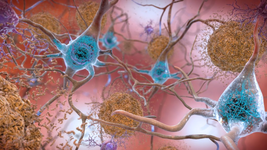
In the Alzheimer’s affected brain, abnormal levels of the beta-amyloid protein clump together to form plaques (seen in brown) that collect between neurons and disrupt cell function. Abnormal collections of the tau protein accumulate and form tangles (seen in blue) within neurons, harming synaptic communication between nerve cells.
Credit: National Institute on Aging, NIH

Researchers  at Tohoku University in Japan have identified a new treatment candidate that seems to not only halt but partially reverse neurodegenerative symptoms in mouse models of dementia and Alzheimer’s disease.

Kohji Fukunaga, professor emeritus in Tohoku University’s Graduate School of Pharmaceutical Sciences and paper author, said: “There are currently no disease-modifying therapeutics for neurodegenerative disorders such as Alzheimer’s disease, Lewy body dementia, Huntington disease and frontotemporal dementia in the world. We discovered the novel, disease-modifying therapeutic candidate SAK3, which, in our studies, rescued neurons in most protein-misfolding, neurodegenerative diseases.”

In previous work, the team found that the SAK3 molecule – the base structure of which is found in the enhancement of T-type Ca2+ channel activity – apparently improved memory and learning in a mouse model of Alzheimer’s disease.

SAK3 enhances the function of a cell membrane channel thereby promoting neuronal activity in the brain. Typically, SAK3 promotes neurotransmitter releases of acetylcholine and dopamine — neurotransmitters which are lowered in Alzheimer’s disease and Lewy body dementia. The Ca2+ channel enhancement is thought to trigger a change from resting to active in neuronal activity. When the Ca2+ channel is dysregulated in the brain, less acetylcholine and dopamine is released. Cognitive confusion and uncoordinated motor function arises from this dysregulated system.

SAK3 binds directly  to the subunit of this channel, enhancing neurotransmission and so improving cognitive deficits. The researchers found that the same process also seemed to work in a mouse model of Lewy body dementia, which is characterised by a buildup of proteins known as Lewy bodies.

“Even after the onset of cognitive impairment, SAK3 administration significantly prevented the progression of neurodegenerative behaviors in both motor dysfunction and cognition,” Prof Fukunaga said.

In comparison, Aduhelm, the Alzheimer’s drug recently approved by the US Food and Drug Administration, reduces the number of amyloid plaques in the brain, but whether the amyloid reduction actually prevents further cognitive or motor decline in patients is not yet known. According to Prof Fukunaga, SAK3 helps destroy amyloid plaque – at least in mice.

SAK3 also helps destroy misfolded alpha-synuclein, which normally helps regulate neurotransmitter transmission in the brain. The misfolded protein can aggregate, contributing to what researchers suspect may be an underlying cause of neurodegenerative symptoms. This aggregation can also cause loss of dopamine neurons, which are associated with learning and memory.

“We found that chronic administration of SAK3 significantly inhibited the accumulation of alpha-synuclein in the mice,” Prof Fukunaga said, noting that the mice received a daily oral dose of SAK3.

According to Prof Fukunaga, SAK3 enhances the activity of the system that identifies and destroys misfolded proteins. In neurodegenerative diseases, this system is often dysfunctional, leaving misfolded proteins to wreak havoc in the cell’s machinery.

“SAK3 is the first compound targeting this regulatory activity in neurodegenerative disorders,” Fukunaga said. “SAK3 administration promotes the destruction of misfolded proteins, meaning the therapeutic has the potential to solve the problems of diverse protein misfolding diseases such as Parkinson’s disease, Lewy body dementia and Huntington disease, in addition to Alzheimer’s disease.”

The team published their results in the International Journal of Molecular Sciences. This treatment candidate has been declared safe by Japan’s governing board, and the researchers are planning to start human clinical trials in the next year.

Source: Tohoku University

Journal information: Xu, J., et al. (2021) T-Type Ca2+ Enhancer SAK3 Activates CaMKII and Proteasome Activities in Lewy Body Dementia Mice Model. International Journal of Molecular Sciencesdoi.org/10.3390/ijms22126185.

Are We Wrong About Amyloid Plaques in Alzheimer’s?

A recent study sheds new light on the disease and the highly debated aducanumab, a new drug recently approved by the FDA that treats the amyloid plaques.

Led by the University of Cincinnati and conducted in collaboration with the Karolinska Institute in Sweden, the study claims that the treatment of Alzheimer’s disease might lie in normalising levels of a brain protein called amyloid-beta peptide. This protein is needed in its original, soluble form to keep the brain healthy, but it sometimes hardens into ‘brain stones’ or clumps, called amyloid plaques.

“It’s not the plaques that are causing impaired cognition,” said senior author Alberto Espay, professor of neurology at UC. “Amyloid plaques are a consequence, not a cause,” of Alzheimer’s disease, stated Prof Espay, who is also a member of the UC Gardner Neuroscience Institute.

Since its discovery, scientists have focused on treatments to eliminate the plaques. But the UC team, he said, viewed it differently: Cognitive impairment could be due to a decline in soluble amyloid-beta peptide instead of the corresponding accumulation of amyloid plaques. 
To test their hypothesis, they analyzed the brain scans and spinal fluid from 600 individuals enrolled in the Alzheimer’s Disease Neuroimaging Initiative study, who all had amyloid plaques. From there, they compared the amount of plaques and levels of the peptide in the individuals with normal cognition to those with cognitive impairment. They found that individuals with high levels of the peptide were cognitively normal, despite the numbers of plaques in their brains.

They also found that higher levels of soluble amyloid-beta peptide were associated with a larger hippocampus, the area of the brain most important for memory.

According to the authors, as we age most people develop amyloid plaques, but few people develop dementia. In fact, by the age of 85, 60% of people will have these plaques, but only 10% develop dementia.

“The key discovery from our analysis is that Alzheimer’s disease symptoms seem dependent on the depletion of the normal protein, which is in a soluble state, instead of when it aggregates into plaques,” said co-author Kariem Ezzat from the Karolinska Institute.

The most relevant future therapeutic approach for the Alzheimer’s program would then be to restore these brain soluble proteins to their normal levels, said Prof Espay.

The research team is now working to test their findings in animal models. If successful, future treatments may be very different from those tried over the last two decades. Treatment, says Espay, may consist of increasing the soluble version of the protein in a manner that keeps the brain healthy while preventing the protein from hardening into plaques.  

Source: University of Cincinnati 

Journal information: Andrea Sturchio et al, High cerebrospinal amyloid-β 42 is associated with normal cognition in individuals with brain amyloidosis, EClinicalMedicine (2021). DOI: 10.1016/j.eclinm.2021.100988

Intense Training Results in Temporary Mitochondrial Impairment

Photo by Quino Al on Unsplash

Elite athletes have temporary mitochondrial impairment following intense workouts, according to new research, which suggests they may need to be mindful about overtraining. 

Mitochondria are organelles that are responsible for most of the useful energy derived from the breakdown of carbohydrates and fatty acids, which is converted to ATP by the process of oxidative phosphorylation. Mitochondrial capacity is a term used to describe the body’s ability to generate energy, and is one factor associated with increased athletic performance during endurance exercise. Previous research found that untrained recreational athletes had a decrease in mitochondrial capacity after sprinting exercises.

In this study, the researchers worked with a small group of male elite athletes, many of whom were national title holders or had international recognition for their performance in cycling and triathlon. The athletes participated in a four-week training programme in their primary sport, which consisted of two to four days of low-to-moderate–intensity endurance workouts, followed by three days of more intense training. These intense workouts included high-intensity interval training in the morning, followed by a seven-hour break and then a moderate-intensity cycling session in the afternoon. Each volunteer did between 12 and 20 hours of activity per week. The athletes, though used to heavy training, were not accustomed to this specific workout schedule.

The researchers were surprised to observe that the highly trained participants’ mitochondrial capacity was impaired after the month-long training period. “We thought that elite athletes should be more resistant against [these] kind of alterations,” said Filip Larsen, PhD, of the Swedish School of Sport and Health Sciences and corresponding author of the study.

However, elite athletes may be able to prevent temporary mitochondrial impairment by listening to their bodies, the researchers suggested. By paying attention to changes such as “mood disturbances, reductions in maximal heart rate [during exercise] and muscles that feel heavy and unresponsive” top athletes may be able to pull back and avoid overtraining situations that could contribute to reduced mitochondrial content and function, Larsen explained. “Exercise is good for you, but too much unaccustomed training might have mitochondrial consequences.”

The study also found that reduced mitochondrial capacity did not affect exercise performance, suggesting that oxygen delivery from the heart to the muscles plays a more important role than mitochondrial function in performance. Expression of three proteins with strong antioxidant properties were also found to be increased in the muscles after intense training.

Source: American Physiological Society

Journal information: Daniele A. Cardinale et al, Short term intensified training temporarily impairs mitochondrial respiratory capacity in elite endurance athletes, Journal of Applied Physiology (2021). DOI: 10.1152/japplphysiol.00829.2020