Tag: medical research

A Novel Anticoagulant That can be ‘Deactivated’

Source: NCI

A new biomolecular anticoagulant platform reported in Nano Letters holds promise as a revolutionary advancement over the anticoagulants currently used during surgeries and other procedures. The technology is based around injectable fibre structures which can be quickly dissolved and excreted by the kidneys.

“We envision the uses of our new anticoagulant platform would be during coronary artery bypass surgeries, kidney dialysis, and a variety of vascular, surgical and coronary interventions,” said Kirill Afonin, leader of the team which invented the technology. “We are now investigating if there are potential future applications with cancer treatments to prevent metastasis and also in addressing the needs of malaria, which can cause coagulation issues.”

The team’s technology turns to programmable RNA-DNA anticoagulant fibres that, when injected into the bloodstream, form into modular structures that communicate with thrombin. The technology allows the structures to prevent blood clotting as it is needed and then be quickly eliminated via the renal system once their job is done.

The fibre structures use aptamers, short sequences of DNA or RNA designed to specifically bind and inactivate thrombin.

“Instead of having a single small molecule that deactivates thrombin,” Afonin said, “we now have a relatively large structure that has hundreds of the aptamers on its surface that can bind to thrombin and deactivate them. And because the structure becomes larger, it will circulate in the bloodstream for a significantly longer time than traditional options.”

The extended circulation in the bloodstream allows for a single injection, instead of multiple doses. The design also decreases the concentration of anticoagulants in the blood, resulting in less stress on the body’s renal and other systems, Afonin said.

This technology also introduces a novel “kill-switch” mechanism, which reverses the fibre structure’s anticoagulant function with a second injection. This lets makes the fibres able to be metabolised into materials that are tiny, harmless, inactive and easily excreted by the renal system.

The entire process takes place outside the cell, through extracellular communication with the thrombin. The researchers note that this is important as immunological reactions do not appear to occur, based on their extensive studies.

The team has tested and validated the platform in computer models, human blood and various animal models“We conducted proof-of-concept studies using freshly collected human blood from donors in the US and in Brazil to address a potential inter donor variability,” Afonin said.

The technology may provide a foundation for other biomedical applications that require communication via the extracellular environment in patients, he said. “Thrombin is just one potential application,” he said. “Whatever you want to deactivate extracellularly, without entering the cells, we believe you can. That potentially means that any blood protein, any cell surface receptors, maybe antibodies and toxins, are possible.”

The technique permits the design of structures of any shape desired, with the kill switch mechanism intact. “By changing the shape, we can have them go into different parts of the body, so we can change the distribution,” Afonin said. “It gets an extra layer of sophistication of what it can do.”

While the application is sophisticated, production of the structures is relatively easy. “The shelf life is amazingly good for these formulations,” Afonin said. “They’re very stable, so you can dry them, and we anticipate they will stay for years at ambient temperatures, which makes them very accessible to economically challenged areas of the world.”

Source: University of North Carolina

Patient Doing Well after World-first Pig Heart Transplant

Photo by Piron Guillaume on Unsplash

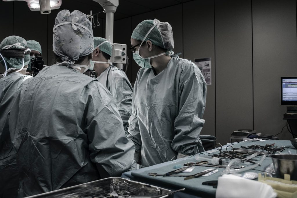
David Bennett, a 57 year old US man, is doing well after being the world’s first human transplant of a pig heart, according to the man’s son, David Bennett Jr.

When his father first told him of the pig heart option, his son was incredulous, telling the BBC: “I didn’t believe him, I thought he was suffering from delirium at first.”

However, when he did some research on the work done, he realised it was a reality and that they were “walking into the unknown”.

He added that according to Dr Bartley Griffith, who performed the surgery, his father has a good prognosis of 6–9 months. The experimental seven-hour procedure at the University of Maryland Medical Center in Baltimore was considered the last hope of saving Mr Bennett’s life, though it is currently unclear what his long-term chances of survival are. 

“It was either die or do this transplant,” Mr Bennett explained a day before the surgery, adding that it was his “last choice”.

Dr Griffith said heart failure and an irregular heartbeat made him ineligible for a human heart transplant or a heart pump.

Xenotransplantation, as these inter-species transplants are called, have failed, largely because patients’ bodies quickly rejected the animal organ. Notably, in 1984, Baby Fae, a dying infant, lived 21 days with a baboon heart. 

What makes this attempt different is that the Maryland surgeons used a heart from a pig that had been genetically modified to remove a sugar in its cells that’s responsible for that hyper-fast organ rejection. Many biotech companies are working on adapting pig organs for xenotransplantation.

“I think you can characterise it as a watershed event,” Dr David Klassen, chief medical officer at the United Network for Organ Sharing (UNOS), which oversees the US transplant system.

Dr Klassen nevertheless cautioned that it’s only a first tentative step into exploring whether xenotransplantation might finally work this time.

The Food and Drug Administration, which oversees such experiments, allowed the surgery under what’s called a “compassionate use” emergency authorisation, available when a patient with a life-threatening condition has no other options.

Surgeon Bartley Griffith said the surgery would bring the world “one step closer to solving the organ shortage crisis”. At present, 17 people die every day in the US waiting for a transplant, with more than 100 000 reportedly on the waiting list. A record 3800 heart transplants were done last year, according to the UNOS.

Source: BBC News

Space Could be Ideal Place for Stem Cell Production

Astronaut Raja Chari sequences DNA from bacteria samples to understand the microbial environment on the International Space Station. Credit: NASA

The lack of gravity in outer space could be the key to the efficient production of large quantities of stem cells. Scientists at Cedars-Sinai have found that the microgravity environment in space stations can potentially aid life-saving advances on Earth by facilitating the rapid mass production of stem cells.

A new paper in Stem Cell Reports outlines key opportunities discussed at a space biomanufacturing symposium to expand the manufacture of stem cells in space.

With new rocket technology, the cost of access to space has plummeted, opening up new opportunities for research and industry, as well as spaceflight by private citizens. Biomanufacturing of therapeutic and research biomaterials can be more productive in microgravity conditions.

“We are finding that spaceflight and microgravity is a desirable place for biomanufacturing because it confers a number of very special properties to biological tissues and biological processes that can help mass produce cells or other products in a way that you wouldn’t be able to do on Earth,” said stem cell biologist Arun Sharma, PhD, head of a new Cedars-Sinai research laboratory.

“The last two decades have seen remarkable advances in regenerative medicine and exponential advancement in space technologies enabling new opportunities to access and commercialise space,” he said.

Attendees at the virtual space symposium in December identified more than 50 potential commercial opportunities for conducting biomanufacturing work in space, according to the Cedars-Sinai paper. The most promising fell into three categories: Disease modelling, biofabrication, and stem-cell-derived products.

Scientists use disease modelling, to study diseases and possible treatments by replicating full-function structures – whether using stem cells, organoids or other tissues.

Decades of spaceflight experience has shown that when the body is exposed to low-gravity conditions for extended periods of time, it experiences accelerated bone loss and ageing. By developing disease models based on this accelerated ageing process, research scientists can better understand the mechanisms of the ageing process and disease progression.

“Not only can this work help astronauts, but it can also lead to us manufacturing bone constructs or skeletal muscle constructs that could be applied to diseases like osteoporosis and other forms of accelerated bone ageing and muscle wasting that people experience on Earth,” explained Dr Sharma.

Biofabrication, another major topic of discussion at the symposium, produces materials like tissues and organs with 3D printing a core technology.

A major issue with biofabrication on Earth involves gravity-induced density, which makes it hard for cells to expand and grow. This requires the use of scaffolding structures, but it generally cannot support the small, complex shapes found in vascular and lymphatic pathways. With the lack of gravity in space, scientists are hopeful that they can use 3D printing to print unique shapes and products, like organoids or cardiac tissues, in a way that can’t be replicated on Earth. This technology is being tested on the International Space Station.

The third category has to do with the production of stem cells and understanding how some of their fundamental properties are influenced by microgravity. Some of these properties include potency, or the ability of a stem cell to renew itself, and differentiation, the ability for stem cells to turn into other cell types.

Understanding some of the effects of spaceflight on stem cells can potentially lead to better ways to manufacture large numbers of cells in the absence of gravity. In coming months, Cedars-Sinai scientists will send stem cells into space to test whether it is possible to produce large batches in a low gravity environment.

“While we are still in the exploratory phase of some of this research, this is no longer in the realm of science fiction,” Dr Sharma said. “Within the next five years we may see a scenario where we find cells or tissues that can be made in a way that is simply not possible here on Earth. And I think that’s extremely exciting.”

Source: Cedars-Sinai Medical Center

Biological Research Often Incorrectly Reports Sex Differences

Photo by Tim Mossholder on Unsplash

An analysis of published studies from a range of biological specialties shows that when data are reported by sex, critical statistical analyses are often missing and the findings are likely to be reported in misleading ways.

The analysis was published in the journal eLife.

“We found that when researchers report that males and females respond differently to a manipulation such as a drug treatment, 70% of the time the researchers have not actually compared those responses statistically at all,” said senior author Donna Maney, a professor of neuroscience in Emory’s Department of Psychology. “In other words, an alarming percentage of claims of sex differences are not backed by sufficient evidence.”

In the articles lacking the proper evidence, she added, sex-specific effects were claimed almost 90% of the time. In contrast, authors that tested statistically for sex-specific effects only reported them 63% of the time.

”Our results suggest that researchers are predisposed to finding sex differences and that sex-specific effects are likely over-reported in the literature,” Prof Maney said.

The problem is so pervasive not even her own work was safe. “Once I realised how prevalent it is, I went back and checked my own published articles and there it was,” she said. “I myself have claimed a sex difference without comparing males and females statistically.”

Prof Maney stressed that the problem should not be discounted; it is becoming increasingly serious, she said, because of mounting pressure from funding agencies and journals to study both sexes, and interest from the medical community to develop sex-specific treatments.

Better training and oversight are needed to ensure scientific rigor in research on sex differences, the authors wrote: “We call upon funding agencies, journal editors and our colleagues to raise the bar when it comes to testing for and reporting sex differences.”

Historically, biomedical research has often included just one sex, usually biased toward males. In recent decades, laws have been passed requiring US medical research to include females in clinical trials and report the sex of human participants or animal subjects.

“If you’re trying to model anything relevant to a general population, you should include both sexes,” Prof Maney explained. “There are a lot of ways that animals can vary, and sex is one of them. Leaving out half of the population makes a study less rigorous.”

As more studies consider sex-based differences, Maney adds, it is important to ensure that the methods underlying their analyses are sound.

For the analysis, Prof Maney and co-author Yesenia Garcia-Sifuentes, PhD candidate, looked at 147 studies published in 2019 to see what is used for evidence of sex differences. The studies ranged across nine different biological disciplines, including field studies on giraffes and immune responses in humans.

The studies that were analysed all included both males and females and separated the data by sex. Garcia-Sifuentes and Prof Maney found that the sexes were compared, either statistically or by assertion, in 80% of the articles. Of those articles, sex differences were reported in 70% of them and of those treated as a major finding in about half.

Statistical errors were seen in some studies, with a significant difference for one sex but not the other counted as a difference between them.  The problem with that approach is that the statistical tests conducted on each sex can’t give “yes” or “no” answers about whether the treatment had an effect.

“Comparing the outcome of two independent tests is like comparing a ‘maybe so’ with an ‘I don’t know’ or ‘too soon to tell,'” Maney explains. “You’re just guessing. To show actual evidence that the response to treatment differed between females and males, you need to show statistically that the effect of treatment depended on sex. That is, to claim a ‘sex-specific’ effect, you must demonstrate that the effect in one sex was statistically different from the effect in the other.”

Conversely, their analysis also encountered strategies that could mask sex differences, such as pooling data from males and females without testing for a difference.

“At this moment in history, the stakes are high,” Maney says. “Misreported findings may affect health care decisions in dangerous ways. Particularly in cases where sex-based differences may be used to determine what treatment someone gets for a particular condition, we need to proceed cautiously. We need to hold ourselves to a very high standard when it comes to scientific rigor.”

Source: EurekAlert!

A New Clue to Disarming C. Difficile’s Toxic Weaponry

C difficile. Source: CDC

Therapeutic interventions for Clostridioides difficile infection (CDI) could make use of a glucosyltransferase domain (GTD) as an ideal molecular target, potentially yielding new, effective treatments for this deadly disease.

The study, published in Science Advancesprovided new insights into TcdB, the toxic molecular weaponry of C. difficile and its hypervirulent strains, creating an opportunity to disarm it.

CDI is the leading cause of antibiotic-associated diarrhoea and gastroenteritis-associated deaths worldwide, accounting for 500 000 cases and 29 000 deaths in the US every year and is classified by the Centers for Disease Control and Prevention as one of the top health threats. The emergence and spread of hypervirulent C. difficile strains is of global concern, resembling as it does the occurrence of new virus variants in current COVID pandemic. TcdB is one of two homologous C. difficile exotoxins, and TcdB alone is capable of causing the full spectrum of CDI diseases.

“We focused on the structure and function of TcdB’s crucial GTD, which is the toxin’s ‘warhead.’ The GTD is delivered by the toxin inside the host cells and causes most of the cytosolic damage to patients,” said corresponding author Rongsheng Jin, PhD, professor in the Department of Physiology & Biophysics at the UCI School of Medicine. “We discovered molecular mechanisms by which the GTD specifically recognises and blocks the physiological functions of the human GTPases Rho and R-Ras enzyme families that are crucial signaling molecules.”

The team also showed that the classic form of TcdB and the hypervirulent TcdB recognise their human targets in different ways, leading to distinct structural changes to the host cells caused by bacterial invasion.

“Once the GTD of TcdB is inside the cells, it is shielded by our cells and becomes inaccessible to passive immunotherapy. But our studies suggest that small molecule inhibitors could be developed to disarm the GTD, which will directly eliminate the root cause of disease symptoms and cellular damage,” Prof Jin explained. “This new strategy can potentially be integrated with and complement other CDI treatment regiments.”

Source: UCI School of Medicine

Muscles are Timekeepers for the Liver

Photo by RF._.studio from Pexels

Researchers have found that skeletal muscles play a large part in regulating the liver’s biological clock. The findings were published in Science Advances.

The circadian rhythm is coordinated by the brain at a general level, but each organ or tissue is also subjected to specific regulation, adjusting to time to optimise their processes. However it was not known how the liver “knows” whether it is day or night.

The liver’s main role is digestion, mainly of fats and sugars: the brain is the main consumer of sugar while skeletal muscle is the main consumer of fat.

Scientists at IRB Barcelona discovered a surprising relationship: that it is skeletal muscle which regulates liver function and determines fat metabolism. Skeletal muscle accomplishes this by secreting a that is transported to the liver through serum is responsible for modulating around 35% of the metabolic functions of the liver. The remaining basal functions of this organ and others related to carbohydrate metabolism are independent of muscle activity and are regulated by the basal circadian rhythm from the brain.

“It’s a very nice discovery because it is the first demonstration of the need for communication between the circadian clocks of tissues and organs outside the brain, and we can see that this communication between muscle and liver is altered by aging,” said study leader Dr Salvador Aznar-Benitah at IRB Barcelona. “When we get older, cells stop obeying the biological clock and begin to perform functions in a non-optimal manner, leading to errors that cause tissues to age.”

The researcher’s results show that the liver does not independently regulate the metabolism of fats and that it is muscle that sends the message that it is time to switch on fatty acid metabolism and how it should go about this. “We didn’t expect to find this connection between the liver and muscle because it wasn’t known previously, but, on second thought, it makes complete sense that fat management is coordinated by one of its main consumers,” said Dr Aznar-Benitah. Carbohydrate metabolism meanwhile is dependent on the basal coordination exercised by the brain.

Source: Institute for Research in Biomedicine (IRB Barcelona)

New Drug Targets for Memory Loss

Image by Falkurian Design on Unsplash

Researchers have identified specific drug targets within memory-encoding neural circuits, opening up possibilities for new treatments of a range of brain disorders.

Memory loss is a main feature of a number of neurological and psychiatric disorders including Alzheimer’s disease and schizophrenia. Presently, there are few, very limited memory loss treatments and the search for safe and effective drug therapies has, until now, borne little fruit.

The research was done in collaboration with colleagues at the international biopharmaceutical company Sosei Heptares. The findings, published in Nature Communications, identify specific receptors for the neurotransmitter acetylcholine that re-route information flowing through memory circuits in the hippocampus. Acetylcholine is released in the brain during learning and is critical for the acquisition of new memories. Until now, the only effective treatment for the symptoms of cognitive or memory impairment seen in diseases such as Alzheimer’s is using drugs that broadly boost acetylcholine. However, this leads to multiple adverse side effects. The discovery of specific receptor targets that have the potential to provide the positive effects whilst avoiding the negative ones is promising.

Lead author Professor Jack Mellor from the University of Bristol’s Center for Synaptic Plasticity, said: “These findings are about the fundamental processes that occur in the brain during the encoding of memory and how they may be regulated by brain state or drugs targeting specific receptor proteins. In the long-term, the discovery of these specific targets opens up avenues and opportunities for the development of new treatments for the symptoms of Alzheimer’s disease and other conditions with prominent cognitive impairments. The academic-industry partnership is important for these discoveries and we hope to continue working together on these projects.”

Dr Miles Congreve, Chief Scientific Officer at Sosei Heptares, added: “These important studies have helped us to design and select new, exquisitely targeted therapeutic agents that mimic the effects of acetylcholine at specific muscarinic receptors, without triggering the unwanted side effects of earlier and less-well targeted treatments. This approach has the exciting potential to improve memory and cognitive function in patients with Alzheimer’s and other neurological diseases.”

“It is fascinating how the brain prioritises different bits of information, working out what is important to encode in memory and what can be discarded. We know there must be mechanisms to pull out the things that are important to us but we know very little about how these processes work. Our future program of work aims to reveal how the brain does this using acetylcholine in tandem with other neurotransmitters such as dopamine, serotonin and noradrenaline,” said Professor Mellor.

Source: University of Bristol

Human Breast Milk Could Yield Antibiotic Secrets

Researchers believe that antibacterial properties of sugars in human breast milk could be harnessed for new antimicrobial therapies.

Group B Streptococcus (GBS) bacteria are a common cause of blood infections, meningitis and stillbirth in newborns, and are becoming resistant to antibiotics. Researchers have now discovered that human milk oligosaccharides (HMOs), short strings of sugar molecules abundant in breast milk, can help prevent GBS infections in human cells and tissues and in mice. This might yield new antibiotic treatments, the researchers believe. 

“Our lab has previously shown that mixtures of HMOs isolated from the milk of several different donor mothers have antimicrobial and antibiofilm activity against GBS,” says Rebecca Moore, who is presenting the work at a meeting of the American Chemical Society (ACS). “We wanted to jump from these in vitro studies to see whether HMOs could prevent infections in cells and tissues from a pregnant woman, and in pregnant mice.” Moore is a graduate student in the labs of Steven Townsend, PhD, at Vanderbilt University and Jennifer Gaddy, PhD, at Vanderbilt University Medical Center.

According to the US Centers for Disease Control and Prevention, about 2000 babies in the U.S. get GBS each year, with 4-6% of them dying from it. The bacteria are often transferred from mother to baby during labour and delivery. An expectant mother who tests positive for GBS is usually given intravenous antibiotics during labor to help prevent early-onset infections, which occur during the first week of life. Notably, late-onset infections (which happen from one week to three months after birth) are more common in formula-fed than breastfed infants, suggesting breast milk has factors which could help protect against GBS. If so, the sugars could be a replacement for current antibiotics which are steadily becoming less effective.

The researchers studied the effects of combined HMOs from several mothers on GBS infection of placental macrophages and of the gestational membrane. “We found that HMOs were able to completely inhibit bacterial growth in both the macrophages and the membranes, so we very quickly turned to looking at a mouse model,” Moore says. They examined whether HMOs could prevent a GBS infection from spreading through the reproductive tract of pregnant mice. “In five different parts of the reproductive tract, we saw significantly decreased GBS infection with HMO treatment,” Moore notes.

To determine which HMOs and other oligosaccharides have these antimicrobial effects and why, the researchers made an artificial two-species microbiome with GBS and the beneficial Streptococcus salivarius species growing in a tissue culture plate, separated by a semi-permeable membrane. Then, the researchers added oligosaccharides that are commonly added to infant formula, called galacto-oligosaccharides (GOS), which are derived from plants. In the absence of the sugar, GBS suppressed the growth of the “good” bacteria, but GOS helped this beneficial species grow. “We concluded that GBS is producing lactic acid that inhibits growth, and then when we add the oligosaccharide, the beneficial species can use it as a food source to overcome this suppression,” Moore explained.
The first HMOs tested did not have this effect, but Townsend says it’s likely that one or more of the over 200 unique sugars in human milk will show activity in the artificial microbiome assay. There are likely two reasons why HMOs can treat and prevent GBS infection: they prevent pathogens from sticking to tissue surfaces and forming a biofilm, and they could also act as a prebiotic by promoting good bacteria growth.

“HMOs have been around as long as humans have, and bacteria have not figured them out. Presumably, that’s because there are so many in milk, and they’re constantly changing during a baby’s development,” Townsend said. “But if we could learn more about how they work, it’s possible that we could treat different types of infections with mixtures of HMOs, and maybe one day this could be a substitute for antibiotics in adults, as well as babies.”

Source: American Chemical Society

Improvement of Cell Culture Reporting Needed

Photo by CDC on Unsplash

There is an urgent need for more standardised and detailed reporting of research on mammalian cells, and for greater control over and measurement of the environmental conditions of cell cultures, according to a recent study. This will improve the precision of human physiology models and contribute to the reproducibility of research.

Researchers analysed 810 randomly selected papers on mammalian cell lines. Fewer than 700 of those, involving 1749 individual cell culture experiments, included relevant data on the environmental conditions of the media in which the cells were cultured. The analysis suggests that the relevance and reproducibility of this type of research needs significant improvement.

“Mammalian cell cultures are fundamental to manufacturing viral vaccines and other biotechnologies,” explained marine scientist, Shannon Klein. “They are used to study basic cell biology, replicate disease mechanisms and investigate the toxicity of novel drug compounds before they are tested on animals and humans.”

Though cells are cultured in controlled incubators in line with standard protocols, cells grow and ‘breathe’ over time and exchange gases with their surrounding environment. This impacts their immediate environment, and even these small changes can affect parameters like culture acidity and dissolved oxygen and carbon dioxide. These changes in turn can affect cell function, causing different conditions to that found in a living human body.

The researchers found that around half of the papers analysed failed to report the temperature and carbon dioxide settings of their cell cultures. Less than 10 percent reported the atmospheric oxygen levels in the incubator and less than 0.01 percent reported the medium’s acidity. No papers reported the dissolved oxygen or carbon dioxide in their media.

“We were very surprised that researchers largely overlooked the maintenance of environmental factors, like culture acidity, at levels relevant to the physiological body over the full course of the cell cultures, despite it being well known that this is important for cell function,” said Ph.D. student Samhan Alsolami.

The team, led by KAUST’s marine ecologist Carlos Duarte and stem cell biologist Mo Li in collaboration with developmental biologist Juan Carlos Izpisua Belmonte from the Salk Institute, who is currently a visiting professor at KAUST, recommends that biomedical scientists develop standard reporting and control and measuring procedures, in addition to employing specialised instruments for controlling the culture environments of different cell types. Additionally, scientific journals should establish reporting standards and require adequate monitoring and control of culture medium acidity and dissolved oxygen and carbon dioxide.

“Better reporting, measurement and control of the environmental conditions of cell cultures should improve how well scientists can repeat and reproduce experimental results,” said Alsolami. “More careful attention could drive new discoveries and increase the relevance of preclinical research to the human body.”

The study is published in Nature Biomedical Engineering.

Source: Medical Xpress

How do Patients Who Exit Clinical Trials Early Feel?

Source: JD Mason on Unsplash

A new study has helped researchers understand the experiences of people who withdraw from clinical cancer trials.

Cancer clinical trials (CCTs) provide patients with an opportunity to receive experimental drugs, tests, and/or procedures that may lead to remissions. Such opportunities can be a great benefit for those who took part, but there is little known of the experiences of participants who withdraw from CCTs.

To address this, a first-of-its-kind study from the University of Pennsylvania School of Nursing (Penn Nursing) was conducted to better understand the post-trial needs of these patients and define responsible transitions when patients exit CCTs.

“Understanding the post-trial needs of patients with cancer and their families represents a measure of ethical respect of the many contributions that patients with cancer make to advancing our scientific knowledge and finding treatments that save lives,” said the study’s lead researcher, Connie M Ulrich, the Lillian S Brunner Chair in Medical and Surgical Nursing, professor of nursing, professor of medical ethics and health policy.

The study revealed three important areas:

  • Patients exiting CCTs feel intense symptoms, emotions, and awareness that their life spans are short and options seem limited.
  • The limited discussions with patients who are exiting on their immediate post-trial care needs can result in many feeling that there is no clear path forward.
  • Good communication that deliberately includes attention to post-trial needs throughout the CCT is needed to help scared and disappointed patients navigate their next steps.

The study is set for publication on the JAMA Network.

Source: University of Pennsylvania