Tag: mammography

How Accurate is Supplemental Ultrasound in Breast Cancer Screening Failures?
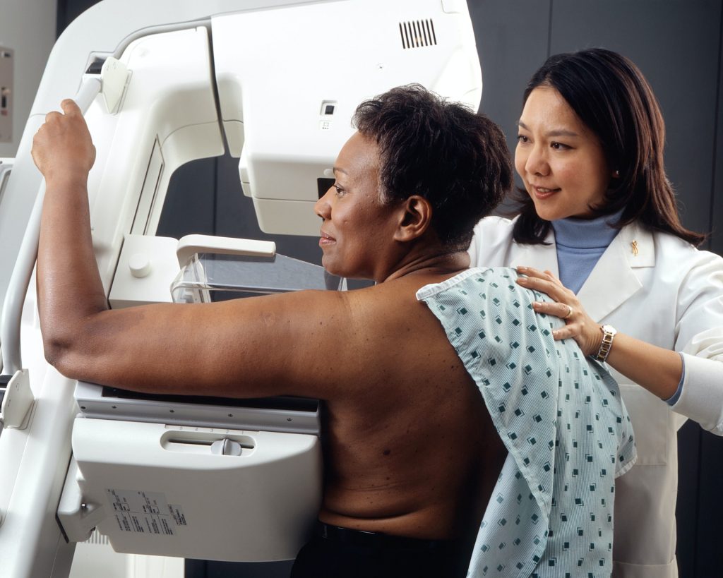
Photo by National Cancer Institute on Unsplash

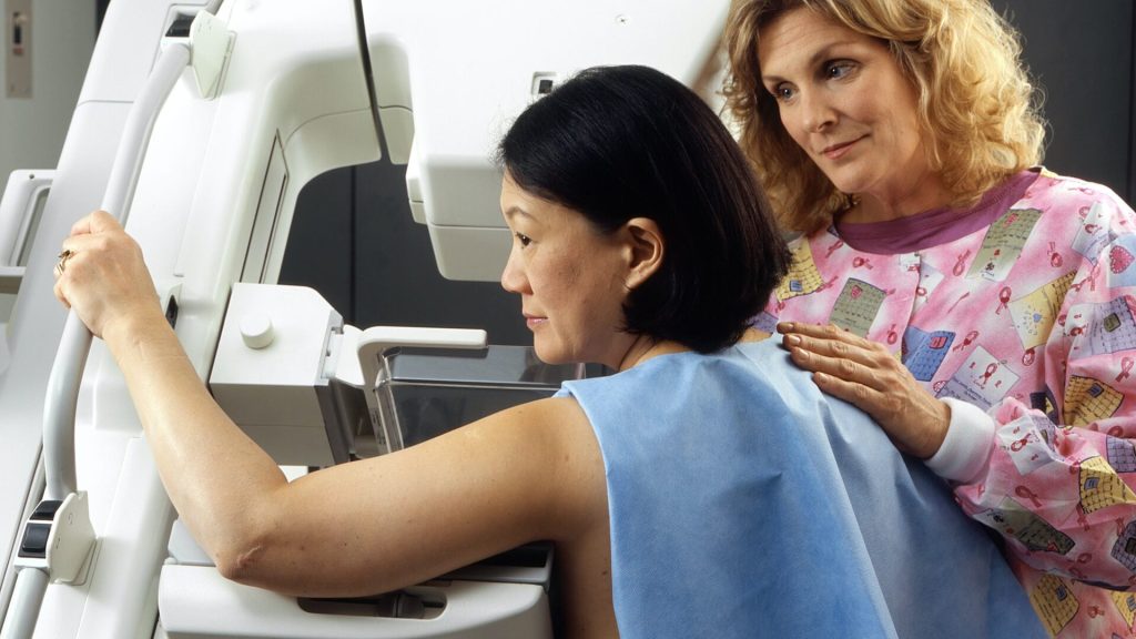
Dense breast tissue, which contains a higher proportion of fibrous tissue than fat, is a risk factor for breast cancer and also makes it more difficult to identify cancer on a mammogram. Many US states have enacted laws that require women with dense breasts to be notified after a mammogram, so that they can choose to undergo supplemental ultrasound screening to improve cancer detection. A recent study published by Wiley online in CANCER, a peer-reviewed journal of the American Cancer Society, evaluated the results of such additional screening to determine its benefits and harms to patients.

Although supplemental ultrasound screening may detect breast cancers missed by mammography, it requires additional imaging and may lead to unnecessary breast biopsies among women who do not have breast cancer. Therefore, it is important to use supplemental ultrasound only in women at high risk of mammography screening failure – in other words, women who develop breast cancer after a mammogram shows no signs of malignancy.

Brian Sprague, PhD, of the University of Vermont Cancer Center, and his colleagues evaluated 38 166 supplemental ultrasounds and 825 360 screening mammograms without supplemental ultrasounds during 2014–2020 at 32 US imaging facilities within three regional registries of the Breast Cancer Surveillance Consortium.

The team found that 95.3% of supplemental ultrasounds were performed in women with dense breasts. In comparison, 41.8% of mammograms without additional screening were performed in women with dense breasts.

Among women with dense breasts, a high risk of interval invasive breast cancer was present in 23.7% of women who underwent ultrasounds, compared with 18.5% of women who had mammograms without additional imaging.

The findings indicate that ultrasound screening was highly targeted to women with dense breasts, but only a modest proportion of these women were at high risk of mammography screening failure. A similar proportion of women who received only mammograms were at high risk of mammography screening failure.

“Among women with dense breasts, there was very little targeting of ultrasound screening to women who were at the highest risk of a mammography screening failure. Rather, women with dense breasts undergoing ultrasound screening had similar risk profiles to women undergoing mammography screening alone,” said Dr Sprague. “In other words, many women at low risk of breast cancer despite having dense breasts underwent ultrasound screening, while many other women at high risk of breast cancer underwent mammography alone with no supplemental screening.”

Clinicians can consider other breast cancer risk factors beyond breast density to identify women who may be appropriate for supplemental ultrasound screening. Publicly available risk calculators from the Breast Cancer Surveillance Consortium are available that also consider age, family history, and other factors (https://www.bcsc-research.org/tools).

Source: Wiley

US Task Force to Recommend Earlier Start to Breast Cancer Screening

Photo by National Cancer Institute on Unsplash

In a move bringing it closer in line with other organisations’ breast cancer screening guidelines, The United States Preventative Task Force (USPSTF) has released a draft statement recommending mammography every other year (biennially) from ages 40 to 74.

These recommendations are not applicable to women with a genetic marker or syndrome linked to increased breast cancer risk, a history of high-dose chest radiotherapy at a young age, or previous breast cancer or a high-risk breast lesion on previous biopsies.

According to the USPSTF, “new and more inclusive science about breast cancer in people younger than 50 has enabled us to expand our prior recommendation and encourage all women to get screened in their 40s. We have long known that screening for breast cancer saves lives, and the science now supports all women getting screened, every other year, starting at age 40.”

South African cancer screening guidelines typically closely follow American ones, according to an article by Lipschitz in the South African Journal of Radiology. Many countries had not recommended screening at the ages of 40–50 due to fears of overdiagnosis.

The UPSTF made particular attention the fact that black women are 40% more likely to die of breast cancer than white women, and have a high rate of aggressive cancers at young ages.

The recommendations are not without criticism. Biennial screenings are not seen as worth it by Desountis et al., as it leaving two years between tests leaves too much time for a tumour to grow.

Debra Monticciolo, MD, of Massachusetts General Hospital in Boston, and a member of the Society of Breast Imaging’s board of directors, told MedPage Today that she was “disappointed” with the decision to recommend biennial scans.

“Even if you look at their own data,” Monticciolo said, “annual screening results in more deaths averted, no matter what type of screening program you put in those models.”

The UPSTF has posted the new recommendations on its website for comment.

Regarding the ongoing debated about continued screening in women ages 75 and older, and supplemental screening for those with dense breasts, the UPSTF found there was not enough evidence for a recommendation.

Humans Still Beat AI in Breast Cancer Screening

Source: National Cancer Institute

A review in The BMJ finds that humans still seem to beat AI when it comes to the accuracy of spotting possible cases of breast cancer during screening.

At this point, there is a lack of good quality evidence to support a policy of replacing human radiologists with artificial intelligence (AI) technology when screening for breast cancer, the researchers concluded.

A leading cause of death for among women the world over, mammography screening is a high volume, repetitive task for radiologists, and some cancers are not picked up.

Previous research has suggested that AI systems outperform humans and might soon be used instead of experienced radiologists. However, a recent review of 23 studies highlighted evidence gaps and concerns about the methods used.

To address this uncertainty, researchers reviewed 12 studies carried out since 2010. They found that, overall, the methods used in the studies were of poor quality, with low applicability to European or UK breast cancer screening programmes.

Three large studies involving nearly 80 000 women compared AI systems with the clinical decisions of the original radiologist. Of these, 1878 had screen detected cancer or interval cancer (cancer diagnosed in-between routine screening appointments) within 12 months of screening.

The majority (34 out of 36 or 94%) of AI systems in these three studies were less accurate than a single radiologist, and all were less accurate than the consensus of two or more radiologists, which is the standard practice in Europe.

In contrast, five smaller studies involving 1086 women reported that all of the AI systems evaluated were more accurate than a single radiologist. However, these were at high risk of bias and not replicated in larger studies.

In three studies, AI used as a pre-screen to triage which mammograms need to be examined by a radiologist and which do not screened out 53%, 45%, and 50% of women at low risk but also 10%, 4%, and 0% of cancers detected by radiologists.

The authors point to some study limitations such as excluding non-English studies, as well as the fast pace of AI development. Nevertheless, stringent study inclusion criteria along with rigorous and systematic evaluation of study quality suggests their conclusions are robust.

As such, the authors said: “Current evidence on the use of AI systems in breast cancer screening is a long way from having the quality and quantity required for its implementation into clinical practice.”

They added: “Well designed comparative test accuracy studies, randomized controlled trials, and cohort studies in large screening populations are needed which evaluate commercially available AI systems in combination with radiologists in clinical practice.”

Source: Medical Xpress