Tag: macular degeneration

Cultured Cells may Restore Vision Lost to Photoreceptor Damage
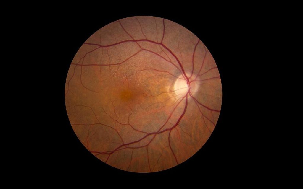
A preclinical study that produced progenitor photoreceptor cells and transplanted them into experimental models of damaged retinas has resulted in significant vision recovery. This finding marks a first step towards potentially restoring vision in eye diseases characterised by photoreceptor loss.

“Our laboratory has developed a novel method that enables the production of photoreceptor progenitor cells resembling those in human embryos,” said Assistant Professor Tay Hwee Goon, first author of the study published in Molecular Therapy. “Transplantation of these cells into experimental models has yielded partial restoration of the retinal function.”

The degeneration of photoreceptors in the eye is a significant cause of declining vision that can eventually lead to blindness and for which there is currently no effective treatment. Photoreceptor degeneration occurs in a variety of inherited retinal diseases, such as retinitis pigmentosa, a rare eye disease that breaks down cells in the retina over time and eventually causes vision loss, and age-related macular degeneration, a leading cause of vision impairment worldwide.

Asst Prof Tay and her team from Duke-NUS Medical School, the Singapore Eye Research Institute and the Karolinska Institute in Sweden, developed a procedure to grow human embryonic stem cells in the presence of purified laminin proteins. These proteins are involved in normal development of human retinas, and in their presence, stem cells could be directed to differentiate into photoreceptor progenitor cells responsible for converting light into signals that are sent to the brain.

When these cells were transplanted into damaged retinas, the preclinical models showed significant recovery of vision. A diagnostic test called electroretinogram also identified significant recovery in the retinas via electrical activity in the retina in response to a light stimulus. The transplanted cells established connections with surrounding retinal cells and nerves in the inner retina. They also survived and functioned for many weeks after transplantation.

Moving forward, the team hopes to refine their method to make it simpler and achieve more consistent results than earlier attempts to explore stem cell therapy for photoreceptor cell replacement.

“It is exciting to find these results, which suggest a promising route towards using stem cells to treat those forms of visual deterioration and blindness caused by the loss of photoreceptors,” said Dr Helder Andre, Head of Molecular and Cellular Research from Karolinska Institute’s Department of Clinical Neuroscience and a senior author of the study.

Associate Professor Enrico Petretto, Director of the Centre for Computational Biology at Duke-NUS and the study’s bioinformatics analysis lead, added: “Our method may also be useful for understanding the molecular and cellular pathways that drive the progression of macular degeneration, perhaps leading to the development of other therapeutic approaches.”

The next challenge for the researchers is to explore the efficacy of their method in models of photoreceptor degeneration that more closely match the human condition.

“If we get promising results in our future studies, we hope to move to clinical trials in patients,” said Professor Karl Tryggvason, from Duke-NUS’ Cardiovascular and Metabolic Disorders Programme, and the corresponding author of the study. “That would be an important step towards for being able to reverse damage of the retina and restore vision.”

Source: Duke-NUS Medical School

Early Warning Signs for Age-related Macular Degeneration

Credit: National Eye Institute

In an important step in treating a major cause of blindness, scientists have successfully identified early signs of age-related macular degeneration (AMD), in which higher number of mast cells are observed. This finding could be exploited by new treatments before symptoms develop. The study is published in PNAS.

Scientists have long known that people with certain genes on chromosomes 1 and 10 have a 2- to 3-fold higher risk of developing AMD, although lifestyle factors also play a role.

The team identified higher numbers of mast cells in the eyes of people when either of the risk genes were present, even when there were symptoms, suggesting an early mechanism in common.

They also showed the mast cells release enzymes in the back of the eye which then damage structures underneath the retina that in time is likely to damage the retina itself.

Mast cells exist in most tissues and are one of the immune system’s first defenses against infection, especially parasitic disease and damage.

Scientists already know there are more mast cells in the choroid in people with established AMD. The current study, however, identified higher levels in people before the disease develops.

The genes on chromosome 1 are linked to a part of the immune system called the complement cascade, which is associated with a risk of AMD.

Though the functional role of genes expressed by chromosome 10 are not known, but increased risk of AMD is.

Dr Richard Unwin, one of the study leaders, said: “What is really exciting about this work is that we are studying tissue from people before they have signs of the disease. This gives us a look into the very earliest stages, and gives us hope that we can intervene to stop the disease developing and ultimately prevent loss of vision”

The scientists used healthy human eye tissue donated post mortem to the Manchester Eye Tissue Repository.

They identified those who are at risk of developing age-related macular degeneration based on their risk genes, and discovered underlying changes in the tissue of the otherwise healthy at-risk individuals.

They collected retinal tissue from the back of donor eyes post mortem, following removal of the cornea for transplantation.

Then they took a small sample from the macula and removed the cells to leave a thin layer of membrane which supports the photoreceptors called rod and cone cells and is where disease begins.

They analysed the proteins present in the membrane from 30 people using mass spectrometry, which identifies protein components based on their mass, to find differences in the tissue make-up between those with and without genetic risk of AMD.

The mass spectrometry, identified a series of enzymes which are made almost exclusively by mast cells. In tissue from an additional 53 people, higher levels of mast cells were found in patients with higher disease risk.

Dr Unwin added: “We next need to look at how mast cells are activated, and whether by preventing, or clearing mast cell activation we can slow or stop disease development. There are several researchers and companies looking at complement mediated-therapies for AMD and while these are promising for Chr1-related disease there is no evidence that they will have an effect on Chr10 disease. A therapy designed to target mast cell activation as a unified mechanism could in theory treat all patients with AMD and prevent sight loss.”

Source: University of Manchester

Can Prozac be Used to Treat Macular Degeneration?

Source: Unsplash

The antidepressant fluoxetine, best known as Prozac, could offer the first treatment for the leading cause of blindness among people over 50, new research from the University of Virginia School of Medicine suggests.

Researchers have found early evidence that the drug fluoxetine may be effective against atrophic (or ‘dry’) age-related macular degeneration, a condition that affects nearly 200 million people worldwide. An analysis based on bench research, mouse models and huge insurance databases concluded that patients taking fluoxetine were less likely to develop atrophic macular degeneration (AMD).

On the strength of their findings, which were published in PNAS, the researchers are urging the investigation of fluoxetine to treat AMD, possibly as an oral pill or slow-release implant in the eye.

“These findings are an exciting example of the promise of drug repurposing, using existing medicines in new and unexpected ways,” said Bradley D. Gelfand, PhD, of UVA’s Center for Advanced Vision Science. “Ultimately, the best way to test whether fluoxetine benefits macular degeneration is to run a prospective clinical trial.”

The researchers believe fluoxetine works by binding with an inflammasome, NLRP3-ASC, which triggers the breakdown of the pigmented layer of the eye’s retina.

After conducting extensive bench research, Dr Gelfand and his team tested fluoxetine and eight other depression drugs in lab mice. Fluoxetine slowed the progression of the disease, but the others did not, the scientists found.

Encouraged by their findings, the researchers looked at fluoxetine use among patients aged over 50 in two enormous insurance databases with over 100 million records. They found that people taking the drug had a “significantly” slower rate of developing dry AMD.

Their approach, which combines bench research with big-data analysis, could lead to faster repurposing of existing drugs.

“Traditional approaches to drug development can be expensive and time-consuming: On average, a new FDA-approved drug takes 10–12 years and costs $2.8 billion (present-day dollars) to develop,” the researchers wrote. “Our identification of the unrecognised therapeutic activity of an existing FDA-approved drug using big data mining, coupled with demonstrating its efficacy in a disease-relevant model, could greatly accelerate and reduce the cost of drug development.”

Dr Gelfand was involved earlier this year in using a similar approach to determine that HIV drugs known as nucleoside reverse transcriptase inhibitors, or NRTIs, may be useful against dry macular degeneration as well.

“While we have had a great deal of success with the approach of using real-world patient data, we may have only begun to scratch the surface of finding new uses for old drugs,” said Dr Gelfand, of UVA’s departments of ophthalmology and biomedical engineering. “It is tempting to think about all the untapped therapeutic potential of medicines sitting on pharmacy shelves.”

Source: University of Virginia

HIV Drugs Could Stop Macular Degeneration

Photo by Victor Freita on Pexels

A new study has found that there is a buildup of damaging DNA in the eyes of patients with geographic atrophy, an untreatable, poorly understood form of age-related macular degeneration that leads to blindness. Based on this, the researchers believe it may be possible to treat the condition with HIV drugs, or even simpler ones.

Dr Jayakrishna Ambati and colleagues had previously discovered that the harmful DNA, known as Alu cDNA, was manufactured in the cytoplasm. This represents the first time toxic Alu cDNA accumulation has been confirmed in patients in any disease.

“Although we’ve known that geographic atrophy expands over time, we didn’t know how or why,” said Dr Ambati, of UVA’s Department of Ophthalmology and Center for Advanced Vision Science. “Our finding in human eyes that the levels of toxic Alu cDNA are highest at the leading edge of the geographic atrophy lesion provides strong evidence that it is responsible for this expansion over time that leads to vision loss.”

Geographic atrophy is an advanced form of age-related macular degeneration, which ultimately destroys vital cells in the retina, resulting in blindness.

Dr Ambati, a leading expert in macular degeneration, and colleagues found that this destruction is brought about by the buildup of Alu DNA. As Alu DNA accumulates in the eye, it triggers harmful inflammation via the inflammasome. The researchers discovered the mechanism involving a previously unknown structural facet of Alu that triggers the immune response that destroys the retinal cells.

HIV drugs called nucleoside reverse transcriptase inhibitors, or NRTIs, could treat this; tests in lab mice suggest these drugs, or safer derivatives known as Kamuvudines, could block the harmful inflammation and protect against retinal cell death.

“Over the last two decades, dozens of clinical trials for geographic atrophy that have targeted other pathways have failed,” Dr Ambati said. “These findings from patient eyes provide a strong impetus for a new direction.”

Dr Ambati says his latest findings support clinical trials testing the drugs in patients with macular degeneration. A prior study of health insurance databases with over 100 million patients found that people taking NRTIs were almost 40% less likely to develop dry macular degeneration.

“Our findings from human eyes show that these toxic molecules, which activate the inflammasome, are most abundant precisely in the area of greatest disease activity,” Dr  Ambati said. “We are very hopeful that a clinical trial of Kamuvudines will be launched soon in geographic atrophy so that we can potentially offer a treatment for this devastating condition.” 

The findings were published in Science Advances.

Source: University of Virginia

Inability to Dispose of Old Cells Leads to Macular Degeneration

Photo by v2osk on Unsplash

Researchers have discovered that failing to dispose of old photoreceptor cells leads to age-related macular degeneration.

The estimated number of people worldwide with age-related macular degeneration in 2020 was 196 million, increasing to 288 million in 2040. Though more than 50 genes are associated with the condition, the precise mechanism is unknown. Most people have a form of the condition, for which there are no known effective treatments.

In order to develop new therapies to treat the disease, University of Maryland School of Medicine (UMSOM) researchers are starting to understand what goes wrong in the disease. Using human and mice tissue, researchers showed that the process which removes the eye’s old, damaged light sensors is disrupted in macular degeneration.

Previously, the lead researcher had found out that many families with hearing disorders had genetic mutations in the gene for the CIB2 protein, and later work also showed that CIB2 was needed for vision in a large human family, as well as in zebrafish. Now, in this latest study, his team built on that previous work to dissect the intricate cell mechanisms behind retinal degeneration.

The team compared healthy mouse eyes to those from a mouse with the CIB2 protein genetically deleted. These CIB2 mutant mice were not disposing of their old light sensor proteins, called photoreceptors, like healthy mouse eyes did.  

“Photoreceptors continue growing in tiny columns in the eye, but over time, light damages the photoreceptors. To combat this, support cells in the eye slowly munch on the old, damaged photoreceptors, keeping the columns the correct length,” explained first author Saumil Sethna, PhD, at the University of Maryland School of Medicine. “If the photoreceptors are not removed, or if the process is backed up due to slow digestion by the support cells, like in the CIB2 mutant mice, the undigested material builds up over time, which may contribute to blindness.”

The researchers then identified several components in this photoreceptor recycling process, including a group of proteins collectively called mTORC1, which is involved in many human diseases, including cancer, obesity, and epilepsy.

Since mTORC1 (part of a family called mTOR) is a decision-maker for many cellular functions including cleaning up cellular debris, the researchers examined mTORC1’s activity in the CIB2 mutant mice and saw that it was overactive. mTORC1 was also found to be overactive in eye tissue of people with a form of age-related macular degeneration. The findings therefore indicate that drugs against mTORC1 may be effective treatments for the most common type of age-related macular degeneration, according to the researcher.
“Researchers have tested many small molecules directed at mTORC1 to treat various diseases, but the problem is that mTOR is needed for so many other cell functions that there are major side-effects when you tinker with it,” said senior author Zubair M Ahmed, PhD, Professor of Otorhinolaryngology-Head & Neck Surgery and Ophthalmology at the University of Maryland School of Medicine. “In our study, we found a backdoor way to regulate mTORC1, which may bypass many of the unpleasant side-effects that normally occur with suppressing mTORC1. We think we may be able to use our new knowledge of this mechanism to develop treatments for age-related macular degeneration and other diseases as well.”

Source: University of Maryland School of Medicine

Anti-HIV Drugs may Combat Macular Degeneration

New research has shown that anti-HIV drugs may fight macular degeneration – overturning a preconception about DNA in the process.

Macular degeneration is the leading cause of blindness in developed countries. Even though HIV does not cause dry macular degeneration, the drugs prevented the loss of vision.

“We are extremely excited that the reduced risk was reproduced in all the databases, each with millions of patients,” said Jayakrishna Ambati, MD, a leading macular degeneration researcher at the University of Virginia School of Medicine. “This finding provides real hope in developing the first treatment for this blinding disease.”

A Big Data Archeology review of four health insurance databases showed that Nucleoside Reverse Transcriptase Inhibitors (NRTIs), a commonly used HIV treatment, reduced the incidence of dry macular degeneration by 40%. The records spanned two decades and covered over 100 million patients. The drugs had also previously been shown to possibly prevent diabetes.

The finding also comes with the discovery that DNA can be produced inside the cytoplasm. Alu DNA (found exclusively in primates), which makes up 10% of the human genome, is transposable and can insert itself into other places on the genome. It was long considered “junk” DNA, but are now believed to have important functions, such as allowing for multiple expressions of proteins from a single Alu element. Since it cannot replicate itself, Alu DNA requires a transposon called L1 to accomplish this, which was now reported to allow the production of Alu DNA outside the chromosome. The buildup of Alu DNA in cells contributes to macular degeneration, by killing off cells that support the retina.
The researchers are urging further investigation into NRTIs or safer derivatives known as Kamuvudines, both of which block a key inflammatory pathway, can be useful in preventing vision loss from dry macular degeneration.

“A clinical trial of these inflammasome-inhibiting drugs is now warranted,” said Ambati. “It’s also fascinating how uncovering the intricate biology of genetics and combining it with big data archeology can propel insights into new medicines.”

Source: Medical Xpress

Journal Information: Shinichi Fukuda el al., “Cytoplasmic synthesis of endogenous Alu complementary DNA via reverse transcription and implications in age-related macular degeneration,” PNAS (2021). www.pnas.org/cgi/doi/10.1073/pnas.202275111