Tag: macrophages

Scientists Discover New Population of Macrophages Aiding Post-virus Lung Repair

Credit: Scientific Animations CC4.0

Researchers in Belgium have discovered a new population of macrophages, important innate immune cells that populate the lungs after injury caused by respiratory viruses. These macrophages are instrumental in repairing the pulmonary alveoli. This groundbreaking discovery promises to revolutionise our understanding of the post-infectious immune response and opens the door to new regenerative therapies.

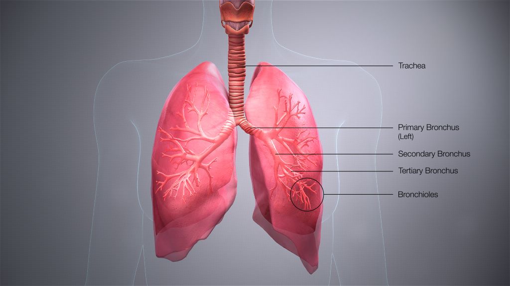
Respiratory viruses, typically causing mild illness, can have more serious consequences, as shown during the COVID pandemic, including severe cases requiring hospitalisation and the chronic sequelae of “long Covid.” These conditions often result in the destruction of large areas of the lungs, particularly the alveoli responsible for gas exchanges. Ineffective repair of these structures can lead to ARDS or a permanent reduction in the lungs’ ability to oxygenate blood, causing chronic fatigue and exercise intolerance.

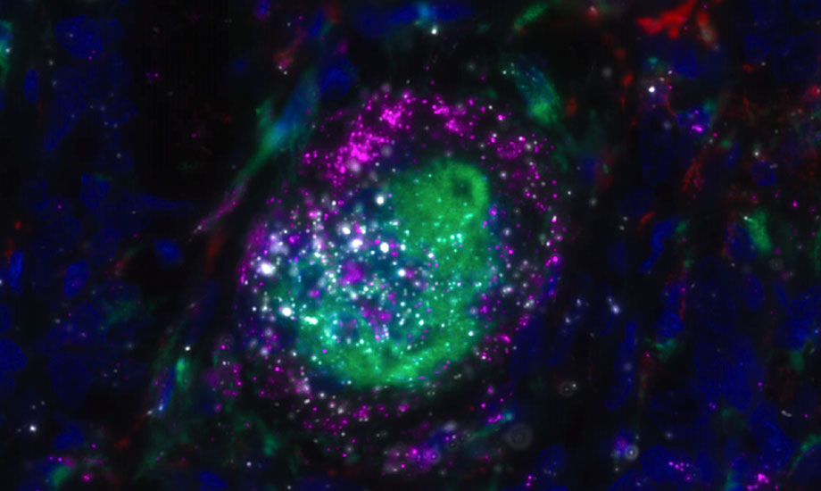
While the role of macrophages during the acute phase of respiratory viral infections is well known, their function in the post-inflammatory period has been largely unexplored. This study by the GIGA Institute at the University of Liège reveals that atypical macrophages, characterised by specific markers and transiently recruited during the early recovery phase, play a beneficial role in regenerating pulmonary alveoli.

Led by Dr Coraline Radermecker and Prof. Thomas Marichal from the Immunophysiology Laboratory, the study was conducted by Dr Cecilia Ruscitti and benefited from the ULiège’s advanced technological platforms, including flow cytometry, fluorescence microscopy, and single-cell RNA sequencing. “Our findings provide a novel and crucial mechanism for alveolar repair by these atypical macrophages,” explains Coraline Radermecker. “We have detailed their characteristics, origin, location in the damaged lung, the signals they require to function, and their role in tissue regeneration, specifically acting on type 2 alveolar epithelial cells, the progenitors of alveolar cells.” The scientific community had overlooked these macrophages because they express a marker previously thought to be specific for another immune cell population, the neutrophils, and because they appear only briefly during the repair phase before disappearing.

“Our study highlights the reparative role of these macrophages, countering the prevailing idea that macrophages following respiratory viral infections are pathogenic,” adds Thomas Marichal. “By targeting the amplification of these macrophages or stimulating their repair functions, we could develop therapies to improve alveolar regeneration and reduce complications from serious respiratory infections and ARDS.”

To illustrate, consider the lungs as a garden damaged by a storm (viral infection). These newly discovered macrophages act like specialised gardeners who clear debris and plant new seeds, enabling the garden to regrow and regain its vitality.

Source: University of Liège

Scientists Discover Macrophages on Standby in the Pleural Cavity

Credit: Scientific Animations CC4.0

Scientists have long thought of the pleural cavity merely as a cushion from external damage. Turns out, it also houses macrophages that rush into the lungs during flu infections.

“We were surprised to find them in the lungs because nobody has seen this before, that these cells go into the lung when there’s an infection,” said UC Riverside virologist Juliet Morrison, who led the discovery team.

A paper published in the Proceedings of the National Academy of Sciences details how during an influenza infection, macrophages leave the exterior cavity and cross into the lungs where they decrease inflammation and reduce levels of disease.

“This study shows it’s not just what happens in the lung that matters, but also what’s outside of the lung. Cell types not normally connected to the lung can have outsized impacts on lung disease and health,” Morrison said.

There are three main cavities in the body: one around the heart, the abdominal cavity, and the pleural cavity surrounding the lungs.

“Because it contains fluid, it prevents the lungs from collapsing. However, people have not thought much about the pleural cavity being a whole organ within itself. This research may change that perception,” Morrison said.

Initially, the researchers set out to understand the more general question of what types of cells are present in the lungs during flu infections. They took existing data on lung-related genes from studies of mice that either died from the flu or survived. They then mined the data using an algorithm to predict cell types that change in the lungs during infections.

“We took big data and broke it down to assign which potential immune cells are in the lung tissues. That’s where I got a hint that maybe we had a previously unknown external source of cells in the lung,” Morrison said.

Next, using a laser-based technique, the team tracked macrophages going into the lungs of mice, and observed what happened if they took these cells out of the equation.

“When you take them out of the mouse you see more disease and more lung inflammation,” Morrison said.

Morrison says she hopes this study will encourage other scientists to reevaluate data sets from older studies.

“Our approach was to take information already out there and put it to new use, and we were able to see something new,” she said.

Moving forward, the research team is hoping to determine which proteins “tell” the macrophages to move into the lungs. Once the protein signals have been identified, it may be possible to create drugs that boost either the number of macrophages, or their activity.

The strategy of boosting human defences to infection, rather than developing another antiviral to which viruses could become resistant, could offer people a flu treatment that would be more effective for much longer.

“If we can boost what resolves infection in us, we probably have a better shot. We’re less likely to have resistance. The immune system is so complicated, but it’s our best bet in the long run to work with what we have rather than chase viruses that continue to escape our therapeutics,” Morrison said.

Scientists Give Macrophages First-aid ‘Backpacks’ to Calm TBI Inflammation

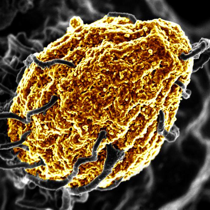
Colourised electron micrograph image of a macrophage. Credit: NIH

Scientists have created a new treatment for traumatic brain injury (TBI). The new approach leverages macrophages, which can increase or decrease inflammation in response to infection and injury. The team attached “backpacks” containing anti-inflammatory molecules directly to the macrophages. These molecules kept the cells in an anti-inflammatory state when they arrived at the injury site in the brain, enabling them to reduce local inflammation and mitigate the damage caused. The research is reported in PNAS Nexus.

“Every year, millions of people suffer from a TBI, but there is currently no treatment beyond managing symptoms. We have applied our cellular backpack technology – which we previously used to improve macrophages’ inflammatory response to cancerous tumours – to deliver localised anti-inflammatory treatment in the brain, which helps mitigate the cascade of runaway inflammation that causes tissue damage and death in a human-relevant model,” said senior author Samir Mitragotri, PhD, in whose lab the research was performed.

Stopping a runaway inflammation train

There is currently no treatment for the damage caused to brain tissue during a traumatic brain injury (TBI), beyond managing a patient’s symptoms. One of the main drivers of TBI-caused damage is a runaway inflammatory cascade in the brain.

As cells die from the impact, they release a cocktail of pro-inflammatory cytokine molecules that attract immune cells to clean up the damage. But the same cytokine molecules can also disrupt the blood-brain barrier, which causes blood to leak into the brain. Blood accumulation in the brain causes swelling, impaired oxygen delivery, and increased inflammation, and creates a vicious cycle of bleeding and damage that drives even more cell death.

The Mitragotri lab saw an opportunity in this problem.

“It’s generally believed anti-inflammatory therapies can be effective for treating TBI, but so far, none of them have proven effective clinically. Our previous work with macrophages has shown us that we can use our backpack technology to effectively steer their behaviour when they arrive at the injury site. Since these cells are already active players in the body’s natural immune response to a TBI, we had a hunch we could augment that pre-existing biology to reduce the initial damage,” said co-first author Rick Liao, Ph.D., a Postdoctoral Fellow at the Wyss Institute and SEAS.

“Body, heal thyself”…with backpacks

Macrophages are very malleable cells and can “switch” between pro-inflammatory and anti-inflammatory states. While the team’s previous work in cancer had been focused on keeping macrophages in a pro-inflammatory state when they arrive at the inflammation-reducing microenvironment of a tumour, this new project would be trying to do the opposite: keep the macrophages “calm” in the inflammation-riddled setting of a brain injury.

To do so, they used a disc-shaped “backpack” they had previously designed to treat multiple sclerosis that contained layers of two anti-inflammatory molecules: dexamethasone, a steroid, and interleukin-4, a cytokine that encourages macrophages to adopt an anti-inflammatory state. They then incubated these microparticles with both human and pig macrophages in vitro and saw that the backpacks stably stuck to the cells without causing any negative effect. They also observed that application of their backpacks decreased the expression of pro-inflammatory biomarkers and increased the expression of anti-inflammatory biomarkers, retaining the pig macrophages in a healing state.

But to prove that this shift would work in the body, they had to test the backpack-bearing macrophages in vivo. They chose pigs as their model organism because their brains’ structures and responses to injury more closely mimic those of humans than mice.

“Probably our biggest challenge in this project was scaling up production to match what we needed to run the experiments. Our previous studies were done in rodents, which required about two million macrophages and four million backpacks administered per subject. For the porcine study, we needed 100 million macrophages and 200 million backpacks per subject – on the scale of what would be administered in humans – and lots of helping hands,” said co-first author Neha Kapate, PhD, a Postdoctoral Fellow at the Wyss Institute and SEAS.

Once they had generated enough backpack-wearing porcine macrophages, they infused them into the pigs’ bloodstreams four hours after a TBI. Seven days later, they analysed the animals’ brains. Pigs that had received the macrophage treatment showed a high concentration of the cells in the area immediately surrounding the injury site, their lesions were 56% smaller, and there was significantly less haemorrhaging than in untreated animals.

Local immune cells also displayed a lower amount of a pro-inflammatory activation marker called CD80, indicating that the macrophages had accomplished their damage control by reducing inflammation in the brain. Corroborating that data, the levels of two soluble biomarkers for inflammation in the blood and cerebrospinal fluid were lower in treated animals than in untreated animals. The macrophage treatment also did not cause any negative effects.

The team plans to conduct future studies that focus on elucidating exactly how their anti-inflammatory macrophage therapy affects the blood-brain barrier’s integrity to prevent bleeding, which could also hold promise for treating other conditions like hemorrhagic strokes.

“Macrophages’ susceptibility to their local environment has historically prevented scientists from taking full advantage of their immune-modulating capabilities. This impressive study describes a truly novel and potentially powerful macrophage-based therapy for treating the inflammation that is the root cause of so many human afflictions in an effective and non-invasive way that works with biology rather than against it,” said Wyss Founding Director Donald Ingber, MD, PhD.

Source: Wyss Institute for Biologically Inspired Engineering at Harvard

Monocytes are Still Capable of Proliferation, Study Shows

Scanning electron micrograph of a macrophage. Credit: NIAID

Cell proliferation from stem cells is vital for organisms to grow and form vital organs. In cancer, however, cell proliferation is no longer controlled and becomes chaotic. Research published in Nature Immunology revealed that monocytes, which are blood immune cells previously thought to be already differentiated, can in fact still proliferate. The study’s researchers at the University of Liège have discovered that, in a healthy individual, monocytes also have this ability to proliferate, in order to replace tissue macrophages.

Monocytes are white blood cells that derive from the bone marrow. A monocyte is part of the innate immune response and functions to regulate cellular homeostasis, especially in the setting of infection and inflammation. They account for approximately 5% of circulating nucleated cells in normal adult blood.

The formation of complex multicellular organisms necessitates billions of cells to be produced from progenitor cells, proliferating and taking on particular morphologies and functions while assembling into tissues and organs. Most of the cells that constitute a living organism are understood to arise from stem cells. When they stop proliferating, they specialise, differentiate and form muscles, brain, bones, immune cells, etc. When proliferation is no longer properly regulated, this can lead to the development of various diseases, chief if which is cancer.

Professor Thomas Marichal (Professor at ULiège, Welbio investigator at the WEL Research Institute) and his team from the GIGA Institute at ULiège discovered that this ability to proliferate is not merely restricted to stem cells, but is also an as-yet-unknown function of blood immune cells, the monocytes.

Indeed, blood monocytes, previously considered as differentiated cells, are capable of proliferating and generating a pool of monocytes in the tissues in order to give rise to macrophages, which are important immune cells that protect us against microbes and support the proper functioning of our organs.

“This is a major fundamental discovery, which changes our conception of the involvement of cell proliferation in the constitution and maintenance of our immune system,” explains Thomas Marichal, director of the study. “Our finding also suggests that the information that can be drawn from an enumeration of blood monocytes, classically carried out during a blood test, would reflect only little of what is happening at the level of the tissues, during ‘infection or inflammation, for example, since monocytes can proliferate when they enter tissues.”

He also adds, “Fortunately, this proliferation is extremely well controlled and does not lead to a humoral process. It has only one goal: to allow, as effectively as possible, the replacement of immune cells that populate our tissues: the macrophages.”

This discovery was possible thanks to the development of new tools and the use of innovative technologies. “This study is a great example of how technological advances can drive breakthrough scientific discoveries. It would have been extremely difficult, if not impossible, to study with such a resolution this population of proliferating monocytes only 10 years ago. This required the use of state-of-the-art equipment recently acquired at the GIGA Institute, the generation of complex genomic data and very sophisticated bioinformatics analyses,” explains Domien Vanneste, first author of the study.

This study paves the way for future investigations that will evaluate the possibility of manipulating or controlling monocyte proliferation for therapeutic purposes, at the benefit of an enhanced health.

Source: University de Liege

Macrophages Explain Vulnerability to Influenza in Old Age

Old man
Source: JD Mason on Unsplash

After being suppressed during the COVID pandemic, influenza is again circulating and threatening the health of over 65s. But why are older people so more susceptible to the flu? A new study from the University of Michigan, published in Nature Communications, offers clues.

The study, led by first author Judy Chen, a PhD candidate, senior author Professor Daniel Goldstein, MD, and their team investigates why cells called alveolar macrophages, the first line of defence in the lungs, appear to be compromised with age.

Macrophages attack pathogens like the flu virus and reside in alveoli. Importantly, these cells appear to be lost with ageing.

Previous research by another group showed that when macrophages from an old mouse were put into a young mouse, and cells looked young again. “This drove us to believe that something in the environment of the lungs is contributing to this,” said Chen.

Signs pointed to a lipid immune modulator known as prostaglandin E2 (PGE2) with wide ranging effects, from labour induction in pregnancy to inflammation with arthritis. The study team discovered there is more PGE2 in the lungs with age. This increase in PGE2, Chen explained, acts on the macrophages in the lung, limiting their overall health and ability to generate.

The team suspects that the buildup of PGE2 is yet another marker of a biological process called senescence, which is often seen with age. Senescence serves as insurance against the runaway division of damaged cells; cells that are senescent are no longer able to replicate.

“One of the interesting things about these cells is they secrete a lot of inflammatory factors,” said Chen.

The study showed that with age, the cells lining the air sacs in the lungs become senescent, and these cells lead to increased production of PGE2 and suppression of the immune response.

To test the link between PGE2 and increased susceptibility to influenza, they treated older mice with a drug that blocks a PGE2 receptor. “The old mice that got that drug actually ended up having more alveolar macrophages and had better survival from influenza infection than older mice that did not get the drug,” said Chen.

The team plans to next investigate the various ways PGE2 affects lung macrophages as well as its potential role in inflammation throughout the body. “As we get older, we become more susceptible not only to influenza, but to other infections, cancers, autoimmune diseases as well.”

Source: Michigan Medicine – University of Michigan

How Macrophages Control an Uncooperative Meal

A macrophage digesting a yeast cell (yellow). Credit: NIH

Certain pathogens such as Salmonella have developed strategies to protect themselves from the macrophages’ digesting attempts, causing severe Typhoid infections and inflammations. Scientists report in Nature Metabolism how the inter-organellar crosstalk between phago-lysosomes and mitochondria restricts the growth of such bacteria inside macrophages.

Signals from the digestion cell organelle

As scavenger cells, macrophages have a very prominent digestion organelle, the phago-lysosome, where engulfed microorganisms are commonly degraded into pieces and become inactivated. “It has long been known that the molecule TFEB (Transcription factor EB) is important for the regulation of the phago-lysosomal system. More recent evidence also suggested that TFEB supports the defense against bacteria,” said Max Planck Institute group leader Angelika Rambold.

She and her team wanted to understand how exactly TFEB mediates its anti-bacterial role in macrophages. They confirmed earlier findings showing that a broad range of microbes, bacterial and inflammatory stimuli activate TFEB and thus the phago-lysosomal system.

“It made sense that pathogen signals trigger TFEB as macrophages need a more active digestion system quickly after they devour a meal of bacteria. But, interestingly, the experiments also revealed an additional strong effect of TFEB activation on another intracellular organelle system — mitochondria. This was completely unexpected and novel to us,” said Angelika Rambold.

Instructing mitochondria to increase anti-microbial activity

Composed of inner and outer membranes, mitochondria are the primary sites of cellular respiration and release energy from nutrients. Moreover, the mitochondria in immune cells were recently identified as sources of anti-microbial metabolites.

By using a broad experimental tool set, the investigators identified the pathway controlling an unexpected crosstalk between lysosomes and mitochondria. “Macrophages make use of extensive inter-organellar communication: the lysosome activates TFEB, which shuttles into the nucleus where it controls the transcription of a protein called IRG1. This protein is imported into mitochondria, where it acts as a major enzyme to produce the anti-microbial metabolite itaconate,” explained Angelika Rambold.

Exploiting organelle communication to control bacterial infections

The researchers investigated whether they could make use of this newly identified pathway to control bacterial growth. “We speculated that activating this pathway could be used to target certain bacterial species, such as Salmonella,” said Angelika Rambold. “Salmonella can escape the degradation by the phago-lysosomal system. They manage to grow inside macrophages, which can lead to the spreading of these bacteria to several organs in an infected body,” explained Alexander Westermann, collaborating scientist from the University of Würzburg.

When the researchers activated TFEB in infected macrophages in mice, the TFEB-Irg1-itaconate pathway inhibited the growth of Salmonella inside the cells. These data show that the lysosome-to-mitochondria interplay represents an antibacterial defense mechanism to protect the macrophage from being exploited as a bacterial growth niche.

In light of the increasing emergence of multi-drug resistant bacteria, with more than 10 million expected deaths per year by 2050 according to the various expert groups, it becomes important to identify new strategies to control bacterial infections that escape immune mechanisms. A promising path could be to use the TFEB-Irg1-itaconate pathway or itaconate itself to treat infections caused by itaconate-sensitive bacteria. According to the researchers from more work is needed to assess whether these new intervention points can be successfully applied to humans.

Source: Max Planck Institute of Immunobiology and Epigenetics

Study Sheds Light on Cardiac Repair after MI

Photo from Olivier Collett on Unsplash

Immune response and the lymphatic system are central to cardiac repair after a heart attack, according to a study published in the Journal of Clinical Investigation. These insights into the basic mechanisms of cardiac repair pave the way towards the development of new treatments to preserve heart function.

“We found that macrophages, or immune cells that rush to the heart after a heart attack to ‘eat’ damaged or dead tissue, also induce vascular endothelial growth factor C (VEGFC) that triggers the formation of new lymphatic vessels and promotes healing,” said co-senior author Edward Thorp, PhD, from the Heart Center at Lurie Children’s and Associate Professor of Pathology and Pediatrics at Northwestern University Feinberg School of Medicine. “Our challenge now is to find a way either to administer VEGFC or to coax these macrophages to induce more VEGFC, in order to speed the heart repair process.”

People who suffer a heart attack are at high risk for heart failure, even with the advances in medications to reduce mortality. This occurs in part because some macrophages that arrive at the site of damage are proinflammatory and do not induce VEGFC.

“It is a Dr. Jekyll and Mr. Hyde scenario, with ‘good’ macrophages that induce VEGFC and the ‘bad’ ones that don’t. We need to prevent the ‘bad’ macrophages from causing further damage,” said co-senior author Guillermo Oliver, PhD, Director of Feinberg Cardiovascular and Renal Research Institute – Center for Vascular and Developmental Biology, and Professor of Medicine at Northwestern University Feinberg School of Medicine. “We are working to understand more about the progression to heart failure after a heart attack, in order to intervene early and reset the course to cardiac repair.”

Source: DICardiology

A Key Immune Function in Red Blood Cells Has Been Discovered

Source: Wikimedia CC0

Red blood cells have been discovered to have a critical function as immune sensors by binding cell-free DNA (nucleic acid) present in the body’s circulation during sepsis and COVID. 

This DNA-binding capability triggers their removal from circulation, driving inflammation and anaemia during severe illness and playing a much larger role in the immune system than previously thought. Scientists have long known that red blood cells also interacted with the immune system, but not whether they directly altered inflammation, until now. The study appears in Science Translational Medicine.

“Anaemia is common, affecting about a quarter of the world’s population. Acute inflammatory anaemia is often seen early after an infection such as parasitic infections that cause malaria,” said senior author Nilam Mangalmurti, MD, an assistant professor of Medicine at Penn. “For a long time we haven’t known why people, when they are critically ill from sepsis, trauma, COVID, a bacterial infection, or parasite infection, develop an acute anaemia. These findings explain one of the mechanisms for the development of acute inflammatory anaemia for the first time.”

Toll-like receptors (TLRs) play a key role in the immune system by activating immune responses like cytokine production. Analysing the red blood cells of about 50 sepsis patients and 100 COVID patients the study found that, during these illnesses, red blood cells express more TLR9 on their surface.

When the red blood cells bind too much inflammation-causing nucleic acid, they lose their normal structure, causing the body to no longer recognise them, prompting macrophages to engulf them. When this happens, it causes the immune system to become activated in otherwise unaffected organs, creating inflammation. The discovery of this mechanism will allow research on blocking this specific receptor and creating targeted therapies for autoimmune diseases, infectious diseases, and various inflammatory illnesses associated with acute anaemia.

“Right now when patients in the ICU become anaemic, which is almost all of our critically ill patients, the standard is to give them blood transfusions, which has long been known to be accompanied by a host of issues including acute lung injury and increased risk of death,” Prof Mangalmurti said. “Now that we know more about the mechanism of anaemia, it allows us to look at new therapies for treating acute inflammatory anaemia without transfusions, such as blocking TLR9 on the red blood cells. Targeting this TLR9 may also be a way to dampen some of the innate immune activation without blocking this receptor in immune cells, which are very important for the host when fighting a pathogen or injury.”

This DNA-binding discovery could also have implications for research into using red blood cells in diagnostics, Prof Mangalmurti said. For example, a physician might be able to take red blood cells from a patient with pneumonia, sequence the nucleic acid absorbed from the infection, and identify the specific kind of pathogen to better determine what kind of antibiotic to prescribe.

Prof Mangalmurti and colleagues are looking at whether this is a valid option in diagnosing infection in critically ill patients and if this DNA-binding mechanism by red blood cells is a universal mechanism of anaemia in parasitic infections.

Source: Perelman School of Medicine at the University of Pennsylvania

Jump-starting Macrophages to Help with IBD Tissue Repair

A novel method which prompts immune cells to aid the repair of damaged intestinal tissues has been developed by researchers at KU Leuven and Seoul National University.

This new approach promises new treatments for inflammatory bowel disease (IBD), including ulcerative colitis and Crohn’s disease. Normally, the immune system defends against pathogens that enter the body. In conditions like IBD, the immune system instead attacks tissues that line the gut, creating ulcers. Some 3.9 million women and 3.0 million women suffer from IBD worldwide.

The origin of IBD is not known, so treatments typically dampen immune response, but at the same time this also obstructs the normal repair of damaged intestinal tissue by other parts of the immune system. Macrophages, for example, consume foreign bodies, clean out debris and direct other steps in inflammatory or repair response through released substances. 

Lead author Professor Gianluca Matteoli, an immunologist at the Translational Research Center for Gastrointestinal Disorders (TARGID) KU Leuven, explained the motivation behind the research. “Our idea is that the migration of macrophages to the damaged tissue in IBD is essential to stimulate its recovery.”

Examining macrophages in the intestines of a handful of people with IBD, the researchers found that a sub-group of cells responding to prostaglandin E2 (PGE2). Prostaglandins are messenger molecules involved in homeostatic functions and mediate pathogenic mechanisms, such as the inflammatory response, and are also involved in tissue repair.

“If the patients had acute disease, they had a lower amount of these beneficial cells, and if they went into remission, then amounts of macrophages went up. This suggests that they are part of the reparative process,” said Professor Gianluca Matteoli, immunologist, Translational Research Center for Gastrointestinal Disorders (TARGID), KU Leuven

In mice with ulcerative colitis (one the main forms of IBD), there were fewer macrophages responsive to prostaglandin than in healthy mice. However, when PGE2 levels were increased, those macrophages still responsive released a substance that stimulated tissue repair. When the researchers knocked out the PGE2 receptors on the macrophages, the level of tissue repair dropped.

Getting the macrophages to absorb a liposome containing a substance to trigger the repair stimulation agent restored the macrophages’ repairing effect. Liposomes are bubbles made of two layers of lipids enclosing an aqueous cavity, often used to hold substances for drug delivery.

“We already knew that prostaglandins were important for inducing proliferation of tissue cells, but this study shows that they are also important for controlling the inflammatory effect, so moving the body from the acute stage where inflammation dominates to the reparative stage,” Professor Matteoli said.

New treatments could involve liposomes being used to prompt macrophages into boosting tissue repair, a well-established experimental tool but which would require considerable work for this new application.

“This is one of the first times it has been used to produce a beneficial, therapeutic effect,” said Professor Seok. 

The next step is to closely examine human macrophages at different stages of IBD. “We want to identify other factors that trip the switch that turns macrophages from inflammatory cells to non-inflammatory cells,” said Professor Matteoli. “Then, using the liposome technology that Professor Seok has developed, these could be used to target the macrophages and so produce very precise drugs.”

Source: News-Medical.Net