Tag: lupus

Kidney Damage in Lupus Comes from an Unexpected Source

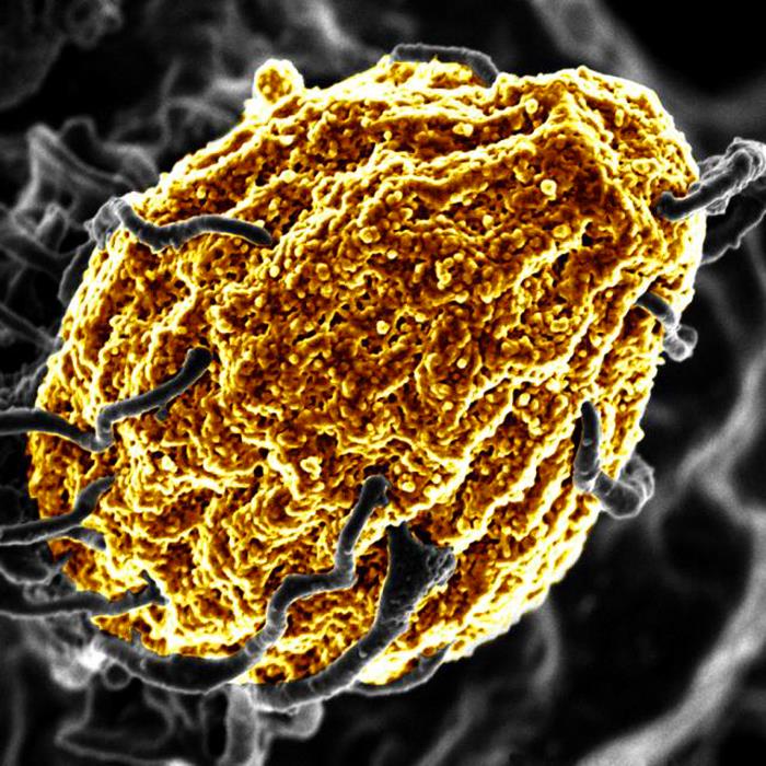
When the NKp46 receptor of the ILCs is blocked (right), the lupus nephritis recedes. Blue: cell nuclei.
Credit: Charité | Frauke Schreiber

A Berlin-led research team has uncovered critical regulators of severe kidney damage in patients with the autoimmune disorder lupus. A small, specialised population of immune cells – called innate lymphoid cells (ILCs) – trigger an avalanche of effects that cause harmful kidney inflammation, also known as lupus nephritis.

The research, published this week in Nature, upends conventional wisdom that autoantibodies are primarily responsible for lupus nephritis.

“While autoantibodies are required for tissue damage, they are by themselves not sufficient. Our work reveals that ILCs are required to amplify the organ damage,” says Dr Masatoshi Kanda, a senior paper author who was a Humboldt Fellow at Max Delbrück Center and is now at the Department of Rheumatology and Clinical Immunology, Sapporo Medical University in Japan.

Lupus, or systemic lupus erythematosus, is most often diagnosed between the ages of 15 and 45. Symptoms can range from mild to severe. But what causes kidney damage in some patients – some to the point of requiring dialysis – has been unclear.

“The role of ILCs in lupus or lupus nephritis was entirely unknown,” says Professor Antigoni Triantafyllopoulou, a senior paper author at the German Rheumatology Research Center (DRFZ), an institute of the Leibniz Association, and at the Department of Rheumatology and Clinical Immunology at Charité – Universitätsmedizin Berlin. “We have now identified most of the circuit controlled by ILCs by looking at the whole kidney at single-cell resolution.”

Unusual immune cells

ILCs are a small group of immune cells that – unlike most other immune cells that circulate throughout the body – live in a specific tissue or organ.

“They are in the tissue all the time, from the time of embryonic development, which makes them very different from other immune cells,” says Professor Andreas Diefenbach, a senior paper author and director of the Institute of Microbiology, Infectious Diseases and Immunology at Charité – Universitätsmedizin Berlin.

Diefenbach’s lab was among those that discovered ILCs in the mid-2000s. Most of his research is focused on ILCs in the gut and how they modify tissue function. In this study, Triantafyllopoulou and Kanda teamed up with his group and Dr Mir-Farzin Mashreghi at the DRFZ to find out whether ILCs were present in the kidney and what role they might play in lupus nephritis.

The whole single-cell picture

To unravel this mystery, the team turned to single-cell RNA sequencing, which identifies genes that are active, or “switched on,” in individual cells and helps researchers understand the cell’s identity and function.

Kanda, a rheumatologist who was studying bioinformatics in Professor Norbert Hübner’s lab at the Max Delbrück Center at the time, developed a specialized protocol for single-cell RNA sequencing of mouse and human kidneys. “Masatoshi’s protocol was very good at pulling out and preserving multiple types of kidney cells, which gave us a much more complete overview of how lupus affects the whole kidney,” explains Triantafyllopoulou. The team sequenced nearly 100 000 individual kidney and immune cells of various types and functions.

The key receptor

Through experiments in mice, the team learned that a subgroup of ILCs with a receptor called NKp46 must be present and activated to cause lupus nephritis. When NKp46 is activated, this subgroup of cells ramped up production of a protein called GM-CSF, which stimulates invading macrophages to multiply. In the kidney, a flood of incoming macrophages caused severe tissue damage and fibrosis.

“These ILCs are really amplifiers in this system,” Diefenbach says. “They are small in population, but they seem to fertilise the whole process.”

When the team blocked NKp46 with antibodies or the receptor was genetically removed, kidney tissue damage was minimal. They also blocked GM-CSF with similar anti-inflammatory effects.

“Critically, autoantibody levels did not change when NKp46 was inhibited, but kidney tissue damage was reduced, which shows autoantibodies are not directly responsible for kidney inflammation,” Triantafyllopoulou explains.

The team also compared the results to sequencing data from tissue taken from human patients with lupus and found ILCs present, though more work is required to fully understand how to target ILCs in human kidneys. Nevertheless, the insights gained through these detailed studies point to new antibody therapies for patients with severe forms of lupus. The hope is to prevent the need for kidney dialysis in these patients.

Source: Max Delbrück Center for Molecular Medicine in the Helmholtz Association

Does Air Pollution Affect Lupus Risk?

Photo by Kouji Tsuru on Pexels

New research published in Arthritis & Rheumatology indicates that chronic exposure to air pollutants may increase the risk of developing lupus, an autoimmune disease that affects multiple organs.

For the study, investigators analysed data on 459 815 participants from the UK Biobank. A total of 399 lupus cases were identified during a median follow-up of 11.77 years. Air pollutant exposure was linked with a greater likelihood of developing lupus. Individuals with a high genetic risk and high air pollution exposure had the highest risk of developing lupus compared with those with low genetic risk and low air pollution exposure.

“Our study provides crucial insights into the air pollution contributing to autoimmune diseases. The findings can inform the development of stricter air quality regulations to mitigate exposure to harmful pollutants, thereby reducing the risk of lupus,” said co–corresponding author Yaohua Tian, PhD, of the Huazhong University of Science and Technology, in China.

Source: Wiley

Interferons Drive Lupus Symptoms and Affect Treatment

A woman with Systemic Lupus Erythematosus. Source: Wikimedia CC0

In a new study, researchers from Johns Hopkins Medicine say they have uncovered insights as to why lupus symptoms and severity present differently in individuals with the autoimmune condition. The team says this is a crucial step forward in understanding biological mechanisms behind lupus, and may also lead to shifts in how clinicians treat patients with the condition.

The full report, published in Cell Reports Medicine, concludes that specific combinations and elevated levels of immune system proteins, known as interferons, are associated with certain lupus symptoms such as skin rashes, kidney inflammation and joint pain. Interferons normally help to fight infection or disease, but are overactive in lupus, causing widespread inflammation and damage. The study also shows that other common lupus-related symptoms cannot be explained by increased interferon levels.

“For years, we have accumulated knowledge that interferons play a role in lupus,” says corresponding author and rheumatologist Felipe Andrade, MD, PhD, associate professor of medicine at the Johns Hopkins University School of Medicine. He says this research began with questions about why certain lupus treatments were ineffective for some patients. “We have seen instances where the patient surprisingly didn’t improve – we wondered if certain interferon groups were involved.”

Some lupus treatments are designed to suppress a specific group of interferons, known as interferon I. In clinical trials for these treatments, the team observed some patients failing to improve, despite genetic tests showing high interferon I levels before treatment, or what experts call a high interferon signature. The team believed that two other interferon groups, interferon II and interferon III, may be to blame for these poor treatment responses.

To investigate, the team looked at how different combinations of interferon I, II or III, and their overactivity, may present in people with lupus. Researchers took 341 samples from 191 participants to determine the activity of the three interferon groups, and used human cell lines engineered to react to the presence of each specific interferon group to analyse the samples. Through this process, researchers determined that the majority of participants fell into four categories: those only with increased interferon I; those with a combination of increased interferons I, II and III; those with a combination of increased interferons II and III; or those with normal interferon levels.

Researchers were able to use these findings to also make several associations between these interferon combinations and lupus symptoms. In those with elevated interferon I, lupus was mainly associated with symptoms affecting the skin, such as rashes or sores. Participants with elevated levels of interferon I, II and III exhibited the most severe presentations of lupus, often with significant damage to organ systems, such as the kidneys.

Not every symptom found in lupus was associated with elevated interferons, though. The formation of blood clots and low platelet counts, which also affect clotting, did not have an association with increased levels of interferon groups I, II or III. Researchers say this indicates that both interferon-dependent and other biological mechanisms are involved in this complex disease. The study also found that genetic testing of genes associated with these interferon groups, or the interferon signature, did not always indicate elevated interferon levels. They plan to investigate this in future studies.

“What we’ve seen in our study is that these interferon groups are not isolated; they work as a team in lupus and can give patients different presentations of the disease,” says rheumatologist Eduardo Gómez-Bañuelos, MD, PhD, assistant professor of medicine at Johns Hopkins and the study’s first and additional corresponding author. Evaluating a patient’s elevated interferon combinations allows for a better understanding of how they may react to treatments, and would allow clinicians to group them into clinical subtypes of lupus, Gómez-Bañuelos explains.

Source: Johns Hopkins Medicine

Monitoring for Foetal Heart Condition in Pregnant Women with Autoimmune Antibodies

Photo by Mart Production on Pexels

Some individuals with anti-Ro/SSA antibodies (anti–Sjögren’s-syndrome–related antigen A autoantibodies, also called anti-Ro antibodies) have autoimmune diseases such as lupus or Sjögren’s syndrome, but many have no symptoms. A clinical trial published in Arthritis & Rheumatology found that high levels of these antibodies in pregnant women are associated with foetal atrioventricular block (AVB), which occurs when inflammation and subsequent scarring prevent electric signals from the heart’s atria from reaching the ventricles. The disease is associated with life-long pacing and can be fatal.

In the trial, called Surveillance To Prevent AV Block Likely to Occur Quickly (STOP BLOQ), the incidence of AVB increased with higher levels of anti-Ro/SSA antibodies, reaching 7.7% for those in the top quartile, which increased to 27.3% in those with a previous child who had AVB, although participant numbers in that category were small.  Antibody titres did not change over time. The trial also revealed that home-based foetal heart rate monitoring reliably detected conduction abnormalities, which may reduce the need for serial echocardiograms.

“Examining the levels of anti-Ro/SSA antibodies is an important advance since for women with low titres, monitoring is probably not necessary and for those with high titres the increased risk supports surveillance,” said corresponding author Jill Buyon, MD, of NYU Langone Health. She added that this study also indicated that titres of antibodies do not change and that additional factors besides antibodies contribute to risk.

“That home monitoring can rapidly and accurately identify early foetal conduction disease is a major step forward that may significantly decrease the need for echocardiograms and hopefully facilitate reversibility,” added senior author and research professor Bettina Cuneo MD, of the University of Arizona-Tucson College of Medicine.

Source: Wiley

Obinutuzumab Reduces Lupus Kidney Flareups and Preserves Function

Photo by Robina Weermeijer on Unsplash

In a post hoc analysis of the phase 2 NOBILITY trial, researchers found that treatment with obinutuzumab was superior to placebo for preserving kidney function and preventing flares in patients with lupus nephritis, a kidney condition associated with the autoimmune disease lupus.

Obinutuzumab is a recombinant, humanised type II anti-CD20 IgG1 monoclonal antibody glycoengineered to enhance antibody-dependent cell-mediated cytotoxicity and phagocytosis.

In the analysis, which is published in Arthritis & Rheumatology, compared with standard-of-care treatment alone, the addition of obinutuzumab to lupus nephritis treatment reduced the risk of developing a composite outcome of death, fall in kidney function, or treatment failure by 60%. Adding obinutuzumab also reduced the risk of lupus nephritis relapses by 57% and significantly decreased the rate of decline in kidney function over the trial’s two years duration.    

Overall, 38% of obinutuzumab-treated patients compared with 16% of placebo-treated patients achieved a complete remission of lupus nephritis by week 76, with the need for fewer glucocorticoids.   

“These data are really important because the ultimate goal of lupus nephritis therapy is to preserve kidney survival so patients never have to face the need for dialysis or transplantation because their kidneys failed,” said corresponding author Brad Rovin, MD, of Ohio State University Wexner Medical Center. “The addition of obinutuzumab to standard lupus nephritis therapy may increase the likelihood of achieving this goal.”

Source: Wiley

Sex-specific DNA Fragment Explains Lupus’s Prevalence in Women

A woman with Systemic Lupus Erythematosus. Source: Wikimedia CC0

The autoimmune condition lupus occurs in women at a rate nine times higher than in men. Some of the factors that cause the disease’s high prevalence in women have eluded discovery, but in a new study published in the Journal of Clinical Investigation Insight, Johns Hopkins Medicine researcher investigated the immune system processes in lupus and the X chromosome, and uncovered answers about the disease’s frequency in females.

A number of dysregulated genetic and biological pathways contribute to the development of lupus and its varied symptoms of muscle and joint pain, skin rashes, kidney problems and other complications throughout the body. One such pathway involves a protein in the immune system called toll-like receptor 7 (TLR7), which, in lupus, reacts to the body’s own RNA, molecules that act as messengers of genetic information. TLR7’s reaction to RNA triggers an immune response that damages healthy tissue.

In the full article, researchers honed in on this TLR7 immune response in lupus, looking specifically at how a piece of genetic material only found in women, known as X-inactive specific transcript (XIST), could trigger TLR7’s immune system response. XIST is a type of RNA that plays a crucial role in inactivating one of the two X chromosomes found in female cells so that females do not have imbalanced gene expression.

“XIST has previously been implicated in autoimmunity, but more as something that could prevent autoimmune conditions like lupus, rather than drive the disease’s development,” says study author and lead researcher Erika Darrah, PhD. “Our findings show the opposite, that XIST actually plays a role in promoting autoimmunity – increasing the susceptibility to lupus and its severity in women.”

The research team first tested whether XIST could bind to TLR7 and initiate the receptor’s immune response using cellular experiments. They observed that XIST could strongly bind to TLR7 and trigger the production of molecules called interferons, an immune system protein seen at high levels in lupus that contributes to tissue damage in this disease. Rather than protect from TLR7 and interferon’s negative effects on the body, these tests illustrated that XIST drove the process of an overactive immune response and therefore contributed to lupus development.

“XIST has now taken on a different role, an alarm signal related to autoimmunity,” says study author Brendan Antiochos, MD. “The immune system activation through XIST and TLR7 is female-specific, helping explain the observation that lupus is so much more common in women compared to men.”

To further study XIST’s role in lupus, researchers also examined XIST levels in patients from two lupus cohorts. The team tested blood samples from patients at the Johns Hopkins Lupus Center for XIST levels, and also used publicly available data from another study that showed XIST and interferon levels in white blood cells taken from the kidneys of people with lupus. They assessed that not only did the levels of XIST in the kidney correlate with higher interferon levels, but also, those with more XIST in their blood cells experienced greater disease severity and worsened lupus symptoms.

Darrah and Antiochos say these findings may implicate XIST in other autoimmune conditions that are more often seen in women, and that more research should be conducted to investigate this female-specific process.

Researchers also say that understanding XIST’s role in lupus development may lead to creative therapies that target the XIST-TLR7 pathway, as well as offer an additional explanation for patients who may wonder about the origins of their disease.

Source: Johns Hopkins Medicine

Researchers Link Lupus Flare-ups to Microbiota Blooms

A woman with Systemic Lupus Erythematosus. Source: Wikimedia CC0

Recurrent bouts of systemic lupus erythematosus, marked by the body’s immune system attack of its own tissues, closely tracked with upticks in growth in the gut of a certain species of bacteria. New research from NYU Grossman School of Medicine shows that bacterial blooms of the gut bacterium Ruminococcus blautia gnavus occurred at the same time as disease flare-ups in five of 16 women with lupus of diverse racial backgrounds studied over a four-year period.

Systemic lupus erythematosus involves damaging inflammation, especially in the kidneys, but also in joints, skin, and blood vessels. Four of these study patients with R. gnavus blooms had severe cases of the most common and kidney-specific form of the disease, lupus nephritis, while one had a severe example of lupus involving inflammation in multiple joints.

Published in the Annals of Rheumatic Diseases, the team’s analysis of these lupus patients’ gut bacterial blooms identified 34 genes that already had established links to the bacterium’s growth in people with inflammation. While the specific causes of lupus remain unknown, many experts suspect that bacterial imbalances trigger inherited genetic factors responsible for the disease.

This study also investigated how tightly these patients’ immune system antibodies bonded to structures in the bacterial wall, much like they would an invading virus. These antibodies showed a strong affinity to specific bacterial lipoglycan molecules that are known triggers of inflammation. These lipoglycans were found to be common in R. gnavus strains in lupus patients but not in healthy people. Antibodies are a major cause of the body damage in this disease, and this diagnostic antibody response, the researchers say, highlights the important role played by R. gnavus in the autoimmune disease.

“Our findings provide the strongest evidence to date that silent growths of Ruminococcus blautia gnavus are tied to active serious renal disease in lupus patients,” said study lead investigator Doua Azzouz, PhD.

“Interestingly, our study also established this common bacterial link among a racially diverse group of females with varying forms of lupus,” said Azzouz, a postdoctoral researcher. Lupus is more common in women than in men, and the disease affects more Blacks, Hispanics, and Asians than Whites.

“Our goal is to use our growing understanding of the biological pathways that underpin the disease to develop new treatments that prevent or treat flares for all forms of lupus,” said study senior investigator and immunologist Gregg Silverman, MD.

“Such future treatments for lupus, especially lupus nephritis, could potentially decrease the use of drugs designed to dampen the immune system and instead promote the use of less-toxic antibacterial agents, probiotics or dietary regimens that prevent imbalances such as Ruminococcal blooms in the local gut bacterial population, or microbiome,” said Silverman.

Previous research by Silverman’s team showed that R. gnavus blooms weaken the gut wall barrier, prompting bacterial leakages that in turn trigger inflammatory and overactive immune responses.

The team plans to extend the research to other medical centre and also plans further experiments in mouse models of lupus to see how R. gnavus colonisation triggers lupus. Using mouse models, they also want see whether if R. gnavus blooms speed up or otherwise affect the severity of flares and inflammation.

The researchers say they also want to conduct experiments on various lipoglycan molecules from different R. gnavus strains to see if any particular part of the molecular structure is key to triggering inflammation or if other lipoglycans also prompt an immune response tied to lupus or other diseases of the gut, including Crohn’s.

For the study, researchers used stool and blood samples from lupus patients being treated at NYU Langone. All study participants were being closely monitored for disease flare-ups. Test results were compared with those of 22 female volunteers of similar age and racial background who did not have lupus and were otherwise healthy.

As an autoimmune disease, systemic lupus erythematosus can lead to widespread inflammation and long-term tissue damage in affected organs. According to researchers, about half of patients develop lupus nephritis, of whom one-quarter are likely to experience end-stage renal disease that may require regular blood dialysis and even kidney transplantation.

Source: NYU Langone Health / NYU Grossman School of Medicine

Macrophage Discovery Could Lead to Treatments for Diseases Such as Lupus and COVID

A macrophage engulfing a yeast cell. Source: CC0

Scientistshave made an important breakthrough in understanding failures during the progression of inflammatory diseases and in doing so unearthed a potential new therapeutic target. The scientists report in Nature that an enzyme called Fumarate Hydratase is repressed in macrophages. These immune cells are already implicated in a range of diseases including Lupus, arthritis, sepsis and COVID.

Lead author Luke O’Neill, Professor of Biochemistry at Trinity said: “No-one has made a link from Fumarate Hydratase to inflammatory macrophages before and we feel that this process might be targetable to treat debilitating diseases like Lupus, which is a nasty autoimmune disease that damages several parts of the body including the skin, kidneys and joints.”

Joint first-author Christian Peace added: “We have made an important link between Fumarate Hydratase and immune proteins called cytokines that mediate inflammatory diseases. We found that when Fumarate Hydratase is repressed, RNA is released from mitochondria which can bind to key proteins ‘MDA5’ and ‘TLR7’ and trigger the release of cytokines, thereby worsening inflammation. This process could potentially be targeted therapeutically.”

Fumarate Hydratase was shown to be repressed in a model of sepsis, an often-fatal systemic inflammatory condition that can happen during bacterial and viral infections. Similarly, in blood samples from patients with Lupus, Fumarate Hydratase was dramatically decreased.

“Restoring Fumarate Hydratase in these diseases or targeting MDA5 or TLR7 therefore presents an exciting prospect for badly needed new anti-inflammatory therapies,” said Prof O’Neill.

Excitingly, this newly published work is accompanied by another publication by a group led by Professor Christian Frezza, now at the University of Cologne, and Dr Julien Prudent at the MRC Mitochondrial Biology Unit (MBU), who have made similar findings in the context of kidney cancer.

“Because the system can go wrong in certain types of cancer, the scope of any potential therapeutic target could be widened beyond inflammation,” added Prof O’Neill.

Source: Trinity College Dublin

Iron Holds a Clue to New Lupus Treatments

Source: Wikimedia CC0

A new approach for treating systemic lupus erythematosus (SLE) could lie in targeting iron metabolism in immune system cells. Researchers found that blocking an iron uptake receptor reduces disease pathology and promotes the activity of anti-inflammatory regulatory T cells in a mouse model of SLE. The findings were published in the journal Science Immunology.

Treatments for lupus aim to control symptoms, reduce immune system attack of tissues, and protect organs from damage. Only one targeted biologic agent has been approved for treating SLE, belimumab in 2011.

“It has been a real challenge to come up with new therapies for lupus,” said Jeffrey Rathmell, PhD, Vanderbilt University professor. “The patient population and the disease are heterogeneous, which makes it difficult to design and conduct clinical trials.”

Rathmell’s group has had a long-standing interest in lupus as part of a broader effort to understand mechanisms of autoimmunity.

When postdoctoral fellow Kelsey Voss, PhD, began studying T cell metabolism in lupus, she noticed that iron appeared to be a “common denominator in many of the problems in T cells,” she said. She was also intrigued by the finding that T cells from patients with lupus have high iron levels, even though patients are often anaemic.

“It was not clear why the T cells were high in iron, or what that meant,” said Voss.

To explore T cell iron metabolism in lupus, Voss and Rathmell drew on the expertise of other investigators at VUMC.

First, Voss used a CRISPR genome editing screen to evaluate iron-handling genes in T cells. She identified the transferrin receptor, which imports iron into cells, as critical for inflammatory T cells and inhibitory for anti-inflammatory regulatory T cells.

The researchers found that the transferrin receptor was more highly expressed on T cells from SLE-prone mice and T cells from patients with SLE, which caused the cells to accumulate too much iron.

“We see a lot of complications coming from that – the mitochondria don’t function properly, and other signalling pathways are altered,” Voss said.

An antibody that blocks the transferrin receptor reduced intracellular iron levels, inhibited inflammatory T cell activity, and enhanced regulatory T cell activity. Treatment of SLE-prone mice with the antibody reduced kidney and liver pathology and increased production of the anti-inflammatory factor, IL-10.

“It was really surprising and exciting to find different effects of the transferrin receptor in different types of T cells,” Voss said. “If you’re trying to target an autoimmune disease by affecting T cell function, you want to inhibit inflammatory T cells but not harm regulatory T cells. That’s exactly what targeting the transferrin receptor did.”

In T cells from patients with lupus, expression of the transferrin receptor correlated with disease severity, and blocking the receptor in vitro enhanced production of IL-10.

Since the transferrin receptor mediates iron uptake in many cell types, the researchers want to develop transferrin receptor antibodies that bind specifically to T cells, to minimise off-target effects. They are also interested in studying the details of their unexpected discovery that blocking the transferrin receptor enhances regulatory T cell activity.

Source: Vanderbilt University Medical Center

New TYK2 Inhibitor Shows Promise for Lupus Treatment

A recent phase 2 clinical trial published  in Arthritis & Rheumatology has reported promising results for deucravacitinib, an oral inhibitor tyrosine kinase 2 (TYK2) inhibitor, in patients with active lupus.

Systemic lupus erythematosus (SLE) is a chronic autoimmune disease characterised by the presence of antinuclear autoantibodies and diverse clinical manifestations. Treatment often lacks efficacy for SLE patients, and current therapies are associated with undesirable side effects.

TYK2 is an intracellular kinase that mediates signalling of key cytokines involved in the pathogenesis of SLE. A biologic agent targeting the type I interferon (IFN) receptor has been approved in SLE. Deucravacitinib is an oral, selective, allosteric inhibitor of TYK2 that binds the regulatory domain and locks the enzyme in an inactive state distinguishing it from JAK inhibitors that bind the highly conserved active domain

For the trial, adults with active SLE were enrolled from 162 sites in 17 countries. Patients (n = 363) were randomised 1:1:1:1 to receive deucravacitinib 3mg twice daily, 6mg twice daily, 12mg once daily, or placebo. The primary endpoint was SRI-4 and secondary outcomes were assessed on a variety of disease activity measures.

At week 32, the percentage of patients experiencing benefits was 34% with placebo compared with 58%, 50%, and 45% with the respective deucravacitinib regimens.

Rates of adverse events were similar across groups, except higher rates of infections and cutaneous events, including rash and acne, with deucravacitinib treatment. Rates of serious adverse events were comparable, with no deaths, opportunistic infections, tuberculosis infections, major adverse cardiovascular events, or thrombotic events reported.