Tag: lung disease

Better Transplant Outcomes from Slightly Warmer Donor Lungs
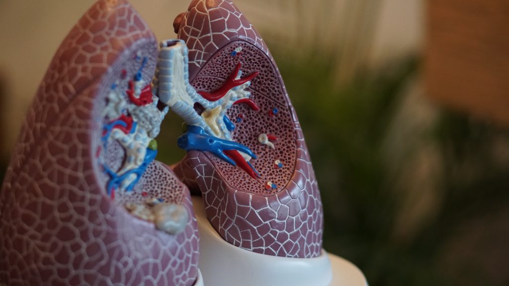
Photo by Robina Weermeijer on Unsplash

Storing donor lungs for transplant at 10°C markedly increases the length of time the organ can live outside the body, according to results of a trial were published in the New England​ Journal of Medicine Evidence. These findings will help reduce the strain on hospitals, reduce waitlists and possibly eliminate the need to bump other surgeries for a lung transplant.

The multicentre, non-randomised clinical trial study of 70 patients demonstrated that donor lungs remained healthy and viable for transplant up to four times longer compared to storage at the current standard of ice cooler preservation of around 4°C. The study was led by a team of scientists at the Toronto Lung Transplant Program in UHN’s Ajmera Transplant Centre.

“The clinical impact of this study is huge,” says lead author Dr Marcelo Cypel, Surgical Director of the Ajmera Transplant Centre and a surgeon within UHN’s Sprott Department of Surgery.

“It’s a paradigm shift for the practice of lung transplant.​ I have no doubt that this will become the gold standard practice of lung preservation for the foreseeable future.”

Lungs available for transplant are currently limited by the length of time a donor organ can be kept viable. Increasing storage time allows for viable donor lungs to come from greater distances, increasing the potential for greater numbers of lungs becoming available for transplant and overcoming many of the hurdles around transplant logistics.

“In transplant, we still see a critical shortage of organs and people dying on the waitlist because there are not enough lungs to be transplanted,” says Dr Cypel, who is also a professor in the Division of Thoracic Surgery, Department of Surgery at the University of Toronto.

“It’s a great accomplishment to see that our research is now having an impact, and that we can actually have more cases done at our centre, with continued outstanding clinical results.

“Better organ preservation also means better outcomes for patients.”

Transplant surgeries could become planned procedures

The trial took place over 18 months at UHN’s Toronto General Hospital, the Medical University of Vienna, and Hospital Universitario Puerta de Hierro-Majadahonda in Madrid.

“The ability to extend the lifespan of the donor organ poses several advantages,” says study first author Dr Aadil Ali, adjunct scientist at the Toronto General Hospital Research Institute.

“Ultimately, these advantages will allow for more lungs to be utilised across farther geographies and the ability to improve recipient outcomes by converting lung transplantation into a planned rather than urgent procedure.”

Some advantages of this new 10°C standard for lung storage include the potential to reduce or eliminate the 24/7 schedule and urgency of lung transplant procedures. By increasing the length of time donor lungs are viable, transplant surgeries could become planned procedures, which avoids bumping scheduled surgeries and overnight transplantation.

The study also suggests the new preservation temperature will allow more time to optimise immunologic matching between donor and recipients, and the possibility of performing lung transplantation in a semi-elective rather than urgent fashion.

For more on the study, watch Dr Marcelo Cypel’s presentation of findings at a recent American Association for Thoracic Surgery event.

Also, watch a video with Drs Cypel and Ali discussing the foundational work leading to this breakthrough.

Source: University Health Network

Modern Ventilators Shown to Overstretch Lung Tissue

Source: Pixabay CC0

In pulmonary medicine, it has long been debated as to whether ventilator overstretches lung tissue, and now new research published in the American Journal of Respiratory and Critical Care Medicine has proven that they do in fact cause overstretching.

The University of California Riverside researchers showed that there were major differences between natural breathing versus the forced breathing from ventilators. These results are critical, particularly in context of the COVID pandemic and the rush to build ventilators.

“Using novel techniques, we observed that ventilators can overextend certain regions of the lungs,” said Mona Eskandari, assistant professor of mechanical engineering, who led the research. These results may explain why lung health declines for patients the longer they spend on the machines, especially in the case of disease.

Eskandari’s bMECH lab pioneered a technique to study lungs as they are made to breathe. On a custom-built ventilator designed in their lab, the researchers imitated both natural and artificial breathing. Then, they observed isolated lungs involved in both types of breathing using multiple cameras collecting fast, high-resolution images, a method called digital image correlation.

“Our setup allows us to imitate both physiological and artificial breathing on the same lung with the switch of a button,” Eskandari said. “The unique combination of our ventilator with digital image correlation gives us unprecedented insights into the way specific regions of the lungs work in concert with the whole.”

Using their innovative method to interface these two systems, UCR researchers collected evidence demonstrating that natural breathing stretches certain parts of the lung as little as 25% while those same regions stretch to as much as 60% when on a ventilator.

Scholars traditionally model the lungs like balloons, or what they refer to as thin-walled pressure vessels, where pushing air in and pulling air out are understood to be mechanically equivalent.

To explain what they observed in this study, the researchers propose moving away from thin-walled pressure vessel models and instead towards thick-walled models. Unlike thin-walled pressure vessels theory, a thick-walled model accounts for the differing levels of stress in airways resulting from ventilators pushing air in versus natural breathing, which pulls air in. This helps to explain how airways are more engaged and air is more evenly distributed in the lung during physiological breathing.

Iron lungs, the gigantic ventilators used during the late 1940s polio outbreak, acted more like a human chest cavity, expanding the lung as it naturally would. This creates a vacuum effect that pulls air into the lungs. Though this action is gentler for the lungs, these bulky systems prevented easy access to monitoring other organs in hospital care.

By contrast, modern ventilators are more portable and easier for caretakers to work with. However, they push air into the lungs that is not evenly distributed, overstretching some parts and causing a decline in lung health over time.

While it is unlikely that hospitals will return to the iron lung models, it is possible that modern machines can be altered to reduce injury.

“Now that we know about excessive strain when air is delivered to the lungs, the question for us becomes about how we can improve ventilation strategies by emulating natural breathing,” Eskandari said.

Source: University of California – Riverside

Link Found Between Telomeres and COVID Lung Damage

Researchers developing a therapy to regenerate lung tissue damaged by severe COVID have postulated that shortened telomeres are associated with the damage.

Telomeres are structures at the ends of chromosomes that maintain their integrity, and a small portion of them are lost with each cell division, such as when regenerating damaged tissue. As the telomere sections shorten, they eventually become unable to divide and are senescent. The team was already working on a way to regenerate lung tissue in pulmonary fibrosis, and adapted their research to the COVID pandemic. In pulmonary fibrosis, lung tissue becomes scarred and rigid, resulting in reduced lung capacity. In previous research, they had shown that telomere damage to the alveolar type II pneumocytes – which happen to be the same cells targeted by SARS-CoV-2.

Maria A Blasco, a researcher at CNIO said, “When I read that type II alveolar pneumocytes were involved in COVID-19, I immediately thought that telomeres might be involved.” The researchers believe short telomeres hinder tissue regeneration after severe COVID.

Blasco explained, “we know that the virus infects alveolar type II pneumocytes and that these cells are involved in lung regeneration; we also know that if they have telomeric damage they cannot regenerate, which induces fibrosis. This is what is seen in patients with lung lesions after COVID-19: we think they develop pulmonary fibrosis because they have shorter telomeres, which limits the regenerative capacity of their lungs.”

To support this, the team analysed the telomeres of 89 COVID patients. Although it might be expected that older patients had shorter telomeres, the researchers found that all of those with severe COVID had shorter telomeres – regardless of age.

The researchers wrote: “These findings demonstrate that molecular hallmarks of aging, such as the presence of short telomeres, can influence the severity of COVID-19 pathologies.”The involvement of shorter telomeres opens up the possibility of using telomerase to lengthen them again, as a potential treatment.The team will now move to an experimental mouse model, infecting mice with short telomeres and no telomerase with COVID, giving telomerase to some to see if the lung tissue can regenerate after severe COVID.

Source:News-Medical.Net

Journal information: Sanchez-Vazquez R, Guío-Carrión A, Zapatero-Gaviria A, Martínez P, Blasco M. Shorter telomere lengths in patients with severe COVID-19 disease. Aging (Albany NY). 2021. doi:10.18632/aging.202463

Gut Microbiome is Linked to Pulmonary Disease

A link has been shown between the gut microbiome and chronic obstructive pulmonary disease (COPD), a lung disease with an often poor prognosis.

Senior author Prof Phil Hansbro, Director of the Centenary University of Technology Sydney Centre for Inflammation, said, “It’s already known that the lung microbiome is a contributing factor in COPD. We wanted to see if the gut environment was also somehow involved–to determine whether the gut could act as a reliable indicator of COPD or if it was connected in some way to the development of the disease.”

Stool samples of COPD patients showed elevated levels of the bacteria Streptococcus and Lachnospiraceae. Additionally a unique metabolite signature was identified in individuals with COPD, created by the chemical by-products of the metabolic process.

First author Dr Kate Bowerman from the University of Queensland said, “Our research indicates that the gut of COPD patients is notably different from healthy individuals. This suggests that stool sampling and analysis could be used to non-invasively diagnose and monitor for COPD,” she said.
“The ‘gut-lung axis’ describes the common immune system of the lung and gastrointestinal tract. This means that activity in the gut can impact activity in the lung. Our COPD findings suggest that the gut microbiome should now also be considered when looking for new therapeutic targets to help treat lung disease,” Prof Hansbro said.

Source: Medical Xpress

Vaping May Increase Respiratory Disease Risk

Research has increasingly been focused on the health effects of vaping, usually touted as a way to quit combustible smoking, and evidence is accumulating for their risks.

Research by Boston University School of Public Health and School of Medicine has revealed that, far from being harmless, vaping may in fact raise the risk for a number of respiratory diseases by 21% for former e-cigarette users and 43% for current users.

Corresponding author Dr Andrew Stokes, assistant professor of global health at Boston University said, “This provides some of the very first longitudinal evidence on the harms associated with e-cigarette products.

“In recent years we have seen dramatic increase in e-cigarette use among youth and young adults which threatens to reverse decades of hard-fought gains,” Stokes said. “This new evidence also suggests that we may see an increase in respiratory disease as youth and young adults age into midlife, including asthma, COPD, and other respiratory conditions.”

Previous research has largely focused on in vitro studies, or short-term studies with human participants. The study used data from 21 618 participants from the nationally representative Population Assessment of Tobacco and Health (PATH) survey. To isolate the effect of e-cigarettes, the researchers adjusted for any form of combustible smoking use, including second-hand smoking.

“With a longitudinal study design and extensive sensitivity analyses, the study adds to a growing body of evidence indicating long-term health risks of e-cigarette use to the respiratory system,” said study lead author Wubin Xie, a postdoctoral associate at Boston University.

Source: Medical Xpress