Tag: liver

TB Alters Liver Metabolism and could Promote Diabetes, Study Shows

Tuberculosis bacteria. Credit: CDC

Scientists from the University of Leicester have discovered that tuberculosis disrupts glucose metabolism in the body. The findings, which have now been published in PLoSPathogens complement the understanding that diabetes worsens the symptoms of tuberculosis. Importantly, they now say, undiagnosed tuberculosis could be pushing vulnerable patients towards metabolic disease such as diabetes.

Tuberculosis (TB) remains one of the most devastating infectious diseases worldwide, killing over 4,000 people every day. Prevention through the development of improved vaccines remains a priority for the World Health Organisation. Currently only one vaccine exists for TB and this is predominantly given to infants and young children to help protect them from severe forms of infection. 

Scientists at the University are researching tuberculosis in the hope of creating improved vaccines and are specifically looking at ways in which undiagnosed and subclinical infection can impact health. This new discovery, they say, could pave the way to define the molecular pathways by which the immune response changes liver metabolism, thereby allowing for the creation of targeted interventions. 

Professor Andrea Cooper from the University’s Leicester Tuberculosis Research Group (LTBRG), is among the authors on the paper.

She said: “Our paper changes the focus from diabetes making TB worse to the possibility that late diagnosis of TB can contribute to disruption of glucose metabolism, insulin resistance and therefore can promote progress towards diabetes in those that are susceptible. 

“As diabetes compromises drug treatment, our paper also supports the idea that metabolic screening should be involved in any drug or vaccine trials.”

The study first used laboratory models of pulmonary TB to examine the changes happening within the liver during the early stages of infection. It found that an immune response was triggered within the liver cells and glucose metabolism was altered. 

First author Dr Mrinal Das then reanalysed published metabolic data from humans, where he found that liver glucose metabolism was also disrupted when people progressed to TB from latent infection.

Professor Cooper added: “Our future aim is to define the molecular pathways by which the immune response is changing liver metabolism, allowing us to potentially create targeted interventions.

 “We will also be investigating how latent TB (which is infection with the bacterial agent of TB without significant symptoms) might be impacting metabolic health in humans.” 

Source: University of Leicester

Microplastics Rapidly Bioaccumulate Everywhere in the Body

Photo by FLY:D on Unsplash

The prevalence of microplastics in the environment is well known, along with their harm to marine organisms, but few studies have examined the potential health impacts on mammals. Now, a new study published in the International Journal of Molecular Sciences has found that in mice, the infiltration of microplastics was as widespread in the body as it is in the environment, leading to behavioural changes, especially in older test subjects.

Study leader University of Rhode Island Professor Jaime Ross and her team focused on neurobehavioural effects and inflammatory response to exposure to microplastics, as well as the accumulation of microplastics in tissues, including the brain.

“Current research suggests that these microplastics are transported throughout the environment and can accumulate in human tissues; however, research on the health effects of microplastics, especially in mammals, is still very limited,” said Ross, an assistant professor of biomedical and pharmaceutical sciences at the Ryan Institute for Neuroscience and the College of Pharmacy. “This has led our group to explore the biological and cognitive consequences of exposure to microplastics.”

Behavioural changes detected

Ross’ team exposed young and old mice to varying levels of microplastics in drinking water over the course of three weeks. They found that microplastic exposure induces both behavioural changes and alterations in immune markers in liver and brain tissues. The study mice began to exhibit behaviours akin to dementia in humans. The results were even more profound in older animals.

“To us, this was striking. These were not high doses of microplastics, but in only a short period of time, we saw these changes,” Ross said. “Nobody really understands the life cycle of these microplastics in the body, so part of what we want to address is the question of what happens as you get older. Are you more susceptible to systemic inflammation from these microplastics as you age? Can your body get rid of them as easily? Do your cells respond differently to these toxins?”

To understand the physiological systems that may be contributing to these changes in behaviour, Ross’ team investigated how widespread the microplastic exposure was in the body, dissecting several major tissues including the brain, liver, kidney, gastrointestinal tract, heart, spleen and lungs. The researchers found that the particles had begun to bioaccumulate in every organ, including the brain, as well as in bodily waste.

“Given that in this study the microplastics were delivered orally via drinking water, detection in tissues such as the gastrointestinal tract, which is a major part of the digestive system, or in the liver and kidneys was always probable,” Ross said. “The detection of microplastics in tissues such as the heart and lungs, however, suggests that the microplastics are going beyond the digestive system and likely undergoing systemic circulation. The brain blood barrier is supposed to be very difficult to permeate. It is a protective mechanism against viruses and bacteria, yet these particles were able to get in there. It was actually deep in the brain tissue.”

Possible mechanism

That brain infiltration also may cause a decrease in glial fibrillary acidic protein (called “GFAP”), a protein that supports many cell processes in the brain, results have shown. “A decrease in GFAP has been associated with early stages of some neurodegenerative diseases, including mouse models of Alzheimer’s disease, as well as depression,” Ross said. “We were very surprised to see that the microplastics could induce altered GFAP signalling.”

She intends to investigate this finding further in future work. “We want to understand how plastics may change the ability for the brain to maintain its homeostasis or how exposure may lead to neurological disorders and diseases, such as Alzheimer’s disease,” she said.

Source: University of Rhode Island

Women’s Lean Body Mass and Age Speed up Blood Alcohol Elimination

Photo from Pixabay CC0

The rate at which women eliminate alcohol from their bloodstream is largely predicted by their lean body mass, although age plays a role, too, scientists found in a new study published in the journal Alcohol Clinical and Experimental Research. Since women with obesity also have more lean body mass, older women with obesity clear alcohol from their systems 52% faster than younger women of healthy weights, the study found.

“We believe the strong relationship we found between participants’ lean body mass and their alcohol elimination rate is due to the association that exists between lean body mass and lean liver tissue – the part of the liver responsible for metabolising alcohol,” said research group leader M. Yanina Pepino, a professor of food science and human nutrition at the University of Illinois Urbana-Champaign.

To explore links between body composition and alcohol elimination rates, the team conducted a secondary analysis of data from a study performed at and another at Indiana University, Indianapolis. Both projects used similar methods to estimate the rate at which alcohol is broken down in the body.

The combined sample from the studies used in the analysis included 143 women who ranged in age from 21 to 64 and represented a wide range of body mass indices – from healthy weights to severe obesity. Among these were 19 women who had undergone different types of bariatric surgery. Lean body mass is total body weight minus fat.

In a subsample of 102 of these women, the researchers had measured the proportions of lean and fat tissue in their bodies and calculated their body mass indices. Based on their BMI, those in the subsample were divided into three groups: normal weight (BMI of 18.5–24.9), overweight BMI (25–29.9) and obese (BMI 30+).

As the researchers expected, women with higher BMI had not only more fat mass than women of healthy weights, they also had more lean mass. On average, the group with obesity had 52.3 kg of lean mass, compared with 47.5 kg for the normal weight group.

The two studies both used an alcohol clamp technique, where participants received an intravenous infusion of alcohol at a rate controlled by a computer-assisted system. The system calculated personalised infusion rates based upon each participant’s age, height, weight and gender and was programmed so they would reach a target blood alcohol concentration of .06% within 15 minutes and maintain that level for about two hours

Using a breathalyser, breath samples were collected at regular intervals throughout the experiments to estimate participants’ blood alcohol concentration and provide feedback to the system.

“We found that having a higher fat-free body mass was associated with a faster alcohol elimination rate, particularly in women in the oldest subgroups,” said Neda Seyedsadjadi, a postdoctoral fellow at the university and the first author of the study.

“The average alcohol elimination rates were 6 grams per hour for the healthy weight group, 7 grams for the overweight group, and 9 grams for the group with obesity,” she said. “To put this in perspective, one standard drink is 14 grams of pure alcohol, which is found in 12 ounces of beer, 5 ounces of table wine or 1.5 ounces shot of distilled spirits.”

The interaction between participants’ age and lean body mass accounted for 72% of the variance in the time required to eliminate the alcohol from their system, the team found.

Pepino, who also holds an appointment as a health innovation professor at Carle Illinois College of Medicine, has conducted several studies on alcohol response in bariatric surgery patients.

The findings also shed light on alcohol metabolism and body composition in women who have undergone weight loss surgery. Researchers have long known that bariatric surgery alters women’s response to alcohol but were uncertain if it affected how quickly they cleared alcohol from their systems.

Some prior studies found that these patients metabolised alcohol more slowly after they had weight loss surgery. The new study’s findings indicate that these participants’ slower alcohol elimination rates can be explained by surgery-induced reductions in their lean body mass. Weight loss surgery itself had no independent effects on patients’ alcohol elimination rates, the team found.

Source: University of Illinois at Urbana-Champaign

A Hormone Injection Sobers Up Drunk Mice

Mouse
Photo by Kanasi on Unsplash

Researchers have found that a simple injection of hormone called fibroblast growth factor 21 (FGF21) protects mice against ethanol-induced loss of balance and righting reflex, effectively sobering them up.

“We’ve discovered that the liver is not only involved in metabolising alcohol but that it also sends a hormonal signal to the brain to protect against the harmful effects of intoxication, including both loss of consciousness and coordination,” says co-senior study author Steven Kliewer of the University of Texas Southwestern Medical Center, regarding the study results published in the journal Cell Metabolism.

“We’ve further shown that by increasing FGF21 concentrations even higher by injection, we can dramatically accelerate recovery from intoxication. FGF21 does this by activating a very specific part of the brain that controls alertness,” says Kliewer.

The consumption of ethanol produced by the natural fermentation of simple sugars in ripening fruits and nectars can cause intoxication, impairing mobility and judgement. Animals that consume fructose and other simple sugars have evolved liver enzymes to break down ethanol.

FGF21 is a hormone that is induced in the liver by a variety of metabolic stresses, including starvation, protein deficiency, simple sugars, and ethanol. In humans, ethanol is by far the most potent inducer of FGF21 described to date. Previous studies showed that FGF21 suppresses ethanol preference, induces water drinking to prevent dehydration, and protects against alcohol-induced liver injury.

In the new study, Kliewer and co-senior study author David Mangelsdorf of the University of Texas Southwestern Medical Center show that FGF21 plays a broader role in defending against the harmful consequences of ethanol exposure than previously thought. In mice, FGF21 stimulated arousal from intoxication without changing the breakdown of ethanol. Mice lacking FGF21 took longer than their littermates to recover their righting reflex and balance following ethanol exposure. Conversely, pharmacologic FGF21 administration reduced the time needed for mice to recover from ethanol-induced unconsciousness and lack of muscle coordination.

Surprisingly, FGF21 did not counteract sedation caused by ketamine, diazepam, or pentobarbital, indicating specificity for ethanol. FGF21 mediated its anti-intoxicant effects by directly activating noradrenergic neurons in the locus coeruleus region in the brain, which regulates arousal and alertness. Taken together, the results suggest that the FGF21 liver-brain pathway evolved to protect against ethanol-induced intoxication. According to the authors, this pathway may modulate a variety of cognitive and emotional functions to enhance survival under stressful conditions.

Whether activation of the noradrenergic system contributes to FGF21’s other effects is yet to be determined. Although both FGF21 and noradrenergic nervous system activity are induced by ethanol in humans, additional studies will also be required to determine whether FGF21’s anti-intoxicant activity translates to humans.

“Our studies reveal that the brain is the major site of action for FGF21’s effects,” Mangelsdorf says. “We are now exploring in greater depth the neuronal pathways by which FGF21 exerts its sobering effect.”

Source: Cell Press

Excellent Outcomes for Shrinking Liver Cancer Tumours Before Transplant

Photo by Piron Guillaume on Unsplash

Shrinking liver cancer tumours in order to allow the patient to qualify for a liver transplant leads to excellent 10-year post-transplant outcomes, according to the findings of a new study published in JAMA Surgery. The results validate current US policies around transplant eligibility.

Selection of patients with hepatocellular carcinoma (HCC), the most common form of liver cancer, for transplant has been guided for more than two decades by standards known as the Milan criteria. The Milan criteria state that transplantation should be performed in those with a single tumour of 5cm or less in diameter or three tumours that are each 3cm or less in diameter, have no macrovascular invasion, and no metastasis. Over time, the rising incidence of HCC and mortality rates in the United States have led to refinements to the selection policy, shifting the focus to guidelines that also incorporate tumour biology, response to bridging therapies, and waiting times for patients within and beyond the Milan criteria.

One aspect of the current criteria is known as downstaging: the process of applying liver directed therapy to tumours too big for the Milan criteria with the hope of reducing them to the suggested size. Downstaging is now an option in selecting suitable liver transplant candidates with initial tumors that exceed the criteria. However, liver cancer can recur after transplantation, either within or outside the liver. The treatment options of patients who have recurrence post transplantation is limited and prognosis is poor.

In this cohort study, a retrospective multicentre analysis of prospectively collected data was conducted for 2645 adults who had undergone liver transplant for HCC at five centres between January 2001 and December 2015. The outcomes of 341 patients whose disease was downstaged to fit within the Milan criteria were compared with those in 2122 patients whose disease always fit within the criteria and 182 patients whose disease was not downstaged.

The 10-year post-transplant survival and recurrence rates were, respectively, 52.1% and 20.6% among those whose disease was downstaged; 61.5% and 13.3% in those always within the criteria; and 43.3% and 41.1% in those whose disease was not downstaged.

“Our study validates national policy on downstaging prior to transplantation and shows the clear utility benefit for transplantation prioritisation decision-making,” said Parissa Tabrizian, MD, co-lead author on the study. “These results can increase the level of recommendations for the downstaging policy on a global basis. It also demonstrates that surgical management of HCC recurrence after transplantation is associated with improved survival in well-selected patients and should be pursued. The study also supports expanding the policy of downstaging applied to guidelines in Europe and Asia.”

“Our study represents a solid confirmation that HCC patients effectively downstaged to Milan criteria have an outstanding median survival of 10 years, thus providing the rationale to adopt this policy on a global basis,” said Josep Llovet, MD, PhD, co-lead author on the study. “With this study clinical practice guidelines of management of HCC can recommend our approach with an acceptable level of evidence.”

Source: The Mount Sinai Hospital

In Liver Cancer, Protective p53 Protein Turns Traitor

Genetics
Image source: Pixabay

p53 is one of the most important proteins in cancer biology. Often referred to as a “guardian of the genome”, p53 activates in response to cellular stressors such as DNA damage. Its activation induces processes such as cell death that prevent cancer development. Because of this, p53 mutations are extremely common in cancers, including hepatocellular carcinoma.

However, a study published in Cancer Research, revealed that the constant activation of p53 in liver cells of patients suffering from chronic liver disease (CLD) could actually promote the development of liver cancer.

CLD can be brought on by different factors including viruses, alcohol use, and fat accumulation, all of which can induce p53 activation. Previous studies have shown that p53 is in a constant state of activation in the liver cells of CLD patients. Yet, it is not clear what role this plays in CLD pathophysiology.

“Clinical data clearly show that p53 is activated in the hepatocytes of individuals with CLD,” says Yuki Makino, lead author of the study at Osaka University. “Because p53 is such a vital part of how the human body prevents tumor formation, its role in CLD became even more intriguing.”

To create a mouse model with p53 accumulation in hepatocytes, researchers deleted Mdm2, the protein responsible for regulating p53 expression by targeting it for degradation. These mice developed liver inflammation with higher amounts of hepatocyte apoptosis and senescence-associated secretory phenotype (SASP), a phenomenon where cells produce signals within the microenvironment that can cause nearby cells to become cancerous. In fact, mice with p53 accumulation did have increased liver tumour development.

“We also observed an expanded population of hepatic progenitor cells (HPCs), which have stem cell-like characteristics,” explained senior author Tetsuo Takehara. “When the HPCs were isolated, grown in culture, and then injected under the skin of lab mice, these animals developed tumors. This suggested that HPCs played a key part in the liver tumor formation seen in the animals with p53 accumulation.”

Interestingly, acceleration of liver tumour development and the other observed phenotypes did not occur when p53 was deleted in addition to Mdm2 in the hepatocytes. These results demonstrated the significance of constant p53 activity in the tumorigenesis.

“We then compared samples from 182 CLD patients with 23 healthy liver samples,” said Dr Makino. “The CLD liver biopsy samples showed activated p53 was positively correlated with apoptosis levels, SASP, HPC-associated gene expression, and later cancer development.”

The authors concluded that constitutively activated p53 in hepatocytes of CLD patients can create a microenvironment that is supportive of tumour formation from HPCs. Their work proposes a novel and paradoxical mechanism of liver tumorigenesis because p53 is one of the most well-known tumor suppressor genes. These data could highlight p53 as a potential cancer-prevention treatment target for CLD patients.

Source: Osaka University

Living Donors Liver Transplant a Viable Option in Colorectal Cancer

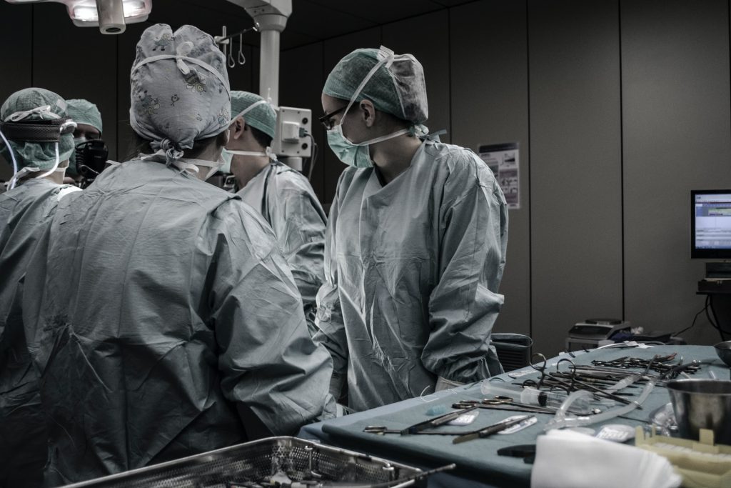
Doctors and nurses performing a surgery
Photo by Piron Guillaume on Unsplash

A recent study published in JAMA Surgery has demonstrated the viability of living-donor liver transplant for patients who have systemically controlled colorectal cancer and liver tumours that cannot be surgically removed.

“This study proves that transplant is an effective treatment to improve quality of life and survival for patients with colorectal cancer that metastasised to the liver,” said senior study author Dr Gonzalo Sapisochin.

The study focused on colorectal cancer partly for its tendency to spread to the liver. Nearly half of all patients with colorectal cancer develop liver metastases within a few years of diagnosis and 70% of liver tumours in these patients cannot be removed without removing the entire liver.

Unfortunately, most of these patients cannot get deceased-donor liver transplants because their liver function is fairly normal in spite of their tumours. This puts them near the bottom of the national organ transplant waiting list.

Thanks to recent advances in cancer treatments, many of these patients are able to get their cancer under systemic control, which means only their liver tumours prevent them getting a ‘cancer free’ label. It also increases the odds that these patients – and their new livers – will remain cancer free, which is crucial when balancing the benefit to the patient with the risk to a living donor.

“I’ve seen so many cancer patients, whose cancers were not spreading, but we couldn’t remove the tumours from their livers and we knew they would die,” said first study author Dr Roberto Hernandez-Alejandro. “We hoped living-donor liver transplant could give them another chance.”

Because it offered a last resort, the study attracted patients from near and far. All patients and donors went through a rigorous screening process to ensure they were good candidates for the procedure, and they were educated about the risks of the surgery and the possibility of cancer recurrence.

Patients and donors underwent staggered surgeries to fully remove patients’ diseased livers and replace them with half of their donors’ livers. Over time, both patients’ and donors’ livers regenerated and regain normal function.

Patient imaging and blood analysis was closely monitored for any signs of cancer recurrence and will continue to be followed for up to five years after their surgery. At the time of study publication, two patients had follow-up of two or more years and both remained alive and well, cancer-free.

“We have seen very good outcomes with this protocol, with 100 percent survival and 62 percent of patients remaining cancer free one year and a half after surgery,” said study author Dr Mark Cattral. “It is very strong data to support that we can offer this treatment safely and make appropriate use of scarce life-saving organs.”

Source: University Health Network

Muscles are Timekeepers for the Liver

Photo by RF._.studio from Pexels

Researchers have found that skeletal muscles play a large part in regulating the liver’s biological clock. The findings were published in Science Advances.

The circadian rhythm is coordinated by the brain at a general level, but each organ or tissue is also subjected to specific regulation, adjusting to time to optimise their processes. However it was not known how the liver “knows” whether it is day or night.

The liver’s main role is digestion, mainly of fats and sugars: the brain is the main consumer of sugar while skeletal muscle is the main consumer of fat.

Scientists at IRB Barcelona discovered a surprising relationship: that it is skeletal muscle which regulates liver function and determines fat metabolism. Skeletal muscle accomplishes this by secreting a that is transported to the liver through serum is responsible for modulating around 35% of the metabolic functions of the liver. The remaining basal functions of this organ and others related to carbohydrate metabolism are independent of muscle activity and are regulated by the basal circadian rhythm from the brain.

“It’s a very nice discovery because it is the first demonstration of the need for communication between the circadian clocks of tissues and organs outside the brain, and we can see that this communication between muscle and liver is altered by aging,” said study leader Dr Salvador Aznar-Benitah at IRB Barcelona. “When we get older, cells stop obeying the biological clock and begin to perform functions in a non-optimal manner, leading to errors that cause tissues to age.”

The researcher’s results show that the liver does not independently regulate the metabolism of fats and that it is muscle that sends the message that it is time to switch on fatty acid metabolism and how it should go about this. “We didn’t expect to find this connection between the liver and muscle because it wasn’t known previously, but, on second thought, it makes complete sense that fat management is coordinated by one of its main consumers,” said Dr Aznar-Benitah. Carbohydrate metabolism meanwhile is dependent on the basal coordination exercised by the brain.

Source: Institute for Research in Biomedicine (IRB Barcelona)

Immunotoxin Treatment Stops Liver Fibrosis

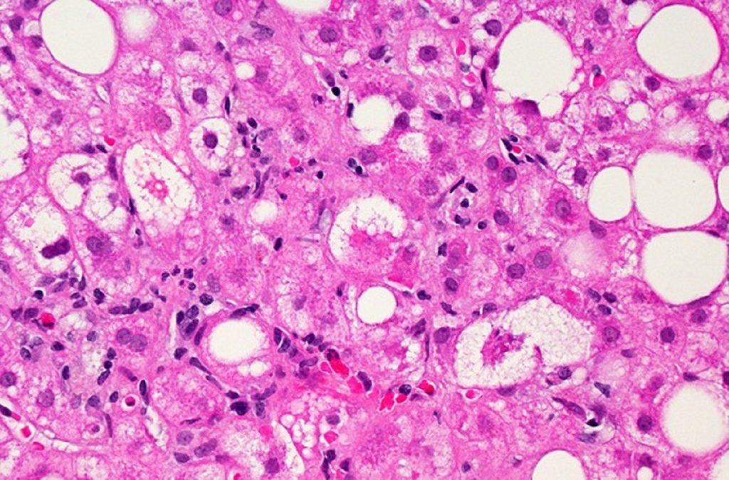
A microscopic image of liver tissue affected by non-alcoholic fatty liver disease (NAFLD). The large and small white spots are excess fat droplets filling liver cells (hepatocytes). Credit: Dr. David Kleiner, National Cancer Institute/NIH
A microscopic image of liver tissue affected by non-alcoholic fatty liver disease (NAFLD). The large and small white spots are excess fat droplets filling liver cells (hepatocytes). Credit: Dr David Kleiner, National Cancer Institute/NIH

A new study successfully used immunotoxins to prevent the progression of liver fibrosis by targeting a protein specific to that disease.

Fibrosis, the buildup of collagen and scar tissue, can be caused by alcohol abuse and disease. University of California San Diego School of Medicine researchers and their collaborators are looking for ways to treat fibrosis by preventing liver cells from producing collagen. 

“So we thought…what if we take immunotoxins and try to get them to kill collagen-producing cells in the liver,” explained team lead Tatiana Kisseleva, MD, PhD, associate professor of surgery at UC San Diego School of Medicine. “If these antibodies carrying toxic molecules can find and bind the cells, the cells will eat up the ‘gift’ and die.”

The study focussed on immunotoxins designed to bind a protein called mesothelin, which is rarely found in the healthy human body. The protein is only produced by cancer cells and collagen-producing liver cells, known as portal fibroblasts.

Kisseleva teamed up with co-author Ira Pastan, MD, at the National Cancer Institute, part of the National Institutes of Health (NIH). Dr Pastan is co-discoverer of mesothelin and an expert on using immunotoxins to target the protein on cancer cells, and he leads several clinical trials using it in treating patients with ovarian cancer, mesothelioma and pancreatic cancer.

Since the immunotoxins specifically recognise human mesothelin, the researchers couldn’t use a traditional mouse model of liver fibrosis. So, they transplanted human liver cells isolated from patients to mice and treated them with the anti-mesothelin immunotoxin. Compared to untreated mice, 60 to 100 percent of human mesothelin-producing cells were killed by the immunotoxins, which also reduced the deposition of collagen.

Liver fibrosis treatment is very limited at present, with weight loss being currently the only known method for reducing liver fibrosis associated with non-alcoholic fatty liver disease. Alcoholic liver disease is most commonly treated with corticosteroids, but they are not highly effective. Early liver transplantation is the only proven cure, but it is rarely available.

“What we want to know now is, can this same strategy be applied to other organs?” Dr Kisseleva said. “Surprisingly enough, the same cells are responsible for fibrosis in the lung and kidneys. This is especially exciting because we already know from Dr Pasten’s cancer clinical trials that anti-mesothelin immunotoxins are safe in humans, potentially speeding up their application in other areas.”

The findings were published in Proceedings of the National Academy of Sciences

Source: University of California San Diego

Journal information:Nishio, T., et al. (2021) Immunotherapy-based targeting of MSLN+ activated portal fibroblasts is a strategy for treatment of cholestatic liver fibrosis. PNAS. doi.org/10.1073/pnas.2101270118

Macrophage Role in Liver Regeneration Identified

A macrophage engulfing a yeast cell. Source: CDC

Researchers have found out what role macrophages play in liver regeneration after resection. The results are published in the journal Biomedicine & Pharmacotherapy.

In mammals, the liver is the most regenerative internal organ. It can restore its original size with as little as 25% of the original tissue remaining. Macrophages play an important role in this process. It is known, for example, that if the liver is affected by foreign substances, including drugs, macrophages migrate to the liver, absorb harmful microorganisms and dead cells, cause inflammation and thus contribute to the restoration of the organ. However, it is still unknown unambiguously how macrophages affect the growth of the liver after its resection, ie when a large part of the organ is removed. RUDN University doctors investigated this issue in an experiment with laboratory mice.

“The role of macrophages in the liver growth after massive resections is uncertain. Some studies reveal the lack of immigration of macrophages to the liver during its recovery from partial resection, whereas other studies demonstrate such possibility. So, we focused our study on the macrophage population dynamics after 70% liver resection in mouse mode”, Andrey Elchaninov, MD, PhD, researcher at the Department of Histology, Cytology and Embryology of RUDN University.

The researchers removed 70% of the liver of a number of lab mice. Immediately after that, then a day later, three days later, and a week later, the scientists took liver samples for analysis. The resulting cells were studied using an immunohistochemical method. The sections were labelled with specific antibodies to the glycoproteins CD68, CD206 and other compounds that are found on the surface of macrophages. To count them, the antibodies are labelled with fluorescent dyes and glow when attached to macrophages. The researchers also measured the rate of reproduction and cell death of macrophages.

The researchers found that after resection, macrophages migrate to the liver in large numbers. A day after surgery, the number of macrophages with CD68 in the liver doubles, which persists after a week. It also turned out that the resection led to significant changes in the ratio of different types of macrophages. For example, the proportion of Ly6C cells in the week after surgery increased 4-fold — from 5% to 22%, and the proportion of CD86 droppedfrom 50% to 15%. The role of macrophages is ambiguous. On the one hand, they release chemicals (chemoattractors) that attract white blood cells responsible for the body’s inflammatory response, but on the other hand, they regulate the reproduction of liver cells and the metabolism in the organ.

“Corresponding profiles of macrophages in the regeneration of the liver cannot be unambiguously defined as pro- or anti-inflammatory,” the researchers said. “Their typical features include elevated expression of leukocyte chemoattractant factors, and many of the differentially expressed sequences are related to the control of cell growth and metabolic processes in the liver. Our findings revealed essential roles of macrophages and macrophages proliferation in the mouse liver during its recovery from a massive resection.”

Source: EurekAlert!

Journal reference: Elchaninov, A., et al. (2021) Macro- and microtranscriptomic evidence of the monocyte recruitment to regenerating liver after partial hepatectomy in mouse model. Biomedicine & Pharmacotherapy. doi.org/10.1016/j.biopha.2021.111516.