Tag: lab testing

Blood Test for Alzheimer’s Proves Highly Accurate
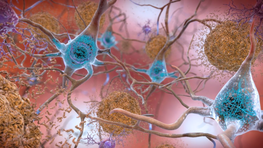
Plaques and neurons. Source: NIAH

A study in the journal Neurology has shown that a less expensive blood test to detect Alzheimer’s is highly accurate at early detection, providing further evidence that the test should be considered for routine screening and diagnosis. 

“Our study shows that the blood test provides a robust measure for detecting amyloid plaques associated with Alzheimer’s disease, even among patients not yet experiencing cognitive declines,” said senior author Professor Randall J. Bateman, MD.

“A blood test for Alzheimer’s provides a huge boost for Alzheimer’s research and diagnosis, drastically cutting the time and cost of identifying patients for clinical trials and spurring the development of new treatment options,” Prof Bateman said. “As new drugs become available, a blood test could determine who might benefit from treatment, including those at very early stages of the disease.”

Developed by Prof Bateman and colleagues, the blood test assesses whether amyloid plaques have begun accumulating in the brain based on the ratio of the levels of the amyloid beta proteins Aβ42 and Aβ40 in the blood.

The gold standard PET scan evaluation requires a radioactive brain scan, at an average cost of $5000–$8000 (R75 000–R120 000) per scan. Another common test, which analyses levels of amyloid-beta and tau protein in cerebrospinal fluid, costs about $1000 (R15 000) but requires a spinal tap process.

This study estimates that prescreening with a $500 (R7500) blood test could halve both the cost and the time it takes to enrol patients in clinical trials that use PET scans. Using only blood testing for screening could be done in under six months, a tenth or less of the cost. The test is currently only available in the US and Europe.

The current study shows that the blood test remains highly accurate, even when performed in different labs following different protocols, and in different cohorts across three continents.

Scientists didn’t know if small differences in sampling methods (such as anticoagulant use) could have a big impact on test accuracy because results are based on subtle shifts in amyloid beta protein levels in the blood. Subtle interfernece in these amyloid protein ratios could have triggered a false negative or positive result.

To confirm the test’s accuracy, researchers tested blood samples from current Alzheimer’s studies in the United States, Australia and Sweden, each of which uses different protocols for the processing of blood samples and related brain imaging.

Findings from this study confirmed that the Aβ42/Aβ40 blood test using a high-precision immunoprecipitation mass spectrometry technique developed at Washington University provides highly accurate and consistent results for both cognitively impaired and unimpaired individuals across all three studies.

When blood amyloid levels were combined with another major Alzheimer’s risk factor – the presence of the genetic variant APOE4 – the blood test accuracy was 88% compared to brain imaging and 93% when compared to spinal tap.

“These results suggest the test can be useful in identifying nonimpaired patients who may be at risk for future dementia, offering them the opportunity to get enrolled in clinical trials when early intervention has the potential to do the most good,” Prof Bateman said. “A negative test result also could help doctors rule out Alzheimer’s in patients whose impairments may be related to some other health issue, disease or medication.”

Source: Washington University School of Medicine

In Vitro Cancer Cells Differ to Those in Body

Photo by Drew Hays on Unsplash

A new study has shown that most cancer cells grown in vitro have little in common genetically with cancer cells in humans.

Human cancer cells grown in culture dishes have the least genetic similarity to their human sources, according to a new computer-based technique developed by researchers at John Hopkins.

According to the researchers, the finding should help shift more resources to cancer research models such as genetically engineered mice and balls of human tissue known as ‘tumouroids’ to better evaluate human cancer biology and treatments, and the genetic errors responsible for cancer growth and progress.

“It may not be a surprise to scientists that cancer cell lines are genetically inferior to other models, but we were surprised that genetically engineered mice and tumouroids performed so very well by comparison,” says Patrick Cahan, PhD, associate professor of biomedical engineering at The Johns Hopkins University and the Johns Hopkins University School of Medicine and lead investigator of the new study.

The new computer modelling technique, CancerCellNet, compares the RNA sequences of a research model with data from a cancer genome atlas to see how closely the two sets match up.

On average, genetically engineered mice and tumouroids have RNA sequences most closely aligned with the genome atlas baseline data in 4 out of every 5 tumour types they tested, including breast, lung and ovarian cancers.

This adds to evidence that cancer cell lines grown in the laboratory have less parity with their human source due to the many differences between a human cell’s natural environment and a laboratory growth environment, the researchers said. “Once you take tumours out of their natural environment, cell lines start to change,” said Prof Cahan.

Around the world, scientists depend on a range of research models to enhance their understanding of cancer and other disease biology, and to develop treatments for conditions. Of these, one of the most widely used is cell lines created by extracting cells from human tumours and growing them with various nutrients in laboratory flasks.

Other methods involve mice that have been genetically engineered to develop cancer, or implanting human tumours into mice, known as xenografting, or use tumouroids.

To investigate the accuracy of these models, scientists often transplant lab-cultured cells or cells from tumouroids or xenografts into mice and see if the cells behave as they should — that is, grow and spread, retaining the genetic hallmarks of cancer. However, the researchers contend that this process is expensive, time-consuming and scientifically challenging and so they developed a more streamlined method. The new technique is based on genetic information about cellular RNA.

“RNA is a pretty good surrogate for cell type and cell identity, which are key to determining whether lab-developed cells resemble their human counterparts,” said Prof Cahan. “RNA expression data is very standardised and available to researchers, and less subject to technical variation that can confound a study’s results.”

To start, Prof Cahan and his team had to choose a standard set of data that acted as a baseline to compare the research models. They used data from The Cancer Genome Atlas as ‘training’ data, which includes RNA expression information of hundreds of patient tumour samples, and other information on the tumour.

They also tested their CancerCellNet tool by applying it to data where the tumour type was already known, such as from the International Human Genome Sequencing Consortium.

The John Hopkins researchers combed through The Cancer Genome Atlas data to select 22 types of tumours for study, and used that data as the baseline for comparing RNA expression data from cancer cell lines, xenografts, genetically engineered mouse models and tumouroids.

Some differences observed included prostate cancer cells from a line called PC3 that started to look genetically more like bladder cancer, Prof Cahan noted. It’s also possible, he said, that originally  the cell line was simply labelled incorrectly, or else it could have in fact been derived from bladder cancer. But, from a genetic standpoint, the prostate cancer cell line was not a representative surrogate for what happens in a typical human with prostate cancer.

According to a 0-1 scoring method, cell lines had, on average, lower scoring alignment to atlas data than tumouroids and xenografts.

Prof Cahan said he and his team will be improving the reliability of CancerCellNet by adding additional RNA sequencing data.

Source: John Hopkins Medicine

Journal information: Da Peng et al, Evaluating the transcriptional fidelity of cancer models, Genome Medicine (2021). DOI: 10.1186/s13073-021-00888-w

Training Humans to Spot Abnormalities in 50 Milliseconds

Photo by Kony Xyzx from Pexels

After looking for just one-twentieth of a second, experts in camouflage breaking can accurately detect not only that something is hidden in a scene, but precisely identify the camouflaged target, with great potential in medical diagnostic settings as well for the military.

Medical College of Georgia neuroscientist Dr Jay Hegdé and his colleagues developed a relatively rapid method for training civilian novices to become expert camouflage breakers, a skill that even allowed them to sense that something was amiss even when there was no specific target to identify.

Experienced radiologists also have this intuitive sense, detecting subtle changes in mammograms, sometimes years before there is a detectable lesion. One of the main goals of radiology education is training novices to develop advanced or ‘expert’ search methods to improve their recognition of abnormalities, While artificial intelligence may significantly improve diagnosis, there is also the potential to improve the skills of humans. 

The researchers behind the camouflage breaking technique wanted to know if trainees could detect the actual camouflaged target or just sense that something is out of place, an issue that is highly significant in real world circumstances.

They already knew that they could train most nonmilitary individuals with good vision to break camouflage in as little as an hour daily for two weeks, which could benefit the military.

“The potential for rapid training of novices in the camouflage-breaking paradigm is very promising as it highlights the potential for application to a wide variety of detection and localisation tasks,” said Dr Frederick Gregory, programme manager, US Army Combat Capabilities Development Command Army Research Laboratory. “Results in experts highlight an opportunity to extend the training to real world visual search and visualisation problems that would be of prime importance for the Army to solve.“

This sort of enhanced ability to spot something amiss could have great applications in medical diagnosis and in search and rescue situations, to name a few.

For this study, six adult volunteers with normal or corrected-to-normal vision were trained to break camouflage using Hegdé’s deep-learning method, but received no specific  training on how to pinpoint the target. Participants viewed digitally synthesised camouflage scenes such as foliage or fruit and each scene had a 50-50 chance of containing no target versus a camouflaged target like a human head or a novel, 3D digital image. Similar to how computer scientists ‘trained’ self-driving cars, the idea is to get viewers to get to know the lay of the land that is their focus. “If it turns out there is something that doesn’t belong there, you can tell,” he said.

Trainees could then either look at the image for 50 milliseconds or as long as they wanted, then proceed to the next step where they quickly viewed a random field of pixels, that work like a visual palate cleanser, before acknowledging whether the camouflage image contained a target then using a mouse to show where the target was. “You have to work from memory to say where it was,” he notes.

When the participants could look at the image for as long as they wanted, the reported target location was not much different from when they only had 50 milliseconds — which is not a lot of time for their eyes to move around, Dr Hegdé said.

Again, participants had no subsequent training on identifying precisely where the target was. Yet even without that specific training, they could do both equally well. “This was not a given,” Dr Hegdé noted.

In a second experiment with seven different individuals they used a much-abbreviated training process, which basically ensured participants knew which buttons to push when, using a clearly more pronounced ‘pop-out’ target with scenarios like a black O-shaped target among a crowd of black C shapes. Both the longer and shorter viewing times yielded similar results to the more extensively trained camouflage-breakers both in accuracy and reaction time.

Camouflage is used extensively by the military, from deserts to jungles, with the visual texture changing to blend with the natural environment. “You often are recognised by your outline, and you use these patterns to break up your outline, so the person trying to break your camouflage doesn’t know where you leave off and the background begins,” he said. Animals have also used camouflage for millions of years to evade predators, or to sneak up on prey.

Context is another important factor for recognition, he pointed out, giving the example of not recognising a person whose face you have seen several times when you see them in a different setting. His current Army-funded studies aim to further explore the importance of context, and the ramifications of ‘camouflage breaking’ in identifying medical problems.

He noted that even with his training, some people are inherently better at breaking camouflage than others (he is really bad at it, he admitted) and the reason why is a goal for future research.

Source: Augusta University

An Easy to Swallow Detection Method for Oesophageal Cancer

Image by Natural Herbs Clinic from Pixabay

In the UK, a “game-changer” method to sample cells for the detection of oesophageal cancer is being trialled in a mobile unit.

The cytosponge, a pill containing a sampling sponge, was developed at the and collects cells which are tested at a laboratory. Details on its development were published in The Lancet. In a previous trial with more than 13 000 participants receiving either the cytosponge or usual care from a GP, the odds of detecting oesophageal cancer were ten times higher than with usual care.

It is hoped the test will be much more efficient and quicker than the current detection method, requiring an endoscopy in hospital.

Prof Rebecca Fitzgerald from the University of Cambridge, which developed the test, said it was “really simple and straightforward”.

Early signs of cancer of the oesophagus are often mistaken for heartburn. It is the sixth most common cause of death from cancer worldwide.

A mobile unit will perform the test at GP surgeries at different locations around the UK.

Prof Fitzgerald, who specialises in cancer prevention, said the cytosponge “can diagnose cancer of the oesophagus really early”.

“Usually you would have to go to the hospital and get an endoscopy, with all that entails, and our idea was could you make something that was so simple you could go to a mobile unit or GP surgery,” Prof Fitzgerald said.

“The simplicity is the absolute key of this – we know the power of diagnosis is in the cells you collect.”

She added that due to COVID, “some endoscopy has been completely on hold so you might have to wait months” for the procedure, where a long, thin tube with a camera is sent down the patient’s mouth and throat.

Prof Fitzgerald explained: “You swallow the capsule on a string with water and it will go down to the top of the stomach.

“The capsule will dissolve in five to seven minutes, and as it dissolves out pops a sponge which has been compressed in that capsule. The nurse simply pulls the sponge out with the string and it will collect about a million cells on its way out.

“We put that sponge into a preservative, send it to the laboratory where it is tested to see whether there are Barrett cells or not and whether the cells look like they are turning to pre-cancer. Then we can let the patient know and if there is anything to worry about they can have an endoscopy and treatment.”

The procedure takes about 10 minutes to perform in total.

Source: BBC News

Wastewater Analysis Shows KZN in Third Wave

Image source: CDC/Unsplash

Viral load analysis of wastewater suggests that KwaZulu-Natal may already have entered the third wave of COVID infections, according to research by DUT.

The Institute for Water and Wastewater Technology, based at DUT, has been monitoring viral loads of wastewater at the central treatment plant in eThekwini since July 2020, and found a clear correlation between clinical cases and viral loads detected in wastewater.

While clinical cases were reported to be on the increase in KZN since April 20 this year, they had found an increase in wastewater viral load some three weeks earlier.

The Institute for Water and Wastewater noted that the peak of the COVID second wave in South Africa occurred in January with an average of 40 000 cases in KwaZulu-Natal.

Over this period, the researchers measured average viral loads of 4.72 log copies per 100 millilitres at the central wastewater treatment plant. However, over the last four weeks, viral loads have averaged 5.57 log copies per 100 millilitres.

This has led the institute to suggest that there are far more cases than have been reported clinically, with a significant presence of asymptomatic individuals.

A report [PDF] on the third wave by the National Institute for Communicable Diseases indicated that there was a seroprevalence for SARS-CoV-2, a proxy for previous infection, of 30% to 40% after the third wave. This indicates that COVID infections were already widespread, and lends credence to the institute’s notion of extremely widespread asymptomatic cases. Projections for KZN showed a much lower peak for hospital admissions.

Source: Durban University of Technology

Lab Finds Benzene in Many Sunscreen Products

Some sunscreen products have been found to contain benzene, a known carcinogen. Photo by RF._.studio from Pexels

An online pharmacy company that also conducts independent testing of consumer products has detected benzene in several sunscreen products.

The company, Valisure LLC, has issued a petition to the Food and Drug Administration in the US to enact stricter rules regarding the presence of benzene in sunscreen products. 

Benzene is a colourless or light-yellow liquid chemical at room temperature. A widely used chemical, it has been used primarily as a solvent in the chemical and pharmaceutical industries and is a known carcinogen. Trace levels of benzene may be found in cigarette smoke, gasoline, glues, cleaning products, and paint strippers.

The FDA has forbidden the intentional introduction of the chemical into commercial products due to its toxic properties. The agency does, however, allow benzene-containing products to be sold if the product provides a “substantial therapeutic advance”, on the condition that levels in the product are at or below 2% and that the introduction of benzene into the product is unavoidable. Currently the agency has no guidelines regarding benzene levels in sunscreen products.

Over the past several years, Valisure has become a respected name in product testing—they were behind efforts to have the carcinogen NDMA removed from heartburn medications in 2018, and more recently led the effort to recall hand sanitizers that contained benzene. In 2020, they detected NDMA in metformin, leading to widespread product recalls.
In this new effort, the company tested 294 unique batches from 69 different companies. They found significant variability from batch to batch, even within a single company. Fourteen lots of sunscreen and after-sun care products from four different brands contained between 2.78 – 6.26 ppm of benzene; 26 lots from eight brands contained detectable benzene between 0.11 – 1.99 ppm; and 38 lots from 17 brands contained detectable benzene at < 0.1 ppm. 
There was no detection of benzene in an additional 217 batches of sunscreen from 66 different brands through initial analysis of at least one sample. 
The company also noted that some of the products they tested had levels that were higher than the 2% cap mandated by the FDA. They also noted that since most of the products they tested did not have any detectable amounts of benzene, it clearly is not an unavoidable byproduct of production. The FDA recently discovered that sunscreen chemicals can be readily absorbed through the skin, they added.

Their petition asks the FDA to ban any amount of benzene in sunscreen and after-tanning care products and issue a recall for those that have measurable levels of benzene that have already been sold. They have also published a table [PDF] that lists sunscreens brands with no detectable levels of benzene in them.

Source: Medical Xpress

Geology Helps Medicine to Understand Kidney Stones

Image by photochur from Pixabay
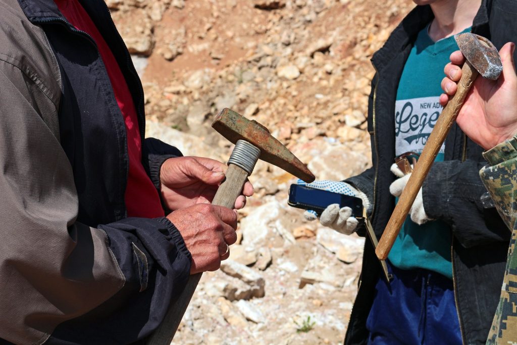
Geologists with the tools of their trade. Image by photochur from Pixabay 

Geology studies stones to help find minerals, predict earthquakes and more, but now their expertise has been tapped to understand kidney stones — how they form, why are some people more susceptible to them and can they be prevented?

In a new paper published in the journal Nature Reviews Urology, researchers described the geological nature of kidney stones, outlined the arc of their formation, introduced a new classification scheme and suggested possible clinical interventions.

“The process of kidney stone formation is part of the natural process of the stone formation seen throughout nature,” Illinois geology professor Bruce Fouke said. “We are bringing together geology, biology and medicine to map the entire process of kidney stone formation, step by step. With this road map in hand, more effective and targeted clinical interventions and therapies can now be developed.”

Kidney stones affect in 10 adults in their lifetime and send half a million people in the United States to emergency rooms annually, according to the National Kidney Foundation. Yet little is understood about the geology behind how kidney stones form, Fouke said.

The team’s previous  research found that kidney stones form in the same way as regular stones do: they don’t crystallise all at once, instead going through cycles of partial dissolution and reformation. Doctors had previously believed that they form suddenly and intact.

The research team described in detail the multiple phases kidney stones go through in forming, dissolving and re-forming, using high-resolution imaging technologies. Their findings defy the typical classification schemes doctors use, which are based on bulk analyses of the type of mineral and the presumed location of formation in the kidney. Instead, the researchers drew up a new classification scheme based on the phase of formation the stone is in, and the chemical processes it is undergoing.

“If we can identify these phase transformations, what makes one step to go to another and how it progresses, then perhaps we can intervene in that progression and break the chain of chemical reactions happening inside the kidney tissues before a stone becomes problematic,” said lead author Mayandi Sivaguru, assistant director of core facilities at the Carl R Woese Institute for Genomic Biology at Illinois.

One particularly revelatory finding was in the very beginnings of kidney stone formation. The stones start off as microspherules, tiny droplets of mineral, which merge to form larger crystals throughout kidney tissues. They are normally flushed out, but when they merge together and form larger stones that continue to grow, they can become excruciatingly painful and even deadly in some cases, Fouke said.

“Stone formation is part of a natural, healthy process within kidneys where these tiny mineral deposits are shuttled away and excreted from the body,” Fouke explained. “But then there is a tipping point when those same mineral deposits start to grow together too rapidly and are physically unable to leave the kidney.”

Image source: Leon Macapagal on Unsplash
An example of agate, which shows similar formation characteristics to kidney stones. Image source: Leon Macapagal on Unsplash

As the stone goes through the formation process, more microspherules merge, lose their rounded shape and transform into much larger, perfectly geometric crystals. Stones go through multiple cycles of partially dissolving—shedding up to 50% of their volume—and then growing again, creating a signature pattern of layered crystals much like those of agates, coral skeletons and hot-spring deposits seen around the world.

“Looking at a cross-section of a kidney stone, you would never guess that each of the layers was originally a bunch of little balls that lined up and coalesced. These are revolutionary new ways for us to understand how these minerals grow within the kidney and provide specific targets for stone growth prevention,” Fouke said.

The researchers listed a number of possible clinical interventions and treatment targets derived from this extra knowledge on kidney stone formation. They hope that these options can be tried out, from drug targets to changes in diet or supplements that could disrupt the cascade of kidney stone formation, Sivaguru said.

To aid in this testing, Fouke’s group developed the GeoBioCell, a microfluidic cartridge that mimics the intricate internal structures of the kidney. The team hopes the device can contribute to research as well as clinical diagnostic testing and the evaluation of potential therapies, particularly for the more than 70% of kidney stone patients with recurring stones.

“Ultimately, our vision is that every operating room would have a small geology lab attached. In that lab, you could do a very rapid diagnostic on a stone or stone fragment in a matter of minutes, and have informed and individualized treatment targets,” Fouke said.

Source: University of Illinois

Journal information: Mayandi Sivaguru et al, Human kidney stones: a natural record of universal biomineralization, Nature Reviews Urology (2021). DOI: 10.1038/s41585-021-00469-x

‘Chemical Nose’ Sniffs Out Cancer-causing DNA Folds

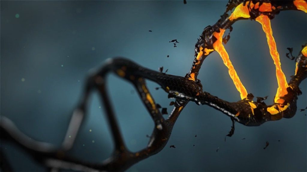
Artistic depiction of DNA double helix. Image by lisichik from Pixabay

Small folds in DNA structure have been implicated in breast cancer and other diseases have been elusive until a team of researchers developed a “chemical nose” to seek them out.

In the journal Nature Chemistry, chemists at  UC Riverside describe the development and testing of a “chemical nose” enabling them to “smell” unusual folds in DNA.

“If a DNA sequence is folded, it could prevent the transcription of a gene linked to that particular piece of DNA,” explained study author and UCR chemistry professor Wenwan Zhong. “In other words, this could have a positive effect by silencing a gene with the potential to cause cancer or promote tumors.”

DNA folding could also have a negative effect, however.

“DNA folds could potentially keep viral proteins from being produced to minimize immune response,” Prof Zhong said.

DNA folds have also been examined as potential targets for chemotherapy.   

To date, scientists have been unable to easily determine the effects of DNA folding on living organisms, as they lacked the proper tools to study them. In order to create one that can study the tiny structures, TUCR organic chemistry professor Richard Hooley and colleagues modified an existing concept that has previously been used to detect other things, such as chemical components in different vintages of wine.

The chemical system could be configured to seek out any kind of molecule, but it could not detect DNA in the way it was currently used. The addition of nonstandard components Prof Hooley’s group enabled the nose to sniff out its DNA target.

“Humans detect smells by inhaling air containing odor molecules that bind to multiple receptors inside the nose,” explained Prof Hooley. “Our system is comparable because we have multiple receptors able to interact with the DNA folds we’re looking for.”

The chemical nose consists of three parts: host molecules, fluorescent guest molecules, and DNA, which is the target. The guest molecules glow when the sought-after folds are present in a sample.

DNA is made of four nucleic acids: guanine, adenine, cytosine and thymine which are combined into the familiar double helix of DMA. Sections that are rich in guanine sometimes fold differently and create what’s called a G-quadruplex.

The parts of the genome that make these quadruplex structures are extremely complex, though the researchers have found that their folds are known to regulate gene expression, and aid in maintaining cell health.

The researchers sought to demonstrate that they could detect a single type of quadruplex composed of four guanines. Prof Zhong said that having accomplished this, the research team will try to build on their success.

“Now we think we can do more,” she said. “There are other three-dimensional structures in DNA, and we want to understand those as well.”

For their next step, the researchers will examine how effects that damage DNA influence the ways they fold. In addition, they also plan to study RNA folding since RNA is also important for cellular functioning.

“RNA has even more complex structures than DNA, and is more difficult to analyze, but understanding its structure has great potential for disease research,” Prof Zhong said. 

Source: UC Riverside News

Novel Magnetic Technique Detects Malaria in Blood

A new magnetic method has been developed that can detect malaria, leading to faster, accurate and cheap diagnosis of the deadly disease.

An international study field-tested this new tool in Papua New-Guinea, in the hopes of helping the fight against this disease, which had 229 million reported cases in 2019, with 700 000 deaths a year.

“Malaria is easily treated but it is actually hard to diagnose, and because of that there can be over-treatment, which we have seen can lead to the spread of drug-resistant malaria,” said Dr Stephan Karl, a Senior Research Fellow in Malaria and Vector Biology at James Cook University’s Australian Institute of Tropical Health and Medicine.

“Improving malaria diagnosis, especially through the development of practical methods for resource-limited places, is important and timely,” he said.

An international team including the University of Augsburg’s Professor Istvan Kezsmarki, with the PNG Institute of Medical Research and the Burnet Institute, came up with the magnetic detection method, called rotating-crystal magneto-optical detection (RMOD).

When malaria parasites break down blood, the haeme molecules are aggregated by the parasites into biocrystals called haemezoin, which contain magnetic iron. This iron can is detectable by the RMOD method.

“I’ve studied the magnetic properties of malaria infected blood since 2006, and we engaged with Professor Kezsmarki’s team in 2013 to demonstrate the sensitivity of this test using human malaria parasites,” Dr Karl said.

A field study was successfully conducted, involving almost 1000 suspected malaria patients in a high-transmission area of Papua New-Guinea.

“After years of in-lab optimisation of the device, in collaboration with Dr. Karl we demonstrated the great potential of RMOD in fast and reliable malaria field tests performed in Papua New-Guinea,” Prof Kezsmarki said.

“We showed that RMOD performs well in comparison to the most reliable existing method..It’s very promising, as RMOD testing can be conducted after a short training session and provides test results within 10 minutes. From a funding perspective the cost is very low since no expensive reagents are used,” said Dr Karl.

Dr Karl said the aim was to refine the design until a test could be done by a simple button push.

Source: Medical Xpress

Journal information: L. Arndt et al, Magneto-optical diagnosis of symptomatic malaria in Papua New Guinea, Nature Communications (2021). DOI: 10.1038/s41467-021-21110-w

COVID Variants May Increase False Negative Results, FDA Warns

The UK variant of the SARS-CoV-2 virus, known as B.1.1.7 could affect the accuracy of certain molecular COVID tests, causing increased false negative results, the US Food and Drug Administration (FDA) has warned.

A false negative from the UK variant could happen if the variant’s mutation happened to be in a part of the virus’ genome that was assessed by that test. However, genetic tests that target multiple parts of the virus genome are less likely to be affected by the new variants, the FDA advises.

Although the possibility of false negative results are to be expected from almost all tests, the effect of the COVID variants could impact the ability to properly monitor the disease, as well as complicating diagnosis.  

The FDA issued an alert on Friday, saying that they monitor tests which have received an Emergency Use Authorisation (EUA). They highlight three tests as being affected, with potentially less sensitivity to the mutations:

  • Accula SARS-Cov-2 Test, from Mesa Biotech
  • TaqPath COVID-19 Combo Kit, from Thermo Fisher Scientific)
  • Linea COVID-19 Assay Kit, from Applied DNA Sciences

However, the warning for Accula test was a result of “an abundance of caution”, and the latter two assess multiple genetic targets, so they are not likely to be impacted. The FDA issued recommendations for the users of these devices.The FDA’s warning was prompted by computer simulations showing reduced efficacy in certain tests for the virus variant.

In their press release, the FDA did not specifically mention whether any tests may be affected by mutations in the South African virus variant.

Source: MedPage Today