Tag: kidneys

The Resilience of Females’ Kidneys is Down to Hormones
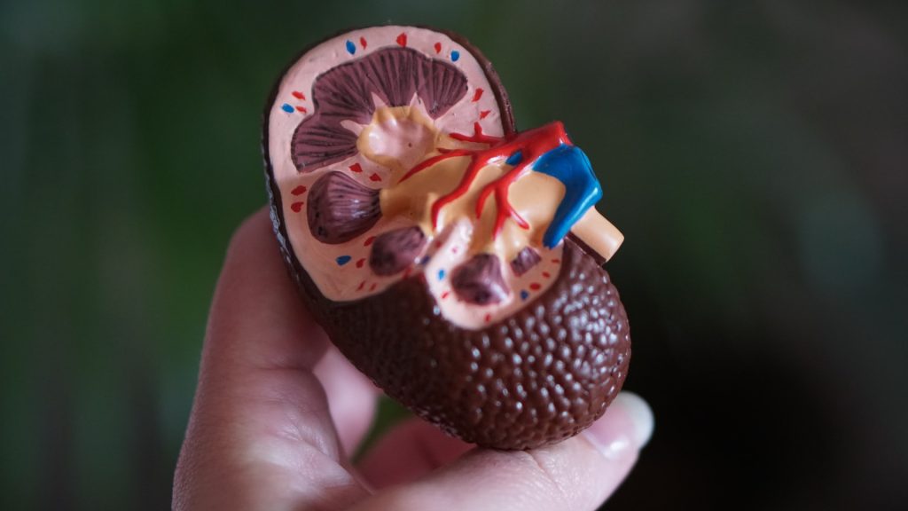
Photo by Robina Weermeijer on Unsplash

Females’ kidneys are known to be more resilient to disease and injury, so what about them can be applied to treat males’ kidneys? A new USC Stem Cell-led study published in Developmental Cell describes not only how sex hormones drive differences in male and female mouse kidneys, but also how lowering testosterone can “feminise” this organ and improve its resilience.

“By exploring how differences emerge in male and female kidneys during development, we can better understand how to address sex-related health disparities for patients with kidney diseases,” said Professor Andy McMahon, the study’s corresponding author, and the director of the Eli and Edythe Broad Center for Regenerative Medicine and Stem Cell Research at the Keck School of Medicine of USC.

First authors Lingyun “Ivy” Xiong and Jing Liu from the McMahon Lab and their collaborators identified more than 1000 genes with different levels of activity in male and female mouse kidneys, in a study supported by the National Institutes of Health. The differences were most evident in the section of the kidney’s filtering unit known as the proximal tubule, responsible for reabsorbing most of the nutrients such as glucose and amino acids back into the blood stream. Most of these sex differences in gene activity emerged as the mice entered puberty and became even more pronounced as they reached sexual maturity.

Because female kidneys tend to fare better in the face of disease or injury, the researchers were interested how the gene activity of kidneys becomes “feminised” or “masculinised” – and testosterone appeared to be the biggest culprit.

To feminize the kidneys of male mice, two strategies worked equally well: castrating males before puberty and thus lowering their natural testosterone levels, or removing the cellular sensors known as androgen receptors that respond to male sex hormones.

Intriguingly, three months of calorie restriction – which is an indirect way to lower testosterone – produced a similar effect. Accordingly, calorie restriction has already been shown to mitigate certain types of kidney injuries in mice.

To re-masculinize the kidneys of the castrated males, the researchers only needed to inject testosterone. Similarly, testosterone injection masculinised the kidneys of females who had their ovaries removed before puberty.

The scientists performed some similar experiments with mouse livers. Although this organ also displays sex-related differences, the hormones and underlying factors driving these differences are very different than those at play in the kidney. This suggests that these sex-related organ differences emerged independently during evolution.

To test whether the same genes are involved in sex-related kidney differences in humans, the scientists analysed a limited number of male and female donor kidneys and biopsies. When it came to genes that differed in their activity between the sexes, there was a modest overlap of the human genes with the mouse genes.

“There is much more work to be done in studying sex-related differences in normal human kidneys,” said McMahon. “Given the divergent outcomes for male and female patients with kidney disease and injury, this line of inquiry is important for making progress toward eventually closing the gap on these sex-related health disparities.”

Source: Keck School of Medicine of USC

    Ultrasound to the Kidneys can Treat Resistant Hypertension

    Credit: Thirdman on Pexels

    A device that uses ultrasound to calm overactive nerves in the kidneys may be able to help some people get their blood pressure under control, according to successful test results published in JAMA Cardiology.

    Led by researchers at Columbia University and Université de Paris, the study has found that the device consistently reduced daytime ambulatory blood pressure by an average of 8.5 points among middle-aged people with hypertension.

    Lifestyle changes, such as cutting salt intake or losing weight, along with medications are often prescribed to lower blood pressure in patients with hypertension. Yet about one-third of hypertensive patients have resistant hypertension.

    “Many patients in our clinical practice are just like the patients in our study, with uncontrolled blood pressure in the 150s despite some efforts,” says Ajay Kirtane, MD, professor of medicine at Columbia University Vagelos College of Physicians and Surgeons and co-leader of the study.

    Leaving blood pressure uncontrolled for too long can lead to heart failure, strokes, heart attacks, and irreversible kidney damage.

    “Renal ultrasound could be offered to patients who are unable to get their blood pressure under control after trying lifestyle changes and drug therapy, before these events occur,” says Kirtane, who is also an interventional cardiologist and director of cardiac catheterisation laboratories at NewYork-Presbyterian/Columbia University Irving Medical Center.

    The study tested the device, which is used in an outpatient procedure called ultrasound renal denervation. The device is still investigational and has not yet been approved by the FDA for use outside of clinical trials.

    Kidney nerves and hypertension

    Hypertension in middle age is thought to be caused in part by overactive nerves in the kidneys, which trigger water and sodium retention and release hormones that can raise blood pressure. (In older people, hypertension often occurs as blood vessels stiffen). Antihypertensive drugs work in different ways to lower blood pressure, by dilating blood vessels, removing excess fluid, or blocking hormones that raise blood pressure. But none target the renal nerves directly.

    Ultrasound therapy calms overactive nerves in the renal artery, disrupting signals that lead to hypertension. The therapy is delivered to the nerves via a thin catheter that is inserted into a vein in the leg or wrist and threaded to the kidney.

    Study results

    The new study pooled data from three randomised trials encompassing more than 500 middle-aged patients with varying degrees of hypertension and medication use.

    Twice as many patients who received the ultrasound therapy reached their target daytime blood pressure (less than 135/85 mmHg) compared to patients in the sham groups.

    “The result was almost identical across the different study groups, which definitively shows that the device can lower blood pressure in a broad range of patients,” Kirtane says.

    The procedure was well-tolerated, and most patients were discharged from the hospital the same day. According to Kirtane, improvements in blood pressure were seen as soon as one month after the procedure.

    The treatment will be evaluated by the FDA in the coming months.

    Bottom line for patients with resistant hypertension

    The investigators expect the treatment could be offered as an adjunct to medication therapy and lifestyle changes for patients with uncontrolled hypertension.

    “Once the device is available, we envision recommending it to patients who have tried other therapies first. The hope is that by controlling blood pressure, we might be able to prevent kidney damage and other effects of uncontrolled blood pressure,” Kirtane adds.

    Source: Columbia University Irving Medical Center

    New Mathematical Model for Potassium Homeostasis

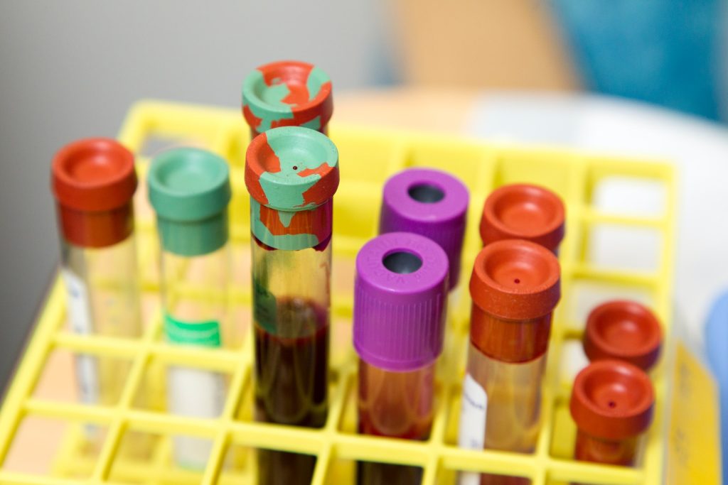
    Blood samples
    Photo by National Cancer Institute on Unsplash

    Potassium is essential to normal cellular function, helping the cardiac muscle work correctly and aids in the transmission of electrical signals within cells. A new mathematical model published in PLOS Computational Biology sheds light on the often mysterious process of potassium homeostasis.

    Using existing biological data, researchers at the University of Waterloo built a mathematical model that simulates how an average person’s body regulates potassium, both in times of potassium depletion and during potassium intake. Because so many foods contain abundant potassium, the body is continually storing, deploying, and disposing of potassium to keep it in a healthy range, ie the process of potassium homeostasis. Understanding potassium homeostasis is essential in helping diagnose the source of the problem when something goes wrong, for example, when kidney disease or medication leads to dysregulation.

    “Too much potassium in the body, or hyperkalaemia, can be just as dangerous as hypokalaemia, or too little,” said study lead author Melissa M. Stadt, a PhD student in applied mathematics. “Dysregulation of potassium can lead to dangerous and potentially fatal consequences.”

    The model could be used for a virtual patient trial, allowing researchers to generate dozens of patients and then predict which ones would have hyper- or hypokalaemia based on different controls.

    “A lot of our models are pieces of a bigger picture,” said Anita Layton, professor of applied mathematics and Canada 150 Research Chair in mathematical biology and medicine. “This model is one new and exciting piece in helping us understand how our incredibly complex internal systems work.”

    The model is especially exciting because it allows scientists to test the muscle-kidney cross-talk signal hypothesis. Scientists have hypothesised that skeletal muscles, which store most of the body’s potassium, can directly signal to the kidneys to dump potassium when there’s too much stores, and vice versa. When the mathematical researchers tested the hypothesis in their model, it more accurately reflected existing biological data regarding potassium homeostasis, suggesting that muscle-kidney cross talk might be an essential piece in the puzzle of potassium regulation.

    Source: University of Waterloo

    Why Women Have the Edge in Recovery from Kidney Injury

    Anatomic model of a kidney
    Photo by Robina Weermeijer on Unsplash

    Women have a better ability to recover from kidney injury than men, but the reasons are not well understood. A study in Cell Reports may provide answers, as researchers found that females have an advantage at the molecular level that protects them from a newly discovered form of cell death that occurs in injured kidneys. This protection could be exploited as a potential therapeutic.

    “Kidney disease afflicts more than 850 million people worldwide every year, so it’s important to understand why female kidneys are more protected from these acute and chronic injuries,” said Tomokazu Souma, MD, PhD, assistant professor at Duke University School of Medicine. “Our study is a step toward identifying the causes and suggests that this female resilience could be therapeutically harnessed to improve kidney repair in both sexes.”

    Souma and colleagues conducted studies in mice focusing on a form of cell death called ferroptosis, which was only recently discovered. This form of cell death is dependent on iron and oxidative stress. It has been identified as a key player in kidney diseases.

    Using genetic and single-cell RNA transcriptomic analysis in mice, the researchers found that being female confers striking protection against ferroptosis through a particular pathway called nuclear factor erythroid 2-related factor 2, or NRF2.

    In females, NRF2 is highly active, keeping cell death in check. In males, however, the sex hormone testosterone reduces the activity of NRF2, thus promoting ferroptosis and undermining cell resiliency in kidney injury.

    Further experiments showed that chemically activating NRF2 protected male kidney cells from ferroptosis, demonstrating that NRF2 could be a potential therapeutic target to prevent failed renal repair after acute kidney injury.

    “By identifying the mechanism in which the female hormonal environment protects and the male hormonal environment aggravates acute and chronic kidney injuries, we believe there is strong potential to boost the resilience of kidneys,” Souma said.

    Source: Duke University Medical Center

    Paracetamol May Protect Against Kidney Damage in Malaria

    Anatomic model of a kidney
    Photo by Robina Weermeijer on Unsplash

    Paracetamol may help protect against kidney damage in patients with malaria, according to a study recently published in Clinical Infectious Diseases.

    The study found that for patients with severe malaria caused by the malaria parasite Plasmodium knowlesi (the most common cause of malaria in Malaysia), taking paracetamol regularly for 3 days led to improvements in kidney function when tested one week later.

    The findings are important because they will help provide the best possible treatment to patients with severe malaria, said study leader Dr Daniel Cooper.

    “Even minor kidney injury can have long-term effects, so anything we can do to minimise kidney injury from malaria will be beneficial for these patients’ long-term outcomes,” Dr Cooper said.

    In collaboration with international partners, the study involved 396 people with knowlesi malaria in Sabah, Malaysia.

    Assistant Professor Bridget Barber said that in severe malaria, red blood cells can rupture, releasing haemoglobin which can have a toxic impact on kidneys, and it is now believed that paracetamol can help to mitigate these toxic effects.

    “These results are consistent with other studies conducted in patients with other forms of malaria, including in adults in Bangladesh, and in children in Africa. Importantly, these findings also suggest that paracetamol may help to protect the kidneys in other conditions that are also associated with rupture of red blood cells,” A/Prof Barber said.

    Source: MedicalXpress

    Long-term Use of RAS Inhibitor Drugs Could Damage Kidneys

    Photo by Robina Weermeijer on Unsplash

    New research is raising concerns that long-term use of renin-angiotensin system (RAS) inhibitor drugs such as ACE inhibitors could be contributing to kidney damage.

    The researchers stress that patients should continue taking the medications. But the scientists are urging studies to better understand the drugs’ long-term effects.

    “Our studies show that renin-producing cells are responsible for the damage. We are now focusing on understanding how these cells, which are so important to defend us from drops in blood pressure and maintain our well-being, undergo such transformation and induce kidney damage,” said UVA’s Dr Maria Luisa Sequeira Lopez. “What is needed is to identify what substances these cells make that lead to uncontrolled vessel growth.”

    A billion people around the world are affected by chronic hypertension. In a study published in JCI Insight, University of Virginia (UVA) researchers were seeking to better understand why severe forms of the condition are often accompanied by atherosclerosis in the kidney, leading to organ damage.

    They found that renin cells, which help regulate blood pressure through renin production, play an important role. Harmful changes in the renin cells can cause the cells to invade the walls of the kidney’s blood vessels. The renin cells then trigger a buildup of another cell type, smooth muscle cells, that cause the vessels to thicken and stiffen, resulting in impeded kidney blood flow.

    Long-term use of RAS inhibitor drugs, such as ACE inhibitors, or angiotensin receptor blockers, have a similar effect. But the study found that long-term use of the drugs was associated with hardened kidney vessels in both lab mice and humans

    The researchers note that the medications can be lifesaving for patients, so they stress the importance of continuing to take them. But they say additional studies are needed to better understand the drugs’ long-term effects on the kidneys.

    “It would be important to conduct prospective, randomised controlled studies to determine the extent of functional and tissue damage in patients taking medications for blood pressure control,” said UVA’s Dr Ariel Gomez. “It is imperative to find out what molecules these cells make so that we can counteract them to prevent the damage while the hypertension is treated with the current drugs available today.”

    Source: University of Virginia

    Geology Helps Medicine to Understand Kidney Stones

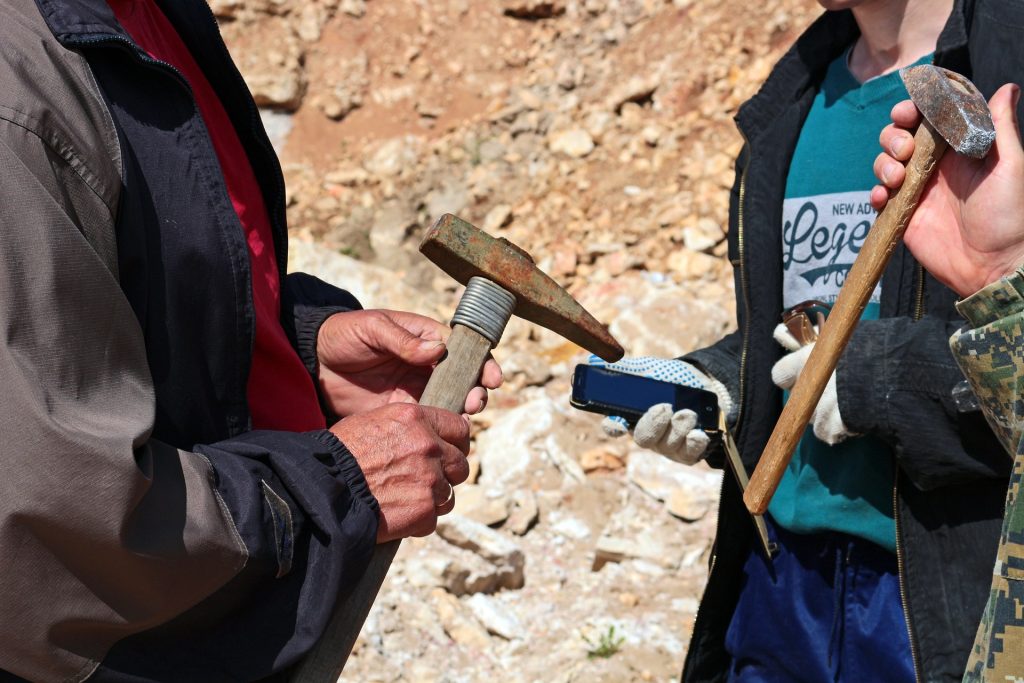
    Image by photochur from Pixabay
    Geologists with the tools of their trade. Image by photochur from Pixabay 

    Geology studies stones to help find minerals, predict earthquakes and more, but now their expertise has been tapped to understand kidney stones — how they form, why are some people more susceptible to them and can they be prevented?

    In a new paper published in the journal Nature Reviews Urology, researchers described the geological nature of kidney stones, outlined the arc of their formation, introduced a new classification scheme and suggested possible clinical interventions.

    “The process of kidney stone formation is part of the natural process of the stone formation seen throughout nature,” Illinois geology professor Bruce Fouke said. “We are bringing together geology, biology and medicine to map the entire process of kidney stone formation, step by step. With this road map in hand, more effective and targeted clinical interventions and therapies can now be developed.”

    Kidney stones affect in 10 adults in their lifetime and send half a million people in the United States to emergency rooms annually, according to the National Kidney Foundation. Yet little is understood about the geology behind how kidney stones form, Fouke said.

    The team’s previous  research found that kidney stones form in the same way as regular stones do: they don’t crystallise all at once, instead going through cycles of partial dissolution and reformation. Doctors had previously believed that they form suddenly and intact.

    The research team described in detail the multiple phases kidney stones go through in forming, dissolving and re-forming, using high-resolution imaging technologies. Their findings defy the typical classification schemes doctors use, which are based on bulk analyses of the type of mineral and the presumed location of formation in the kidney. Instead, the researchers drew up a new classification scheme based on the phase of formation the stone is in, and the chemical processes it is undergoing.

    “If we can identify these phase transformations, what makes one step to go to another and how it progresses, then perhaps we can intervene in that progression and break the chain of chemical reactions happening inside the kidney tissues before a stone becomes problematic,” said lead author Mayandi Sivaguru, assistant director of core facilities at the Carl R Woese Institute for Genomic Biology at Illinois.

    One particularly revelatory finding was in the very beginnings of kidney stone formation. The stones start off as microspherules, tiny droplets of mineral, which merge to form larger crystals throughout kidney tissues. They are normally flushed out, but when they merge together and form larger stones that continue to grow, they can become excruciatingly painful and even deadly in some cases, Fouke said.

    “Stone formation is part of a natural, healthy process within kidneys where these tiny mineral deposits are shuttled away and excreted from the body,” Fouke explained. “But then there is a tipping point when those same mineral deposits start to grow together too rapidly and are physically unable to leave the kidney.”

    Image source: Leon Macapagal on Unsplash
    An example of agate, which shows similar formation characteristics to kidney stones. Image source: Leon Macapagal on Unsplash

    As the stone goes through the formation process, more microspherules merge, lose their rounded shape and transform into much larger, perfectly geometric crystals. Stones go through multiple cycles of partially dissolving—shedding up to 50% of their volume—and then growing again, creating a signature pattern of layered crystals much like those of agates, coral skeletons and hot-spring deposits seen around the world.

    “Looking at a cross-section of a kidney stone, you would never guess that each of the layers was originally a bunch of little balls that lined up and coalesced. These are revolutionary new ways for us to understand how these minerals grow within the kidney and provide specific targets for stone growth prevention,” Fouke said.

    The researchers listed a number of possible clinical interventions and treatment targets derived from this extra knowledge on kidney stone formation. They hope that these options can be tried out, from drug targets to changes in diet or supplements that could disrupt the cascade of kidney stone formation, Sivaguru said.

    To aid in this testing, Fouke’s group developed the GeoBioCell, a microfluidic cartridge that mimics the intricate internal structures of the kidney. The team hopes the device can contribute to research as well as clinical diagnostic testing and the evaluation of potential therapies, particularly for the more than 70% of kidney stone patients with recurring stones.

    “Ultimately, our vision is that every operating room would have a small geology lab attached. In that lab, you could do a very rapid diagnostic on a stone or stone fragment in a matter of minutes, and have informed and individualized treatment targets,” Fouke said.

    Source: University of Illinois

    Journal information: Mayandi Sivaguru et al, Human kidney stones: a natural record of universal biomineralization, Nature Reviews Urology (2021). DOI: 10.1038/s41585-021-00469-x

    Cells in the Centres of Kidney Tumours are The Most Aggressive

    Researchers have found that cells from different parts of kidney tumours behave differently, and cells within the centre of a tumour are the most aggressive and most likely to spread around the body.

    Metastasis, where cancer cells from tumours spread to other parts of the body, is the main cause of death in cancer patients. 

    In this multidisciplinary study published in Nature Ecology and Evolution, scientists analysed 756 cancer biopsy samples from different regions within tumours from the TRACERx Renal study.

    They discovered that, in contrast to the cells at the outside of tumours, the cells in the centres of tumours have more unstable genomes, and a higher potential for metastasis. The cells on the outside had lower growth rates and had less genetic damage.

    “Cancer cells in the central zone of the tumour face harsh environmental conditions, as there’s a lack of blood supply and oxygen. They have to adapt to survive, which makes them stronger and more aggressive. This also means they are more likely to successfully evolve into cells that can disseminate and take hold in distant organs,” explained Kevin Litchfield, paper author and group leader at the UCL Cancer Institute.

    These findings show that it is important to focus on the tumour centre for a better understanding of how cancer spreads, and identify the most dangerous cells. Also, in order to wipe out the most aggressive tumour cells, treatment development must target the unique environmental conditions found within the tumour core.

    The scientists also examined how genetically different populations of cancer cells grow within a tumour. With a unique mapping tall that reconstructed the growth of tumour cells, they discovered that, while tumours tend to follow a pattern where populations of cells grow in the local area, in two cases, cells took hold in a new region of the tumour by seemingly ‘jumping’ over other populations of tumour cells.

    For their next steps, the researchers aim to reconstruct 3D tumour maps, providing even better visualisation of the tumours’ internal structure.

    Samra Turajlic, head of the Crick’s Cancer Dynamics Laboratory, Consultant Medical Oncologist at the Royal Marsden NHS Foundation Trust and the Chief Investigator of TRACERx Renal, said: “Cancer spread is one of the biggest barriers to improving survival rates. In the context of the TRACERx Renal study we previously resolved the genetic make up of different tumour areas, but until now, there has been no understanding of how these differences relate spatially. The most critical question is the part of the tumour from which cancer cells break away and migrate making cancer incurable.

    “Using this unique clinical cohort and a multidisciplinary approach, including mathematical modeling, we identified with precision the place in the tumour where genetic chaos emerges to give rise to metastases. Our observations shed light on the sort of environmental conditions that would foster emergence of aggressive behaviour. These findings are a critical foundation for considering how we target or even prevent distinct populations of cells that pose the biggest threat.”

    Source: Francis Crick Institute

    Cutting Edge Bio-printing Fabricates Tiny Kidneys

    Researchers from the Murdoch Children’s Research Institute (MCRI) and biotech company Organovo have successfully bio-printed miniature human kidneys with unparalleled speed and quality to be used for toxicity screening of medications known to cause kidney damage. 

    A world leader in modeling the kidney, Professor Melissa Little of the MRCI said, “Drug-induced injury to the kidney is a major side effect and difficult to predict using animal studies. Bioprinting human kidneys are a practical approach to testing for toxicity before use.”

    The new study involved testing the toxicity of aminoglycosides, a class of antibiotics that commonly damage the kidney. The study revealed deaths of certain types of kidney cells when exposed to aminoglycosides.

    Organovo first began bio-printing kidneys in 2015, but their new processes are much faster, allowing 200 mini-kidneys to be produced in 10 minutes. The improvement in speed and quality has opened the doorway for bioprinting entire organs for transplant. “3-D bioprinting can generate larger amounts of kidney tissue but with precise manipulation of biophysical properties, including cell number and conformation, improving the outcome.”

    Professor Little said that prior to this study, the possibility of using such technology for transplantation was too complicated to consider. “The pathway to renal replacement therapy using stem cell-derived kidney tissue will need a massive increase in the number of nephron structures present in the tissue to be transplanted,” she said.

    “By using extrusion bioprinting, we improved the final nephron count, which will ultimately determine whether we can transplant these tissues into people.”

    Source: Medical Xpress