Tag: ischaemic stroke

In Severe Stroke, Mechanical Clot Removal Leads to Improved Outcomes

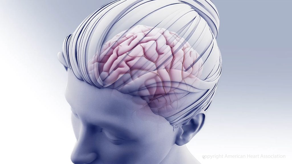
A clot within a blood vessel interrupting blood flow to the brain.
Copyright American Heart Association

Ischaemic stroke patients previously considered unlikely to survive without severe disability may regain far more function if the blood clots are mechanically removed in addition to standard medical therapy, according to preliminary late-breaking research presented today at the American Stroke Association’s International Stroke Conference 2022.

In 2018, the American Heart Association’s stroke treatment guidelines were updated to recommend endovascular therapy (mechanical clot removal) for select stroke patients to improve the odds of functional recovery. This new study in Japan is the first randomised, controlled trial to demonstrate the effectiveness of endovascular therapy in patients with severe strokes involving clots in one or more large brain arteries, causing a large blood flow interruption in the brain. This approach had worked for patients with fewer areas of the brain disrupted, however, clinical experience was mixed for patients with more severe strokes.

Infarction area, or core area, estimates the volume of brain affected and describes the blockage location as seen on a brain CT. A lower number translates to a stroke affecting more core areas of the brain: 8-10=small core, 6-7=moderate core and 0-5=large core. Current US stroke guidelines recommend endovascular therapy for core areas 6-9. This study examined blockages that scored as 3-5. Strokes with blockages measuring 0-2 core areas are considered too severe and highly unlikely the patient would return to ambulatory independence.

“I have often encountered a dramatic improvement in a patient just after the mechanical clot removal procedure, even when the infarction area was large. Yet, patients sometimes also experienced severe haemorrhagic transformation [a life-threatening complication that occurs when blood from outside the brain crosses the blood-brain barrier and worsens stroke outcome] after the artery was reopened. So, in Japan, our stroke physicians are always cautious about endovascular therapy when the infarction area is large,” said Professor Shinichi Yoshimura, lead author of the study.

This randomised study included 203 stroke patients (average age of 76 years; 44% women). Most (71%) were examined and had MRI or a CT scan of the brain within 6 hours after stroke symptoms were first noticed, when patients are generally considered eligible for endovascular therapy. The other patients were seen between 6-24 hours after symptoms were noticed, and additional imaging showed areas of the brain that might benefit from prompt treatment.

On imaging, all patients were found to have clots blocking a large cerebral artery – either the internal carotid artery, the proximal middle cerebral artery or both. The strokes were rated as severe (median 22 on the National Institutes of Health (NIH) Stroke Scale,) and involved disrupted blood flow to large areas of the brain (about 7 out of 10 regions).

After imaging, the patients were randomly selected to receive either standard medical care for stroke (intravenous fluids, controlling blood pressure and other risk factors, and thrombolytics for lower bleeding risk patients) or standard medical care plus endovascular therapy performed within an hour after imaging to mechanically remove the clots. Due to bleeding concerns, intravenous thrombolytics were sparingly administered to select patients in a similar proportion in both treatment groups (27 of those who received endovascular therapy and 29 who received standard care).

Comparing the 100 patients who received endovascular therapy with 102 on standard therapy alone, the analysis found:

  • Patients who received endovascular therapy were 2.43 times more likely (31% vs 13%) to be able to walk unassisted and to have a residual disability rated as none to moderate 90 days later.
  • After 90 days, more of the patients (14% vs. 6.9%) who received endovascular therapy were considered functionally independent, meaning they were either able to carry out all their pre-stroke activities or to have a slight disability that did not require daily assistance.
  • At 48 hours after treatment, more of the patients (31% vs. 8.8%) who received endovascular therapy had major early neurological improvement.

“Our findings confirm that anyone who suffers from stroke should be transferred to a medical facility capable of endovascular therapy as soon as possible. The benefit of endovascular therapy is not limited by the severity or region of a stroke. These patients may have the chance to more fully recover from stroke and go back to their previous lives and activity levels,” said Professor Takeshi Morimoto, senior author of the study.

Several outcomes were compared to evaluate the safety of adding endovascular therapy to medical treatment, with researchers reporting:

  • Within 48 hours, scans revealed that more of the patients who received endovascular therapy had experienced some bleeding within the brain (with or without symptoms), 58% vs. 31%, respectively.
  • However, the number of patients who experienced other adverse outcomes was similar in the two treatment groups. The adverse events included brain bleeding within 48 hours that caused a worsening of neurological status (4 points or greater worsening on the NIH Stroke Scale); the need for surgery to relieve pressure on the brain in the first week; death within 90 days; or the recurrence of ischaemic stroke within 90 days.

“The finding of more intracranial bleeding in the patients who received endovascular therapy is very important. However, there were haemorrhages with symptoms and some that caused no symptoms. The haemorrhages with no symptoms were detected on imaging conducted for this study in the endovascular treatment group, not in the standard practice group. Symptomatic intracranial haemorrhage still occurred more commonly among patients in the endovascular group, however, it was not a statistically significant difference from the standard care group,” Morimoto said.

Due to different treatment protocols in Japan, where there is less use of intravenous thrombolysis than in the US and other western countries, and where more strokes are imaged with MRI than CT, this study’s results may over- or underestimate the effectiveness of endovascular therapy.

The researchers are currently performing sub-analyses to help identify factors that might signal which patients are more likely to have a greater return of function after the treatment. “In addition, tools, devices or rehabilitation methods that could potentially improve the likelihood for similar patients to recover with less disability should be investigated,” Morimoto said.

Source: American Heart Association

Having Better Conversations about Post-stroke Prognosis

Photo by cottonbro from Pexels

Though conversations with stroke survivors and their loved ones about possible lasting impairment can be traumatic, they might also be therapeutic, according to research from The University of Queensland, published in the American Journal of Speech-Language Pathology.

PhD candidate Bonnie Cheng from UQ’s School of Health and Rehabilitation Sciences said that prognosis conversations can trigger mixed emotions of hope and grief, so knowing  how people would prefer for them to happen is important.

“When stroke is encountered for the first time, it’s hard to know what’s important and relevant to ask about, especially during that time of crisis immediately after such a serious health event,” Ms Cheng said.

“During this time, there’s also an immense sense of gratitude for the survival of their loved one that seems to stop significant others from asking for more information.

“Conversations about prognosis and lasting impairments, like speech difficulties, need to be an ongoing dialogue between health professionals, the patient, and their support network.

“It’s important for these conversations to be based on a mutual understanding of what improvements are personally meaningful to the patient and their significant others.”

Aphasia is a common condition after a stroke, diagnosed in one-in-three people after a stroke.

The researchers interviewed people who identified as a significant other of someone with aphasia between three and 12 months after stroke, including spouses, close friends, adult children and parents of someone with aphasia.

“In the interviews, we talked in-depth about their experience of finding out about the prognosis for aphasia, the impact these experiences had on them, and how they would want to get information about prognosis in a perfect world,” Ms Cheng said.

“What we found was significant others need to be included in prognosis conversations so that they too can be informed and supported, alongside the patient.

“The prognosis of aphasia is a sensitive issue to address because it often involves having to adjust to long-term difficulties and changes.

“Recovery needs to be looked at holistically in terms of everyday activities that affect the individual, rather than just scores on a language impairment test.

“Even though we can’t yet ‘cure’ aphasia, this research brings us one step closer to talking about recovery in a way that’s as informative and as compassionate as possible, so that people living with aphasia can be supported to live successfully with the condition.”

Source: University of Queensland

About 1% of Hospitalised COVID Patients Develop Neurological Complications

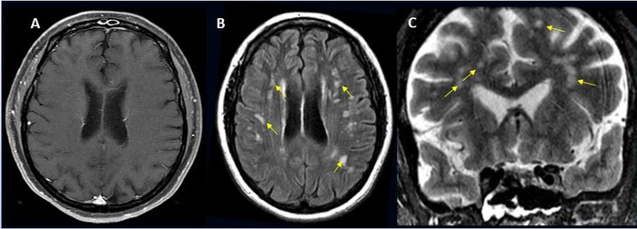
49-year-old female with past medical history of mitral valve disease and tricuspid valve regurgitation who developed headache followed by cough and fever presented to the ER with right upper eyelid ptosis (drooping). Credit: Radiological Society of North America and Scott H. Faro, M.D.

Approximately one in 100 patients hospitalised with COVID will likely develop complications of the central nervous system, according to a large international study. These can include stroke, haemorrhage, and other potentially fatal complications. The study was presented at the annual meeting of the Radiological Society of North America (RSNA).

“Much has been written about the overall pulmonary problems related to COVID, but we do not often talk about the other organs that can be affected,” said study lead author Scott H. Faro, MD, FASFNR, professor of radiology and neurology at Thomas Jefferson University. “Our study shows that central nervous system complications represent a significant cause of morbidity and mortality in this devastating pandemic.”

Dr Faro initiated the study after finding that only a small number of cases informed existing literature on central nervous system complications in hospitalised COVID patients.

To build a more complete picture, he and his colleagues analysed nearly 40 000 cases of hospitalised COVID patients, admitted between September 2019 and June 2020. Their average age was 66 years old, and two thirds were men.

Confusion and altered mental status were the most common causes of admission followed by fever. Comorbidities such as hypertension, cardiac disease and diabetes were common.

There were 442 acute neuroimaging findings most likely associated with the viral infection, with central nervous system complications in 1.2% of this large patient group.

“Of all the inpatients who had imaging such as MRI or a CT scan of the brain, the exam was positive approximately 10% of the time,” Dr Faro said. “The incidence of 1.2% means that a little more than one in 100 patients admitted to the hospital with COVID are going to have a brain problem of some sort.”

Ischaemic stroke, with an incidence of 6.2%, was the most common complication, followed by intracranial haemorrhage (3.72%) and encephalitis (0.47%).

A small percentage of unusual findings was uncovered, such as acute disseminating encephalomyelitis, an inflammation of the brain and spinal cord, and posterior reversible encephalopathy syndrome, a syndrome that mimics many of the symptoms of a stroke.

“It is important to know an accurate incidence of all the major central nervous system complications,” Dr Faro said. “There should probably be a low threshold to order brain imaging for patients with COVID.”

Source: EurekAlert!

Study Links OCD to Increased Risk of Stroke

Photo by Annie Spratt on Unsplash

Adults with obsessive-compulsive disorder (OCD) were more than three times as likely to have an ischaemic stroke later in life compared to adults who do not have OCD, according to new research.

OCD involves intrusive thoughts which may be accompanied by actions to alleviate the stress brought on by these thoughts; these are called obsessions and compulsions, respectively. Potentially debilitating, OCD typically manifests in adolescence and is under-reported and under-treated. The World Health Organization ranks OCD as one of the ten most disabling conditions worldwide, with a lifetime prevalence of 1% to 3%.

Clinical research has shown that patients with OCD often have stroke-related metabolic disorders, such as obesity and diabetes. 

While a growing body of evidence suggests an association between OCD and stroke risk, with some patients developing OCD after a stroke, OCD may also influence stroke risk. OCD raises the risk for ischaemic stroke more than 3-fold, especially among older adults
Drawing on health records from 2001-2010 from the Taiwan National Health Insurance Research Database, researchers compared stroke risk between 28 064 adults diagnosed with OCD and 28 064 adults without an OCD diagnosis. The average age at diagnosis was 37 years old, with equal representation of men and women, with a maximum data coverage of 11 years.

Analysis showed that even after controlling for factors known to increase stroke risk such obesity and high blood pressure, OCD remained an independent risk factor for ischaemic stroke. However, no differences were found when considering hemorrhagic strokes, and medications to treat OCD were linked to increased risk of stroke.
Ya-Mei Bai, MD, PhD, Professor, Department of Psychiatry, Taipei Veterans General Hospital, said: “The results of our study should encourage people with OCD to maintain a healthy lifestyle, such as quitting or not smoking, getting regular physical activity and managing a healthy weight to avoid stroke-related risk factors.”

Worldwide, stroke is the second-leading cause of death after heart disease. Cerebrovascular diseases often present complex cases, and this study was the first to consider how OCD and strokes may be associated on a longer time scale. 

Limitations include the observational nature of the study being unable to establish cause and effect, as well as the inclusion in the data of only stroke patients who sought health care. Additionally, information on disease severity and outcome was not included along with family medical history or environmental influences, which can further affect both the extent of OCD and severity of strokes.

“For decades, studies have found a relationship between stroke first and OCD later,” said Prof Bai. “Our findings remind clinicians to closely monitor blood pressure and lipid proles, which are known to be related to stroke in patients with OCD. More research is needed to understand how the mental processes connected to OCD may increase the risk of ischaemic stroke.”

Source: News-Medical.Net

Journal information: Chen, M-H., et al. (2021) Increased Risk of Stroke in Patients With Obsessive-Compulsive Disorder: A Nationwide Longitudinal Study. Stroke: Journal of the American Heart Association. doi.org/10.1161/STROKEAHA.120.032995.