Tag: ischaemic stroke

Intensive Blood Pressure Reduction after Ischaemic Stroke Increases Disability

Credit: American Heart Association

The largest ever randomised controlled trial of intensive blood pressure lowering after thrombectomy in ischaemic stroke patients found that it led to deterioration in surrounding brain tissue and higher rates of disability, compared to less intensive treatment.

The results of the ENCHANTED2/MT trial were presented in a late-breaking session at the World Stroke Congress and simultaneously published in The Lancet. The trial was stopped early due to the significance of the findings.

Professor Craig Anderson, Director of Global Brain Health at The George Institute for Global Health, said the rapid emergence of this effect suggested the more aggressive approach was compromising the return of blood flow to the affected area.

“Our study provides a strong indication that this increasingly common treatment strategy should now be avoided in clinical practice,” he said.

Endovascular thrombectomy is an increasingly used non-surgical treatment for ischaemic stroke, in which x-ray guided microcatheters are inserted into the blood clot to dissolve it.

“A potential downside of this now widely used and effective treatment is that the rapid return of blood supply to an area that has been deprived of oxygen for a while can cause tissue damage known as reperfusion injury,” said Professor Anderson.

“This has resulted in a shift in medical practice towards more intensive lowering of blood pressure after clot removal to try and minimise this damage, but without evidence to support the benefits versus potential harms.”

To this end, researchers recruited 816 adults with acute ischaemic stroke who had elevated blood pressure after clot removal from 44 centres in China between July 2020 and March 2022. They had an average age of 67 and just over a third were female.

Of these, 407 were assigned to more-intensive (target < 120mmHg) and 409 to the less-intensive (target 140–180mmHg) systolic blood pressure control, with the target to be achieved within one hour of entering the study and sustained for 72 hours.

Researchers looked at how well the patients in both groups recovered according to a standard measure of disability, ranging from 0–1 for a good outcome without or with symptoms but no disability, scores of 2–5 reflecting increasing disability levels, and 6 being death.

Patients in the more-intensively treated group had significantly worse scores on the scale compared to those allocated to those treated less intensively.

Compared to the less-intensive group, they had more early brain tissue deterioration and major disability at 90 days but there were no significant differences in brain bleeds, mortality, or serious adverse events.

Patients who had their blood pressure more intensively controlled also rated their quality of life as significantly worse due to limitations on their physical abilities resulting from their stroke.

Prof Anderson said that after scouring the medical literature the research team had been unable to find strong enough evidence to recommend the ideal target for blood pressure control after blood clot removal in patients with acute ischaemic stroke.

“While our study has now shown intensive blood pressure control to a systolic target of less than 120mmHg to be harmful, the optimal level of control is yet to be defined,” he said.

Source: George Institute for Global Health

Sex Differences in Post-stroke Biomarkers

Credit: American Heart Association

Although males and females are equally affected by stroke Since oestrogen and progesterone have known neuroprotective effects, it is important to compare the size, severity and biochemical composition of the brain tissue following stroke in female and male animal models. In a paper published in IBRO Neuroscience Reports, researchers have discovered that certain biomarkers were different in the brains of male and female mice.

Stroke neurosurgeon and research coordinator Prof Nicole Sylvain and her colleagues are looking at treatments for post-stroke recovery that help supplement these energy losses. Using the Canadian Light Source (CLS) at the University of Saskatchewan (USask), the team was able to identify energy biomarkers in the brain, which could eventually inform clinicians about the effects of potential stroke treatments on brain recovery after a stroke.

The group’s recent study examined post-stroke differences between male and female mice, and found that female mice have higher amounts of glycogen in their brains. Glycogen is a sugar-like substance that circulates in our blood and nourishes our cells. When the supply of glycogen is disrupted by stroke, the brain is severely impacted.

Most pre-clinical stroke research has been performed using male lab animals, with results usually generalised to both sexes. In clinical stoke cases, females have a higher incidence of ischaemic stroke and poorer outcomes, compared to males.

“We found that, for the most part, male data can be generalised for females, however, some of the metabolic markers we measured were actually different,” Sylvain said. “It’s really important to do the research on both sexes.”

It would be impossible for the team to detect the biomarkers without to the Mid-IR beamline.

“The only way to detect them in such an accurate way across the brain is with infrared imaging, so the CLS has been absolutely vital to our research.”

Source: University of Saskatchewan

Switch to Tenecteplase for Ischaemic Strokes Improves Outcomes and Lowers Costs

Credit: American Heart Association

A newer, faster-administration clot-busting drug called tenecteplase outperforms the traditional treatment for ischaemic strokes in several key areas, including better health outcomes and lower costs, according to a new study published in the journal Stroke.

The 15-month study was led by a team of neurologists at Dell Medical School at The University of Texas at Austin.

“The Dell Med Neurology Stroke Program was one of the first in the United States to make this change,” said Steven Warach, MD, lead author of the study . “Based on even the earliest results from this study, other experts across the country were convinced and made the switch from alteplase to tenecteplase at their own stroke centres, including at Ascension hospitals nationwide.” 

The vast majority of strokes of the 800 000 strokes in the US (about 87%) each year are ischaemic.

Both tenecteplase and alteplase are federally approved for use in dissolving clots in blocked heart arteries. But the newer drug tenecteplase is also being used by clinicians, off-label, to treat ischaemic strokes, because clinical trials in stroke suggest that it may be at least as good as alteplase and it is easier to administer. Tenecteplase is administered by a single five- to 10-second intravenous injection. The researchers compared its performance with the standard drug for stroke, alteplase, which is injected over 60 minutes.

“When it comes to treating patients with a stroke, every second matters,” said Warach, who is also a professor of neurology at Dell Med. “The shorter preparation and injection time with tenecteplase not only eliminates a lot of dosing errors related to alteplase, but it’s also more efficient. We were able to deliver the clot-busting medicine more quickly after patients arrived in the emergency department, and for patients who needed to be transferred to another hospital for more advanced care after receiving the clot buster, we were able to initiate the transfer sooner in those treated with tenecteplase.”

For patients who come into the emergency department after a stroke, Warach’s study found that the “door-to-needle” time (from patients’ arrival to treatment) was on average six minutes quicker with tenecteplase. And for patients who also required a thrombectomy, the surgical removal of a blood clot causing the stroke, tenecteplase slashed to the time to transferring the patient to a thrombectomy-capable stroke centre by 25 minutes.

Researchers also saw improvements in clinical outcomes for patients given tenecteplase, including:

  • A 5% increase in patients who were able to walk independently at time of hospital discharge to home.
  • A 4% decrease in occurrences of bad events such as brain haemorrhages, discharge to hospice care or death.

The third major improvement: cost. The research team found that tenecteplase treatment cost the hospitals about US$ 2500 less than alteplase per patient.

Source: EurekAlert!

Flu Vaccine May Reduce Ischaemic Stroke Risk

Source: Pixabay CC0

Receiving an annual flu vaccination may be linked to a reduction in risk of ischaemic stroke, according to a study which appears online in the journal Neurology.

“Studies have shown that getting the flu increases your risk of having a stroke, but research is still being collected on whether getting the flu vaccine can help protect against a stroke,” said study author Francisco J. de Abajo, MD, MPH, PhD, of the University of Alcalá in Madrid. “This observational study suggests that those who have a flu shot have a lower risk of stroke. To determine whether this is due to a protective effect of the vaccine itself or to other factors, more research is needed.”

In their study, the researchers accessed a health care database in Spain, identifying 14 322 participants aged 40 years and over with a first stroke over a 14-year period. Each person who had a stroke was matched to five people of the same age and sex who did not have a stroke.

Then the researchers looked at whether people had received the influenza vaccine at least 14 days before the stroke or before that same date for those who did not have a stroke.

A total of 41.4% of those who had a stroke had received the flu vaccine, compared to 40.5% of those who did not have a stroke – seemingly indicating that the flu jab added to risk. But those vaccinated were more likely to be older and to have other stroke risk factors such as hypertension and high cholesterol. Once these were adjusted for, the researchers found that those who received a flu shot had a 12% reduced stroke risk.

The pneumonia vaccine was also investigated for any effect on the risk of stroke, but none was found.

“These results are yet another reason for people to get their yearly flu shot, especially if they are at an increased risk of stroke,” de Abajo said. “To be able to reduce your risk of stroke by taking such a simple action is very compelling.”

Since the study was observational, it only shows an association and cannot prove a causal link. Other unmeasured factors could also mediate stroke risk,

Source: American Academy of Neurology

Blood Type may Point to Early-onset Stroke Risk

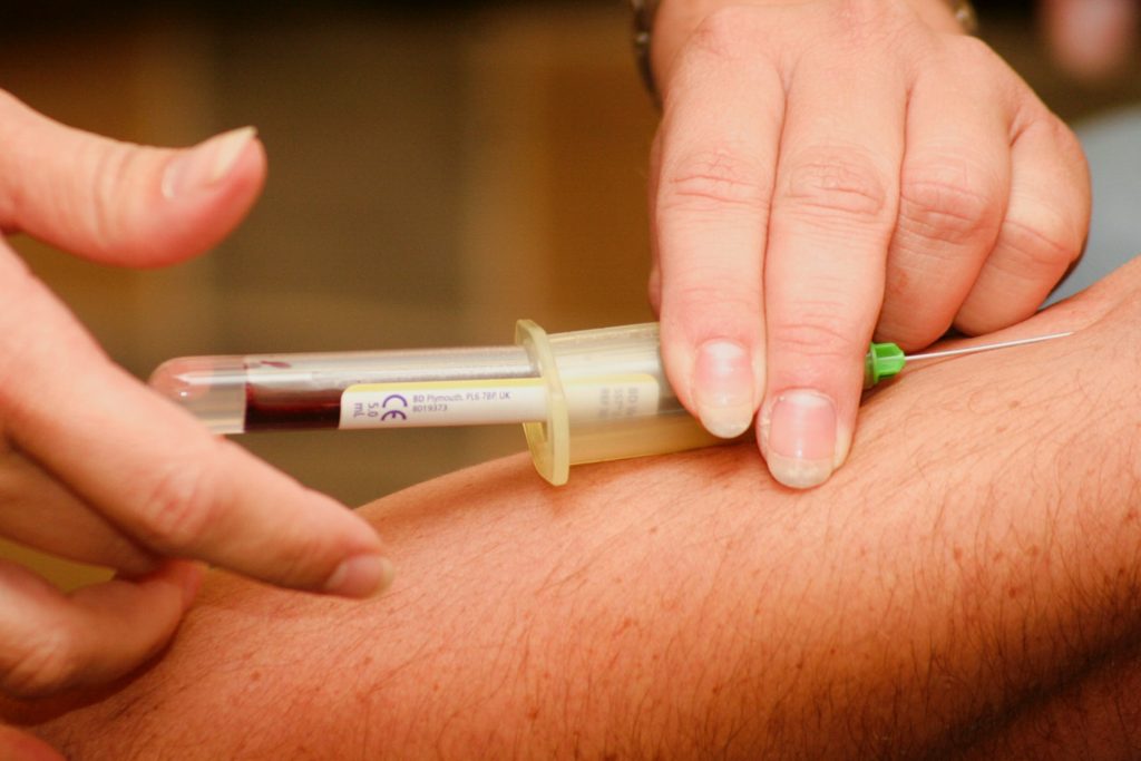
Blood sample being drawn
Photo by Hush Naidoo Jade Photography on Unsplash

Blood type may be associated with early-onset stroke risk, according to a new meta-analysis, which was published in the journal Neurology. The meta-analysis included all available data from genetic studies focusing on ischaemic strokes in adults under age 60.

“The number of people with early strokes is rising. These people are more likely to die from the life-threatening event, and survivors potentially face decades with disability. Despite this, there is little research on the causes of early strokes,” said study co-principal investigator Steven J. Kittner, MD, MPH, Professor of Neurology at University of Maryland School of Medicine (UMSOM).

He and his colleagues performed a meta-analysis of 48 studies on genetics and ischaemic stroke that included 17 000 stroke patients and nearly 600 000 healthy controls. They looked for genetic variants associated with a stroke and found a link between early-onset stroke (before age 60) and the area of the chromosome that includes the gene that determines whether a blood type is A, AB, B, or O.

The study found that people with early stroke were more likely to have blood type A and less likely to have blood type O, compared to people with late stroke and people who never had a stroke. Both early and late stroke were also more likely to have blood type B compared to controls. After adjusting for sex and other factors, researchers found that, compared to those with other blood types, blood type A had a 16% higher risk while blood type O had a 12% lower risk.

“Our meta-analysis looked at people’s genetic profiles and found associations between blood type and risk of early-onset stroke. The association of blood type with later-onset stroke was much weaker than what we found with early stroke,” said study co-principal investigator Braxton D. Mitchell, PhD, MPH, Professor of Medicine at UMSOM.

The researchers emphasised that the increased risk was very modest and that those with type A blood should not worry about having an early-onset stroke or engage in extra screening or medical testing based on this finding.

“We still don’t know why blood type A would confer a higher risk, but it likely has something to do with blood-clotting factors like platelets and cells that line the blood vessels as well as other circulating proteins, all of which play a role in the development of blood clots,” said Dr Kittner. Previous studies suggest that those with an A blood type have a slightly higher risk of developing blood clots in the legs known as deep vein thrombosis. “We clearly need more follow-up studies to clarify the mechanisms of increased stroke risk,” he added.

A limitation of the study was the relative lack of diversity among participants, with only 35% of the participants having non-European ancestry.

Source: University of Maryland School of Medicine

Novel Drug Shown to Repair Damage after Stroke

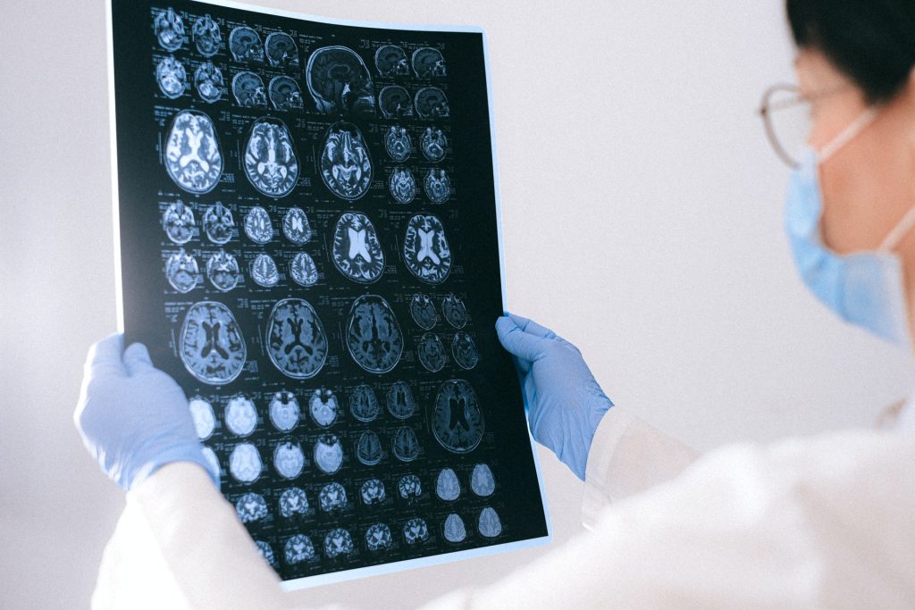
MRI images of the brain
Photo by Anna Shvets on Pexels

A pioneering new study from the University of Cincinnati shows promise that a new drug may help repair damage caused by strokes. The preclinical study appears in the journal Cell Reports.

Currently, there are no FDA approved drugs to repair the damage caused by a stroke. The study found that the new drug, NVG-291-R, enables nervous system repair and significant functional recovery in an animal model of severe ischaemic stroke. Deleting the gene for the drug’s molecular target also shows similar effect on neural stem cells. The drug has also proven to be safe and well-tolerated in volunteers with multiple sclerosis.

“We are very excited about the data showing significant improvement in motor function, sensory function, spatial learning and memory,” said Agnes (Yu) Luo, PhD, associate professor UC and the study’s senior author.

Prof Luo said the drug would be a “substantial breakthrough” if the early results translate into clinical settings. Further study and validation of results from independent groups will be needed to determine if the drug is similarly effective to repair the damage of ischaemic strokes in human patients. Additional studies will be needed to research if NVG-291-R effectively repairs damage caused by haemorrhagic strokes in both animal models and human patients.

“Most therapies being researched today primarily focus on reducing the early damage from stroke,” Assoc Prof Luo said. “However, our group has focused on neurorepair as an alternative and now has shown that treatment with NVG-291-R not only results in neuroprotection to reduce neuronal death but also robust neuroreparative effects.”

The drug proved to be effective even when treatment began as late as seven days after the stroke’s onset.

“The only current FDA-approved drug for treatment of stroke does not repair damage and must be administered within 4.5 hours of stroke onset.” Luo said. “Most therapies being researched need to be applied within 24–48 hours of a stroke’s onset. A product that works to repair damage from stroke even a week after symptom onset would change the paradigm for stroke treatment.”

Jerry Silver, PhD, co-author of the study and professor of neurosciences at CWRU’s School of Medicine, said the study showed the drug repaired damage in at least two ways: creating new neuronal connections and enhancing migration of new neurons derived from neuronal stem cells to the damage site.

“NVG-291-R’s ability to enhance plasticity was demonstrated by using staining techniques that clearly showed an increase in axonal sprouting to the damaged part of the brain,” Prof Silver said. “This enhanced plasticity is an excellent validation of the same powerful mechanisms that we and other researchers were able to demonstrate using NVG-291-R in spinal cord injury.”

Source: University of Cincinnati

A Novel Anticoagulant That can be ‘Deactivated’

Source: NCI

A new biomolecular anticoagulant platform reported in Nano Letters holds promise as a revolutionary advancement over the anticoagulants currently used during surgeries and other procedures. The technology is based around injectable fibre structures which can be quickly dissolved and excreted by the kidneys.

“We envision the uses of our new anticoagulant platform would be during coronary artery bypass surgeries, kidney dialysis, and a variety of vascular, surgical and coronary interventions,” said Kirill Afonin, leader of the team which invented the technology. “We are now investigating if there are potential future applications with cancer treatments to prevent metastasis and also in addressing the needs of malaria, which can cause coagulation issues.”

The team’s technology turns to programmable RNA-DNA anticoagulant fibres that, when injected into the bloodstream, form into modular structures that communicate with thrombin. The technology allows the structures to prevent blood clotting as it is needed and then be quickly eliminated via the renal system once their job is done.

The fibre structures use aptamers, short sequences of DNA or RNA designed to specifically bind and inactivate thrombin.

“Instead of having a single small molecule that deactivates thrombin,” Afonin said, “we now have a relatively large structure that has hundreds of the aptamers on its surface that can bind to thrombin and deactivate them. And because the structure becomes larger, it will circulate in the bloodstream for a significantly longer time than traditional options.”

The extended circulation in the bloodstream allows for a single injection, instead of multiple doses. The design also decreases the concentration of anticoagulants in the blood, resulting in less stress on the body’s renal and other systems, Afonin said.

This technology also introduces a novel “kill-switch” mechanism, which reverses the fibre structure’s anticoagulant function with a second injection. This lets makes the fibres able to be metabolised into materials that are tiny, harmless, inactive and easily excreted by the renal system.

The entire process takes place outside the cell, through extracellular communication with the thrombin. The researchers note that this is important as immunological reactions do not appear to occur, based on their extensive studies.

The team has tested and validated the platform in computer models, human blood and various animal models“We conducted proof-of-concept studies using freshly collected human blood from donors in the US and in Brazil to address a potential inter donor variability,” Afonin said.

The technology may provide a foundation for other biomedical applications that require communication via the extracellular environment in patients, he said. “Thrombin is just one potential application,” he said. “Whatever you want to deactivate extracellularly, without entering the cells, we believe you can. That potentially means that any blood protein, any cell surface receptors, maybe antibodies and toxins, are possible.”

The technique permits the design of structures of any shape desired, with the kill switch mechanism intact. “By changing the shape, we can have them go into different parts of the body, so we can change the distribution,” Afonin said. “It gets an extra layer of sophistication of what it can do.”

While the application is sophisticated, production of the structures is relatively easy. “The shelf life is amazingly good for these formulations,” Afonin said. “They’re very stable, so you can dry them, and we anticipate they will stay for years at ambient temperatures, which makes them very accessible to economically challenged areas of the world.”

Source: University of North Carolina

Blood Vascular Network Retains the ‘Memory’ of a Stroke

Credit: American Heart Association

A study into the structure of blood vascular network structure found that it is dynamic and can adapt to external factors, resulting in a kind of memory of certain events such as an ischaemic stroke. In particular, the study researchers found that rarely used connections incrementally weaken until they disappear eventually.

Researchers from the Max Planck Institute for Dynamics and Self-Organization in Göttingen and the Technical University of Munich used computer simulations to model vascular networks and identified adaptation rules for their connections.

“We found that the strength of a connection within a network depends on the local flow,” explained Karen Alim, corresponding author of the study. “This means that links with a low flow below a certain threshold will decay more and more until they eventually vanish,” she continued. Since the limited amount of material available to build the vascular system needs to be efficiently used, this mechanism offers an elegant way to streamline the vascular system.

Persistent changes in the network

Once a connection has become very weak due to a low flow rate, recovering that connection is very difficult. For example, a blood vessel blockage of the type that could lead to an ischaemic stroke. During an ischaemic stroke, some blood vessels in the affected region are weakened by the blockage.

“We found that in such a case, adaptations in the network are permanent and are maintained after the obstacle is removed. One can say that the network prefers to reroute the flow through existing stronger connections instead of re-growing weaker connections – even if the flow would require the opposite,” explained Komal Bhattacharyya, principal author of the study.

The researchers have thus shown that blood flow permanently changes even after successful removal of the clot. This memory capability of networks can also be found in other living systems: for example, the slime mould Physarum polycephalum uses its adaptive network to navigate its environment based on imprints by food stimuli, as demonstrated previously.

The study was published in Physical Review Letters.

Source: Max Planck Institute for Dynamics and Self-Organization

Portable MRI Machines Extend Stroke Diagnosis Coverage

Credit: American Heart Association

Portable MRI machines, an emerging technology that makes medical imaging accessible even in remote locations, detected ischaemic strokes in 90% of patients scanned, according to a study appearing in the journal Science Advances.

In previous studies, portable MRI devices have demonstrated they can also detect haemorrhagic as well as ischaemic strokes, helping clinicians make crucial life-saving treatment decisions quickly in remote areas for patients who lack ready access to major hospitals with expensive stationary MRI machines, the authors say.

“This is the first systematic evidence you can detect ischaemic strokes using portable, bedside devices,” noted Kevin Sheth, professor of neurology and neurosurgery at Yale School of Medicine and co-corresponding author of the study.

Outcomes for stroke patients improve dramatically the quicker they receive treatment. But access to stationary MRI machines is limited for those who live far away from major hospitals or in developing countries. And even stroke patients who have access to major hospitals often have to wait for scans with stationary MRIs because of heavy demand for the equipment. Portable scans can be used at a patient’s bedside, in ambulances, or in remote clinics, Dr Sheth said.

In addition, quickly differentiating between different types of stroke is crucial for determining proper treatment, the researchers say. Ischaemic strokes are usually treated with blood thinners. But that course of treatment is dangerous for those who experience haemorrhagic strokes or strokes in which there is bleeding in the brain.

Analysing portable MRI scans from 50 patients at Yale New Haven Hospital, the Yale and Harvard researchers found that the results largely confirmed ischaemic stroke diagnoses made by stationary MRIs. For 45 of those patients, the portable MRI detected blood clots as small as 4mm in size.

Source: Yale University

In Severe Stroke, Mechanical Clot Removal Leads to Improved Outcomes

A clot within a blood vessel interrupting blood flow to the brain.
Copyright American Heart Association

Ischaemic stroke patients previously considered unlikely to survive without severe disability may regain far more function if the blood clots are mechanically removed in addition to standard medical therapy, according to preliminary late-breaking research presented today at the American Stroke Association’s International Stroke Conference 2022.

In 2018, the American Heart Association’s stroke treatment guidelines were updated to recommend endovascular therapy (mechanical clot removal) for select stroke patients to improve the odds of functional recovery. This new study in Japan is the first randomised, controlled trial to demonstrate the effectiveness of endovascular therapy in patients with severe strokes involving clots in one or more large brain arteries, causing a large blood flow interruption in the brain. This approach had worked for patients with fewer areas of the brain disrupted, however, clinical experience was mixed for patients with more severe strokes.

Infarction area, or core area, estimates the volume of brain affected and describes the blockage location as seen on a brain CT. A lower number translates to a stroke affecting more core areas of the brain: 8-10=small core, 6-7=moderate core and 0-5=large core. Current US stroke guidelines recommend endovascular therapy for core areas 6-9. This study examined blockages that scored as 3-5. Strokes with blockages measuring 0-2 core areas are considered too severe and highly unlikely the patient would return to ambulatory independence.

“I have often encountered a dramatic improvement in a patient just after the mechanical clot removal procedure, even when the infarction area was large. Yet, patients sometimes also experienced severe haemorrhagic transformation [a life-threatening complication that occurs when blood from outside the brain crosses the blood-brain barrier and worsens stroke outcome] after the artery was reopened. So, in Japan, our stroke physicians are always cautious about endovascular therapy when the infarction area is large,” said Professor Shinichi Yoshimura, lead author of the study.

This randomised study included 203 stroke patients (average age of 76 years; 44% women). Most (71%) were examined and had MRI or a CT scan of the brain within 6 hours after stroke symptoms were first noticed, when patients are generally considered eligible for endovascular therapy. The other patients were seen between 6-24 hours after symptoms were noticed, and additional imaging showed areas of the brain that might benefit from prompt treatment.

On imaging, all patients were found to have clots blocking a large cerebral artery – either the internal carotid artery, the proximal middle cerebral artery or both. The strokes were rated as severe (median 22 on the National Institutes of Health (NIH) Stroke Scale,) and involved disrupted blood flow to large areas of the brain (about 7 out of 10 regions).

After imaging, the patients were randomly selected to receive either standard medical care for stroke (intravenous fluids, controlling blood pressure and other risk factors, and thrombolytics for lower bleeding risk patients) or standard medical care plus endovascular therapy performed within an hour after imaging to mechanically remove the clots. Due to bleeding concerns, intravenous thrombolytics were sparingly administered to select patients in a similar proportion in both treatment groups (27 of those who received endovascular therapy and 29 who received standard care).

Comparing the 100 patients who received endovascular therapy with 102 on standard therapy alone, the analysis found:

  • Patients who received endovascular therapy were 2.43 times more likely (31% vs 13%) to be able to walk unassisted and to have a residual disability rated as none to moderate 90 days later.
  • After 90 days, more of the patients (14% vs. 6.9%) who received endovascular therapy were considered functionally independent, meaning they were either able to carry out all their pre-stroke activities or to have a slight disability that did not require daily assistance.
  • At 48 hours after treatment, more of the patients (31% vs. 8.8%) who received endovascular therapy had major early neurological improvement.

“Our findings confirm that anyone who suffers from stroke should be transferred to a medical facility capable of endovascular therapy as soon as possible. The benefit of endovascular therapy is not limited by the severity or region of a stroke. These patients may have the chance to more fully recover from stroke and go back to their previous lives and activity levels,” said Professor Takeshi Morimoto, senior author of the study.

Several outcomes were compared to evaluate the safety of adding endovascular therapy to medical treatment, with researchers reporting:

  • Within 48 hours, scans revealed that more of the patients who received endovascular therapy had experienced some bleeding within the brain (with or without symptoms), 58% vs. 31%, respectively.
  • However, the number of patients who experienced other adverse outcomes was similar in the two treatment groups. The adverse events included brain bleeding within 48 hours that caused a worsening of neurological status (4 points or greater worsening on the NIH Stroke Scale); the need for surgery to relieve pressure on the brain in the first week; death within 90 days; or the recurrence of ischaemic stroke within 90 days.

“The finding of more intracranial bleeding in the patients who received endovascular therapy is very important. However, there were haemorrhages with symptoms and some that caused no symptoms. The haemorrhages with no symptoms were detected on imaging conducted for this study in the endovascular treatment group, not in the standard practice group. Symptomatic intracranial haemorrhage still occurred more commonly among patients in the endovascular group, however, it was not a statistically significant difference from the standard care group,” Morimoto said.

Due to different treatment protocols in Japan, where there is less use of intravenous thrombolysis than in the US and other western countries, and where more strokes are imaged with MRI than CT, this study’s results may over- or underestimate the effectiveness of endovascular therapy.

The researchers are currently performing sub-analyses to help identify factors that might signal which patients are more likely to have a greater return of function after the treatment. “In addition, tools, devices or rehabilitation methods that could potentially improve the likelihood for similar patients to recover with less disability should be investigated,” Morimoto said.

Source: American Heart Association