Tag: interleukin

A New Insight into the Mechanisms of Epidermal Renewal
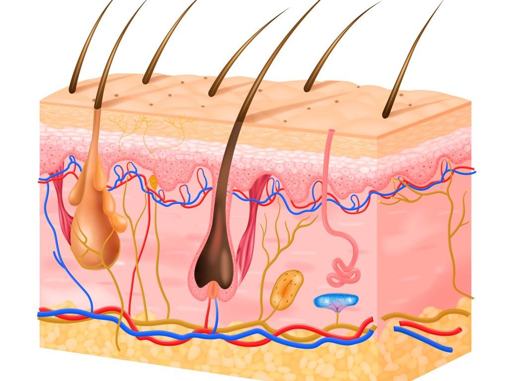
Picture by Macrovector on Freepik

The mechanisms underlying skin renewal are still poorly understood, but interleukin-38 (IL-38), a protein involved in regulating inflammatory responses, could provide insights. Researchers observed it for the first time in the form of condensates in keratinocytes, the cells of the epidermis. The presence of IL-38 in these aggregates is enhanced close to the skin’s surface exposed to atmospheric oxygen. This process could be linked to the initiation of programmed keratinocyte death, a natural process in the epidermis. This study, from University of Geneva (UNIGE) researchers, could bring new perspectives for the study of human epidermis and the illnesses that affect it.

Renewal of the epidermis relies on stem cells located in its lowest layer, which constantly produce new keratinocytes. These new cells are then pushed to the surface, differentiating along the way and accumulating protein condensates. Once they reach the top of the epidermis, they undergo a programmed death, cornification, to create a protective barrier of dead cells.

“The way in which the epidermis constantly renews itself is well documented. However, the mechanisms that drive this process are still not fully understood,” explains Gaby Palmer-Lourenço, associate professor at the Faculty of medicine of UNIGE and principal investigator. The study is published in the journal Cell Reports.

An unexpected role

Interleukin 38 is a small messenger protein that ensures communication between cells. It is known for its role in regulating inflammatory responses and its presence in keratinocytes, the cells of the epidermis, was previously associated with the preservation of the skin’s immune balance. “In keratinocytes in vivo, we found that IL-38 forms condensates, specialized protein aggregates with specific biochemical functions, a behavior that was not known for this protein,” recounts Gaby Palmer-Lourenço. Even more curious, the closer the keratinocytes were to the surface of the skin, the greater the amount of IL-38 within these condensates.

A reaction to oxidative stress

Blood vessels stop in the skin layer located below the epidermis. Therefore, the quantity of oxygen available for the keratinocytes is lower in the basal layers of the epidermis compared to the top layers that are directly exposed to the air that surrounds us. However, even though it is necessary to maintain cell functions, oxygen also causes oxidative stress by forming free radicals, reactive molecules that endanger the cell. “We were able to show that oxidative stress does indeed cause IL-38 condensation under laboratory conditions,” confirms Alejandro Díaz-Barreiro, postdoctoral fellow at the UNIGE Faculty of medicine, and first author of the study.

“Our results lead us to believe that, as we move closer to the epidermal surface, the increasing oxygen concentration promotes the formation of protein condensates, indicating to keratinocytes that they are in the right place to enter cell death,” furthers Gaby Palmer-Lourenço. This hypothesis provides new leads to decipher the mechanisms of epidermal renewal. It could also pave the way for a better understanding of the pathological mechanisms underlying certain skin diseases, such as psoriasis or atopic dermatitis. These questions will be further examined by the research group in future studies.

Contributing to an alternative to animal models

Alejandro Díaz-Barreiro is already working on the next step: “In the model we used previously, the effects of oxidative stress were artificially induced in a single layer of keratinocytes, a scenario that differs from the actual situation in the skin. We are therefore developing a new experimental system to apply oxygen gradients to in vitro reconstituted human epidermis. In this model, only the skin surface will be exposed to ambient air, while the other layers will be protected. This will allow us to study in detail the effect of oxidative stress on epidermal renewal.” By enabling a more precise analysis of human cells, this new system will provide an alternative to animal models often used for the study of skin biology and disease.

Source: Université de Genève

Immunotherapy Blocks Scarring, Improves Cardiac Function in Heart Failure

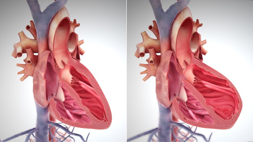
Right side heart failure. Credit: Scientific Animations CC4.0

A new study from Washington University School of Medicine in St. Louis suggests that a type of immunotherapy also may be an effective treatment strategy for heart failure by using an FDA-approved drug to block the signalling protein IL-1 beta. The study is published in Nature.

After a heart attack, viral infection or other injury to the heart, scar tissue often forms in the heart muscle, where it interferes with the heart’s normal contractions and plays a leading role in heart failure, a chronic condition which can only be slowed, not cured.

Studying human tissue samples as part of the new study, the researchers identified a type of fibroblast cell in the heart as the main culprit responsible for the formation of scar tissue in heart failure. To see if they could prevent scar formation, the scientists turned to mouse models of heart failure that have the very same type of fibroblasts. They used a therapeutic monoclonal antibody that blocks the formation of this harmful type of fibroblast, and succeeded in reducing the formation of scar tissue and improving heart function in the mice.

“After scar tissue forms in the heart, its ability to recover is dramatically impaired or impossible,” said cardiologist and senior author Kory Lavine, MD, PhD, a professor of medicine in the Cardiovascular Division at WashU Medicine. “Heart failure is a growing problem in the US and globally, affecting millions of people. Current treatments can help relieve symptoms and slow the progression, but there is a tremendous need for better therapies that actually stop the disease process and prevent the formation of new scar tissue that causes a loss of heart function. We are hopeful our study will lead to clinical trials investigating this immunotherapy strategy in heart failure patients.”

Fibroblasts have many roles in the heart, and parsing out the differences between various populations of these cells has been challenging. Some types of fibroblasts support the heart’s structural integrity and maintain good blood flow through the heart’s blood vessels, while others are responsible for driving inflammation and the development of scar tissue. Only recently, with the wide availability of the most advanced single cell sequencing technologies, could scientists peg which groups of cells are which.

“These various types of fibroblasts highlight newly recognised opportunities to craft treatment strategies that specifically block the type of fibroblasts that promote scarring and protect fibroblasts that maintain the structure of the heart, so the heart doesn’t rupture,” Lavine said. “Our research suggests that the fibroblasts that promote scarring in the injured heart are very similar to fibroblasts associated with cancer and other inflammatory processes. This opens the door to immunotherapies that potentially can stop the inflammation and resulting scar tissue.”

The research team, co-led by Junedh Amrute, a graduate student in Lavine’s lab, used genetic methods to demonstrate that a signaling molecule called IL-1 beta was important in a chain of events driving fibroblasts to create scar tissue in heart failure. With that in mind, they tested a mouse monoclonal antibody that blocks IL-1 beta and found beneficial effects in the mouse hearts. The mouse monoclonal antibody was provided by Amgen, whose scientists were also co-authors of the study. Monoclonal antibodies are proteins manufactured in the lab that modulate the immune system. The treatment reduced the formation of scar tissue and improved the pumping capacity of the mouse hearts, as measured on an echocardiogram.

At least two FDA-approved monoclonal antibodies, canakinumab and rilonacept, can block IL-1 signalling. These immunotherapies are approved to treat inflammatory disorders such as juvenile idiopathic arthritis and recurrent pericarditis, which is inflammation of the sac surrounding the heart.

One of these antibodies also has been evaluated in a clinical trial for atherosclerosis, a buildup of plaque that hardens the arteries. The trial, called CANTOS (Canakinumab Anti-inflammatory Thrombosis Outcome Study), showed a benefit for study participants with atherosclerosis.

“Even though this trial was not designed to test this treatment in heart failure, there are hints in the data that the monoclonal antibody might be beneficial for patients with heart failure,” Lavine said. “Secondary analyses of the data from this trial showed that the treatment was associated with a sizable reduction in heart failure admissions compared with standard care. Our new study may help explain why.”

Even so, the IL-1 antibody used in the CANTOS study had some side effects, such as increased risk of infection, that could perhaps be reduced with a more targeted antibody that specifically blocks IL-1 signaling in cardiac fibroblasts, according to the researchers.

“We are hopeful that the combination of all of this evidence, including our work on the IL-1 beta pathway, will lead to the design of a clinical trial to specifically test the role of targeted immunotherapy in heart failure patients,” Lavine said.

Source: Washington University School of Medicine

Linked Biological Pathways Drive Skin Inflammation

Picture by Macrovector on Freepik

A certain biological pathway involving interleukin-17 drives the inflammation seen in the skin disease psoriasis, according to a new study published in the journal Immunity. The work could lead to improved therapies for all inflammatory skin diseases, including atopic and allergic dermatitis and a type of boil called hidradenitis suppurativa, say the study authors.

Led by researchers at NYU Langone Health, the new study found that the interleukin-17 (IL-17) pathway, whose activity is blocked by existing anti-inflammatory drugs, activates a protein called hypoxia inducible factor 1-alpha (HIF-1-alpha) in psoriasis. Researchers say that IL-17 has long been known to be active in inflammation, but the role of HIF-1-alpha has until now been unclear.

The research team also found that HIF-1-alpha let inflamed skin cells more actively break down sugar for energy, supporting their metabolism and leading to the production of a waste product called lactate. When consumed by inflammatory T cells, lactate triggered production of IL-17, fuelling even more inflammation.

The findings show that in human skin tissue samples from psoriatic patients, measures of gene activity around IL-17 and HIF-1-alpha were similar, suggesting that these factors are interconnected. Experiments in mice treated to develop psoriasis found that subsequent treatment with an experimental drug that blocks the action of HIF-1-alpha, called BAY-87-2243, resolved inflammatory skin lesions.

Further, skin samples from 10 patients successfully treated with anti-inflammatory drug etanercept showed diminished activity for both IL-17 and HIF-1-alpha, suggesting to researchers that when IL-17 is blocked, so is HIF-1-alpha.

“Our study results broadly show that activation of HIF-1-alpha is at the crux of metabolic dysfunction observed in psoriasis and that its action is triggered by IL-17, another key inflammatory-signaling molecule,” said corresponding study author Shruti Naik, PhD, associate professor at NYU Grossman School of Medicine.

Further experiments were performed on skin samples from five patients with psoriasis whose healthy and inflamed skin was separately treated with either BAY-87-2243 or an existing combination of topical drugs (calcipotriene and betamethasone dipropionate). Researchers then compared differences in inflammatory gene activity as a measure of impact and found that the HIF-1-alpha inhibitor had a greater effect than existing topical drugs. Specifically, skin samples that responded to HIF-1-alpha therapy had 2,698 genes that were expressed differently, while standard-of-care-treated samples had 147 differently expressed genes.

Genetic analysis of skin samples from another 24 psoriatic patients treated with the IL-17A-blocking drug secukinumab showed only decreased, not heightened, gene activity connected to HIF-1-alpha when compared to HIF-1-alpha gene activity in nine healthy patients with no psoriatic disease. Researchers say this indicates HIF-1-alpha’s blocked action was codependent on blockage of IL-17.

Additional experiments in mice showed that blocking glucose uptake in the skin slowed psoriatic disease growth by limiting glucose metabolism, or glycolysis. Both the number of immune T cells tied to inflammation and the cell levels of IL-17 also decreased. The researchers found further that levels of lactate, the main byproduct of glycolysis, in psoriatic skin cell cultures dropped once exposed to the glycolysis-inhibiting drug 2-DG.

Directly targeting lactate production in psoriatic mice using a topical skin cream containing lactate dehydrogenase, which breaks down lactate, also slowed disease progression in the skin, with reduced numbers of inflammatory gamma-delta T cells and reduced IL-17 activity. Gamma-delta T cells were shown to take up lactate and use it to produce IL-17.

“Evidence of HIF-1-alpha’s depressed action, or downregulation, could also serve as a biomarker, or molecular sign, that other anti-inflammatory therapies are working,” said study co-senior investigator Jose U. Scher, MD, professor at NYU Grossman School of Medicine.

Scher, who also serves as director of NYU Langone’s Psoriatic Arthritis Center and the Judith and Stewart Colton Center for Autoimmunity, says the team plans to develop experimental drugs that can block HIF-1-alpha and lactate action in the skin “to end the underlying vicious cycle of IL-17-driven inflammation in skin disease. Our research fundamentally expands the scope of feasible therapeutic options.”

Naik points out that while many available therapies for psoriasis, including steroids and immunosuppressive drugs, reduce inflammation and symptoms, they do not cure the disease. She said further experiments are needed to refine which experimental drug works best, with respect to HIA-1-alpha inhibition, before clinical trials could start.

Source: NYU Langone Health / NYU Grossman School of Medicine

Towards Treating Dangerous Immunotherapy Side Effects

Photo by Tima Miroshnichenko on Pexels

While immunotherapy has been shown to greatly improve survival rates for certain types of cancer, in some cases, it can lead to a dangerous over-activation of the immune system. In a recent review published in Journal for ImmunoTherapy of Cancer, potential therapies have been identified, which might make it possible to continue with immunotherapy even when facing severe side effects.

The rare immunotherapy side effect of over-activation was only clinically recognised during regular clinical use rather than in clinical trials or animal experiments. To better understand this over-activation, Lisa LiuMarco Gerling, and colleagues analysed data from all published international reports on this issue after cancer immunotherapy. Their findings indicate that potentially life-threatening inflammation may occur more frequently than previously thought, and might be treatable with existing drugs such as steroids or anti-inflammatory therapies commonly used for rheumatoid arthritis.

“It will be exciting to follow up on the main findings of our systematic review, says Marco Gerling at the Department of Biosciences and Nutrition, Karolinska Institutet and lead author.

“We believe that inhibition of a specific inflammatory molecule, interleukin-6, could allow patients to continue immunotherapy despite strong, systemic activation of the immune system”, he continues. “But we need more data to support the regular use of interleukin-6 inhibitors. We also want to thank Narcisa Hannerz and Sabine Gillsund from Karolinska University Library for their invaluable help with finding articles for this review.“

Source: Karolinska Institutet

A Novel Hydrogel for Treating Spinal Cord Injury

Photo by Kanasi on Unsplash

Researchers at the Chinese Academy of Sciences have developed an innovative scaffold that regulates the immune microenvironment following a spinal cord injury, thereby reduces secondary injury effects. Their work is reported in Biomaterials.

By modifying a hydrogel with a cationic polymer, polyamidoamine, and  interleukin-10 (IL-10; an anti-inflammatory cytokine), the scaffold could enhance tissue remodelling and promote axonal regeneration.

Spinal cord injuries cause axon damage and neural cell death, leading to dysfunction. A secondary stage of injury follows the primary stage and lasts for several weeks. Infiltration and activation of immune cells triggered by a spinal cord injury creates an inflammatory microenvironment characterised with damage-associated molecular patterns (DAMPs) that exacerbates secondary damage and impairs neurological functional recovery.

With the capabilities of effective scavenging of DAMPs and sustained release of IL-10, such a dual-functional immunoregulatory hydrogel not only reduced pro-inflammatory responses of macrophages and microglia, but also enhanced neurogenic differentiation of neural stem cells.

In a mouse model of spinal cord injury, the scaffold suppressed cytokine production, counteracting the inflammatory microenvironment and regulating immune cell activation, resulting in neural regeneration and axon growth without scar formation.

The dual-functional immunoregulatory scaffold with neuroprotection and neural regeneration effects significantly promoted electrophysiological enhancement and motor function recovery after spinal cord injury.

This study suggests that functional scaffold reconstruction of the immune microenvironment is a promising and effective method for treating severe spinal cord injury.  

Source: Chinese Academy of Sciences

Interleukin-12 no Longer the Villain in Psoriasis

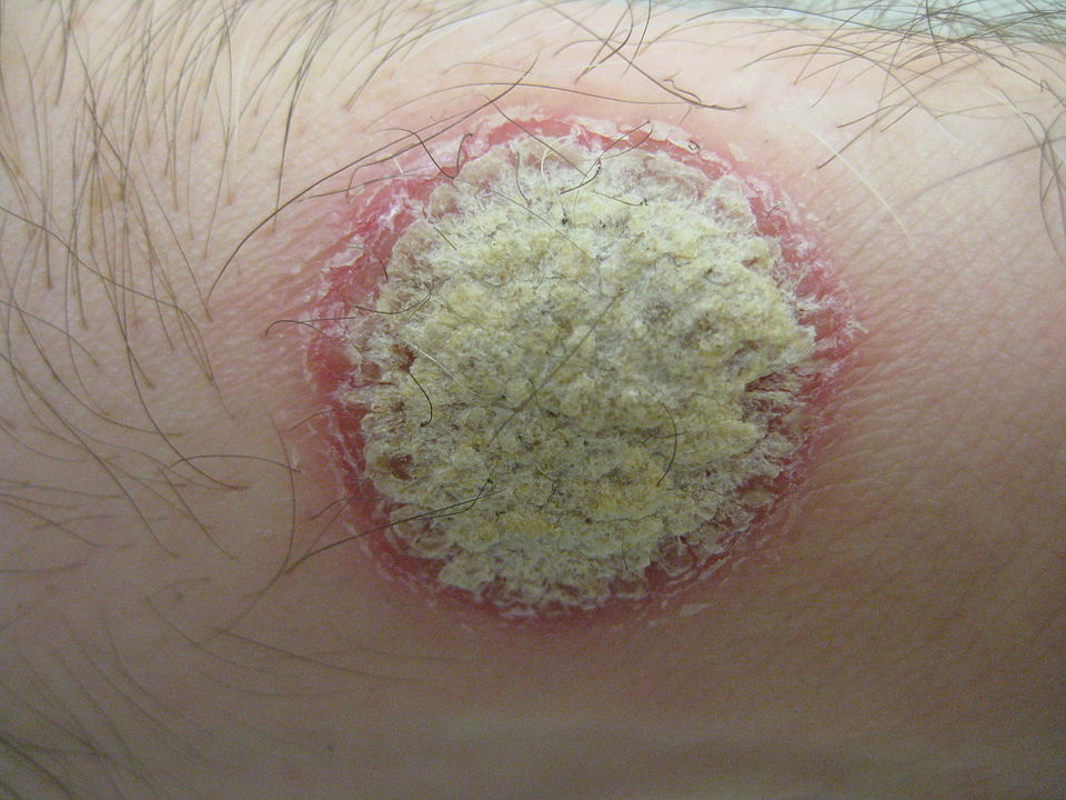
Psoriatic plaque, showing a silvery center surrounded by a reddened border. Source: Wikimedia. By James Heilman, MD – Own work, CC BY-SA 3.0

Considered to be the trigger for psoriais, the immune messenger molecule Interleukin-12 (IL-12) has now been shown to actually cause the skin disease but in fact protects against it. This finding also explains why common psoriasis drugs that block the messenger show insufficient treatment efficacy.

Psoriasis is a chronic inflammatory autoimmune disease that manifests as red, scaly skin patches. No causal treatment for the disease exists, but the symptoms can be significantly alleviated with modern therapies. The development of the skin disease arises from complex changes immune cell networks and the messengers they use for communication. Clinical trials showed that newly developed drugs that blocked only IL-23 are more effective than previous treatments targeting both IL-23 and IL-12 in psoriasis patients, but why this was so was not known. Now, researchers at the University of Zurich (UZH) have uncovered the underlying molecular mechanisms.

From human and mouse studies, they found that various cell types in the skin are also equipped with receptors for IL-12. Not only the T cells of the immune system, but also keratinocytes, horn-forming skin cells that build up the epidermis, can thus recognise the messenger. In fact, the recognition of interleukin-12 by these skin cells was responsible for the protective effect of the messenger, as the researchers found out. “Interleukin-12 is essential for the normal, physiological function of keratinocytes. For example, it prevents the increased cell division observed in psoriasis,” explained group leader Sarah Mundt from the Institute of Experimental Immunology at UZH.

“These results surprised us, because so far drugs for the treatment of psoriasis also aim at blocking interleukin-12,” said immunology professor Burkhard Becher.

“Our findings indicate that blocking IL-12 is not advisable, and such drugs should therefore no longer be used to treat psoriasis patients,” advised first author  Pascale Zwicky, PhD student. Accordingly, psoriasis drugs should only block the messenger substance IL-23, but no longer IL-23 and -12 together.

The UZH researchers’ findings could be important for the treatment of other diseases. “The combined blocking of IL-23 and -12 is also used in the treatment of chronic inflammatory bowel diseases and psoriatic arthritis,” said Prof Becher. “In these diseases, the role of IL-12 has not yet been sufficiently studied. But here, too, a protective role of the messenger substance is possible.”

Source: University of Zurich