Tag: innate immune response

Chronic Activation of the Innate Immune System can Unleash Cancer

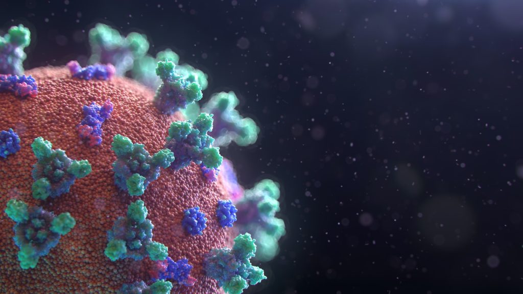
Photo by Sangharsh Lohakare on Unsplash

Along with defending against pathogens, the body’s innate immune system helps to protect the stability of our genomes in unexpected ways that have important implications for the development of cancer, researchers at Memorial Sloan Kettering Cancer Center (MSK) are discovering.

In a pair of recent papers, scientists in the lab of molecular biologist John Petrini, PhD, showed that innate immune signaling plays a key role in maintaining genome stability during DNA replication. Furthermore, the researchers showed that chronic activation of these immune pathways can contribute to tumour development in a mouse model of breast cancer.

Not only do the findings add vital insights to our understanding of fundamental human biology, says Dr Petrini, they may also shed new light on tumour initiation and present potential opportunities for new therapies.

“Living organisms have evolved complex pathways to sense, signal, and repair damaged DNA,” he says. “Here we’re learning new things about the role of the innate immune system in responding to that damage – both in the context of cancer and also in human health more generally.”

How Chronic Activation of the Innate Immune System Can Lead to Cancer

The newest paper, led by first author Hexiao Wang, PhD, a postdoctoral fellow in the Petrini Lab, and published in Genes & Development, reveals a connection between innate immune signaling and tumour development in breast tissue. And, Dr Petrini says, the data suggest that when instability arises in the genome, chronic activation of the innate immune system can greatly increase the chances of developing cancer.

The study focused on a protein complex called the Mre11 complex, which plays a pivotal role in maintaining the stability of the genome by sensing and repairing double-strand breaks in DNA.

To study how problems with the Mre11 complex can lead to cancer, the team manipulated copies of the protein in mammary tissue organoids (miniature lab-grown model organs) and then implanted them into laboratory animals.

When oncogenes (genes known to drive cancer) were activated in these mice, tumors arose about 40% of the time, compared with about 5% in their normal counterparts. And the tumors in the mice with mutant Mre11 organoids were highly aggressive.

The research further showed that the mutant Mre11 led to higher activation of interferon-stimulated genes (ISGs). Interferons are signaling molecules that are released by cells in response to viral infections, immune responses, and other cellular stressors.

They also found that the normally tightly controlled packaging of DNA was improperly accessible in these organoids — making it more likely that genes will get expressed, when they otherwise would be inaccessible for transcription.

“We actually saw differences in the expression of more than 5600 genes between the two different groups of mice,” Dr Petrini says.

And strikingly, these profound effects depended on an immune sensor called IFI205.

When the organoids were further manipulated so they would lack IFI205, the packaging of DNA returned almost to normal, and the mice developed cancer at essentially the same rate as normal mice.

“So what we learned is that problems with Mre11 – which can be inherited or develop during life like other mutations – can create an environment where the activation of an oncogene is much more likely to lead to cancer,” Dr Petrini says. “And that the real lynch pin of this cascade is this innate immune sensor, IFI205, which detects that there’s a problem and starts sending out alarm signals. In other words, when problems with Mre11 occur, chronic activation of this innate immune signaling can significantly contribute to the development of cancer.”

New Understandings Could Pave the Way for Future Treatments

The work builds on a previous study, led by Christopher Wardlaw, PhD, a former senior scientist in the Petrini Lab, that appeared in Nature Communications.

That study focused on the role of the Mre11 complex in maintaining genomic integrity. It found that when the Mre11 complex is inactive or deficient, it results in the accumulation of DNA in the cytoplasm of cells and in the activation of innate immune signaling. This research primarily looked at the involvement of ISG15, a protein made by an interferon-stimulating gene, in protecting against replication stress and promoting genomic stability.

“Together, these studies shed new light on how the Mre11 complex works to protect the genome when cells replicate, and how, when it’s not working properly, it can trigger the innate immune system in ways that can promote cancer,” D. Petrini says.

By shedding light on the interrelationships between these complex systems and processes, the researchers hope to identify new strategies to prevent or treat cancer, he adds, such as finding ways to short-circuit the increased DNA accessibility when Mre11 isn’t working properly.

Source: Memorial Sloan Kettering Cancer Center

New Discovery Explains How SARS-CoV-2 Evades Anti-viral Immunity

Image by Fusion Medical on Unsplash

The novel coronavirus SARS-CoV-2 has an enzyme that can counteract a cell’s innate defence mechanism against viruses, explaining why it is more infectious than the previous SARS and MERS-causing viruses. This discovery, from Kobe University, may point the way to the development of more effective drugs against this and possibly similar, future diseases.

When a virus attacks, the body’s immune response has two basic layers of defence: the innate and the adaptive immune systems. While the adaptive immune system grows stronger against a specific pathogen as the body is exposed to it multiple times and which forms the basis of vaccinations, the innate immune system is an assortment of molecular mechanisms that work against a broad range of pathogens at a basic level. The Kobe University virologist SHOJI Ikuo says, “The new coronavirus, however, is so infectious that we wondered what clever mechanisms the virus employs to evade the innate immune system so effectively.”

Shoji’s team previously worked on the immune response to hepatitis viruses and investigated the role of a molecular tag called “ISG15” the innate immune system attaches to the virus’s building blocks. Having learned that the novel coronavirus has an enzyme that is especially effective in removing this tag, he decided to use his team’s expertise to elucidate the effect of the ISG15 tag on the coronavirus and the mechanism of the virus’s countermeasures.

In a paper in the Journal of Virology, the Kobe University-led team is now the first to report that the ISG15 tag gets attached to a specific location on the virus’s nucleocapsid protein, the scaffold that packages the pathogen’s genetic material. For the virus to assemble, many copies of the nucleocapsid protein need to attach to each other, but the ISG15 tag prevents this, which is the mechanism behind the tag’s antiviral action. “However, the novel coronavirus also has an enzyme that can remove the tags from its nucleocapsid, recovering its ability to assemble new viruses and thus overcoming the innate immune response,” explains Shoji.

The novel coronavirus shares many traits with the SARS and MERS viruses, which all belong to the same family of viruses – which also have an enzyme that can remove the ISG15 tag. But their versions are less efficient at it than the one in the novel coronavirus, Shoji’s team found. And in fact, it has been reported recently that the previous viruses’ enzymes have a different primary target. “These results suggest that the novel coronavirus is simply better at evading this aspect of the innate immune system’s defence mechanism, which explains why it is so infectious,” says Shoji.

But understanding just why the novel coronavirus is so effective also points the way to developing more effective treatments. The Kobe University researcher explains: “We may be able to develop new antiviral drugs if we can inhibit the function of the viral enzyme that removes the ISG15 tag. Future therapeutic strategies may also include antiviral agents that directly target the nucleocapsid protein, or a combination of these two approaches.”

Source: Kobe University

Study Reveals a Possible Secret to Viral Infection Resistance in Humans

Colourised scanning electron microscope image of a natural killer cell. Credit: National Institutes of Health

Studying resistance to viral infections in humans is difficult because it’s virtually impossible to disentangle resisting being infected from simply not being exposed. By studying women who were accidentally exposed to hepatitis C (HCV) over 40 years ago, scientists in Ireland have uncovered a secret that may explain why some people are able to resist viral infections.

The extraordinary work, published in Cell Reports Medicine, has wide-ranging implications from improving our fundamental understanding of viral resistance to the potential design of therapies to treat infected people.

From 1977–79, several thousand women in Ireland were exposed to the hepatitis C virus through contaminated anti-D, a medication made using plasma from donated blood and given to Rhesus negative women who are pregnant with a Rhesus positive foetus. The medication prevents the development of antibodies that could be dangerous in subsequent pregnancies. Some of the anti-D used during the 1977–79 period was contaminated with hepatitis C.

Infected women fell into three groups: those who were chronically infected; those who cleared the infection with an antibody response; and those who appeared protected against infection but yet produced no antibodies against hepatitis C.

“We hypothesised that women who seemed to resist HCV infection must have an enhanced innate immune response, which is the ancient part of the immune system that acts as a first line of defence,” said senior author Cliona O’Farrelly, Professor of Comparative Immunology in Trinity’s School of Biochemistry and Immunology.

“To test this we needed to make contact with women exposed to the virus over forty years ago and ask them to help us by allowing us to study their immune systems to hunt for scientific clues that would explain their differing responses.

“After a nationwide campaign over 100 women came forward and we have gained some unique and important insights. That so many women – many of whom have lived with medical complications for a long time – were willing to help is testament to how much people want to engage with science and help pursue research with the potential to make genuine, positive impacts on society. We are deeply grateful to them.”

The scientists ultimately recruited almost 40 women from the resistant group, alongside 90 women who were previously infected.

In collaboration with the Institut Pasteur in Paris they then invited almost 20 women in each group to donate a blood sample that they stimulated with molecules that mimic viral infection and lead to activation of the innate immune system.

“By comparing the response of the resistant women to those who became infected, we found that resistant donors had an enhanced type I interferon response after stimulation,” said first author Jamie Sugrue, PhD Candidate. Type I interferons are a key family of antiviral immune mediators that play an important role in defence against viruses including hepatitis C and SARS-CoV-2, or COVID.

“We think that the increased type I interferon production by our resistant donors, seen now almost 40 years after the original exposure to hepatitis C, is what protected them against infection.

“These findings are important as resistance to infection is very much an overlooked outcome following viral outbreak, primarily because identifying resistant individuals is very difficult – since they do not become sick after viral exposure, they wouldn’t necessarily know that they were exposed. That’s why cohorts like this, though tragic in nature, are so valuable – they provide a unique opportunity to study the response to viral infections in an otherwise healthy population.”

The lab’s efforts are now focused on leveraging these biological findings to unpick the genetics of viral resistance in the HCV donors. Their work on HCV resistance has already helped ignite international interest in resistance to other viral infections such as SARS-CoV-2.

The O’Farrelly lab has expanded its search for virus-resistant individuals by joining in the COVID human genetic effort and by recruiting members of the public who have been heavily exposed to SARS-CoV-2 but never developed an infection.

Source: Trinity College Dublin

US Army Scientists Develop Novel Anthrax Treatment

Capsule removal from Bacillus anthracis by treatment with Capsule Depolymerase (capsule shown in red). Credit: Photomicrograph by Wilson J. Ribot, USAMRIID

By modifying an enzyme produced by the bacterium that causes anthrax, US Army scientists were able to protect mice from infection with the deadly disease. 

Their findings, published in Science Translational Medicine, suggest a potential therapeutic strategy for treating multidrug-resistant strains of anthrax, and could aid in the development of new treatments for other bacterial infections.

Bacillus anthracis, the bacterium that causes anthrax, is one of the most significant bioterrorism threats, as well as a public health challenge in many places around the world. Its disease-causing capability arised from three main components – lethal toxin, oedema toxin, and the capsule. Researchers in this study developed a method to degrade the capsule surrounding the bacterium, allowing it to be ingested and destroyed by white blood cells, reducing virulence.

There is increasing concern about strains of anthrax that appear to be resistant to treatment with known antibiotics, said Arthur M. Friedlander, MD, the paper’s senior author. He and his team explored alternative treatment approaches that do not rely on the use of antibiotic drugs.

One promising avenue is to make the bacterium more susceptible to the innate immune system. Enzymes known as capsular depolymerases, which are naturally produced by several classes of bacteria, have emerged as a potential new line of antivirulence agents.

“Identification of the capsule depolymerase enzyme within the anthrax bacillus led us to attempt to use that enzyme to remove the capsule,” said Friedlander. “When this proved successful, we utilised recombinant DNA technology and protein engineering methods to engineer and reconfigure the enzyme in new ways.”

Those “engineering changes” included enhancing stability and making production easier, and pegylation, to improve pharmacokinetics. The team then tested the pegylated enzyme, known as PEG-CapD-CPS334C, to be sure it had retained its enzymatic activity.

In the study, 10 out of 10 mice infected with anthrax spores from a nontoxigenic encapsulated strain were completely protected after treatment with PEG-CapD-CPS334C, compared to only 1 of 10 control mice surviving. Similarly, treatment of mice infected with a fully virulent encapsulated strain using PEG-CapD-CPS334C protected 8 of 10, while only 2 of 10 controls survived.

“This strategy renders B. anthracis susceptible to the innate immune responses and does not rely on antibiotics,” the authors concluded. “These findings suggest that enzyme-catalysed removal of the capsule may be a potential therapeutic strategy for the treatment of multidrug-resistant anthrax and other bacterial infections.”

It could also allow the treatment of soldiers exposed to anthrax through natural means or enemy attacks.

Source: EurekAlert!