Tag: injury

The Nagging Pain of Vaccination Shoulder Injuries

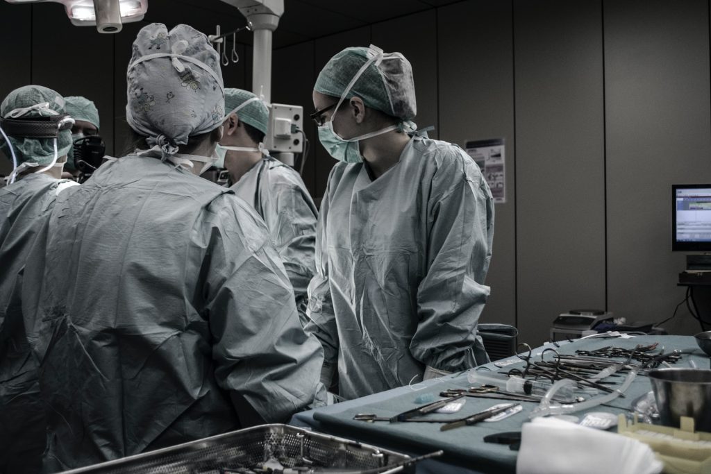
Image source: NCI on Unsplash

Shoulder injury related to vaccine administration (SIRVA) has been documented long before COVID, and most commonly reported after influenza vaccination. The cause is often due to poor administration. 

However, the medical community cautions that currently it’s more of a medicolegal determination rather than a distinct diagnosis. The condition is also plagued by the lack of a solid evidence base, and causality is difficult to pin down.

However, most physicians that MedPage Today interviewed put shoulder injury down to improper injection technique, and that these problems should be taken seriously and treated appropriately. One recent overview noted that SIRVA is a “rare yet increasingly recognised complication of immunisation.”

“We’re certainly not seeing a pandemic of SIRVA” from COVID vaccines, said Dr DJ Kennedy, chair of physical medicine & rehabilitation at Vanderbilt University Medical Center. “It’s really rare and the literature to date is mostly case reports. But I do think it’s possible, absolutely” for vaccine-related shoulder injury to occur.

Dr Laura Keeling, orthopedic surgeon at MedStar Georgetown University Hospital, told MedPage Today that part of the reason SIRVA remains in the medicolegal realm is that it’s “more of a constellation of symptoms and findings” as opposed to a specific diagnosis.

Symptoms can vary depending on where the stray shot landed, resulting in various manifestations such as bursitis, tendonitis, or adhesive capsulitis (aka ‘frozen shoulder’).

Generally, it’s characterised as a “constellation of shoulder pain and reduced range of motion that occurs within 48 hours of vaccination and does not resolve within 1 week,” according to a recent paper co-authored by Dr Keeling. It’s also different from typical post-injection soreness, as the pain is more severe and it can impact mobility and function.

Generally, treatments include anti-inflammatory drugs, corticosteroid injections, and physical therapy. Occasionally surgery is necessary to treat an underlying pathology such as an exacerbated rotator cuff injury. Patients with SIRVA often land in their GP’s office first, and then may be referred to a specialist such as a physiatrist or an orthopedic surgeon.

“It’s the patients who have persistent symptoms who are referred to orthopedic surgeons,” Dr Keeling said. “If physical therapy and injection don’t work, then primary care refers to us.”

Physical medicine & rehabilitation physicians, or physiatrists, also play a large role in treating SIRVA.

“We treat based on a full evaluation including history and physical findings, and imaging if needed,” Dr Kennedy said. “Then we develop a comprehensive rehabilitation plan … that usually involves doing range of motion and strengthening exercises on a daily basis.”

Scott Noren, DDS, an oral surgeon in Ithaca, New York, said after his second COVID shot in early February, he developed shoulder pain: “It went in pretty deep and pretty high,” he told MedPage Today.

An MRI revealed fluid collecting in his joint, as well as adhesive capsulitis, he said. Physical therapy helped improve his range of motion to an extent, but he has lingering pain. It’s difficult to take x-rays and do long procedures as an oral surgeon: “I have pretty good pain even with just normal function now,” he said.

Source: MedPage Today

A Leak-proof, Biocompatible Intestinal Patch

Researchers at Empa have developed a patch that stably seals two sutured pieces of intestine and thus prevents dangerous leaks.

A burst appendix or a life-threatening intestinal volvulus are emergencies that need to be treated by surgeons immediately. However, operations carry risks: highly acidic digestive juices and intestinal bacteria can leak out, causing peritonitis and sepsis.

Sealing sutured tissue with a plaster has already been tried, but the first were not well tolerated or were even toxic. Currently, these plasters are made of biodegradable proteins, which have variable clinical results. These is because they are mainly intended to support the healing process, and dissolve too quickly when in contact with digestive juices and don’t always hold tight. “Leaks after abdominal surgery are still one of the most feared complications today,” explained Empa researcher Inge Herrmann, who is also professor for nanoparticulate systems at ETH Zurich.

Searching for a material that could reliably seal intestinal injuries and surgical wounds, Hermann’s team found a synthetic composite material made up of four acrylic substances that, together, form a chemically stable hydrogel. Additionally, the patch actively cross-links with the intestinal tissue until it is fluid-tight. The quadriga of acrylic acid, methyl acylate, acrylamide and bis-acrylamide works in perfect synergy, as each component conveys a specific feature to the final product: a stable bond to the mucosa, the formation of networks, resistance to digestive juices and hydrophobicity. This new technology is detailed in Advanced Functional Materials.

In lab experiments, the researchers found the polymer system met their expectations. “Adhesion is up to ten times higher than with conventional adhesive materials,” said researcher Alexandre Anthis from Empa’s Particles-Biology Interactions lab in St. Gallen. “Further analysis also showed that our hydrogel can withstand five times the maximum pressure load in the intestine.” The material’s design uses its tailored effect: The rubbery composite selectively reacts with digestive juices that might leak through intestinal wounds, expands and closes all the more tightly. The inexpensive, biocompatible super glue, could thus shorten hospital stays and save healthcare costs, and Anthis is making plans to bring it to market.

Source: Empa

New Wound Dressing Minimises Scarring

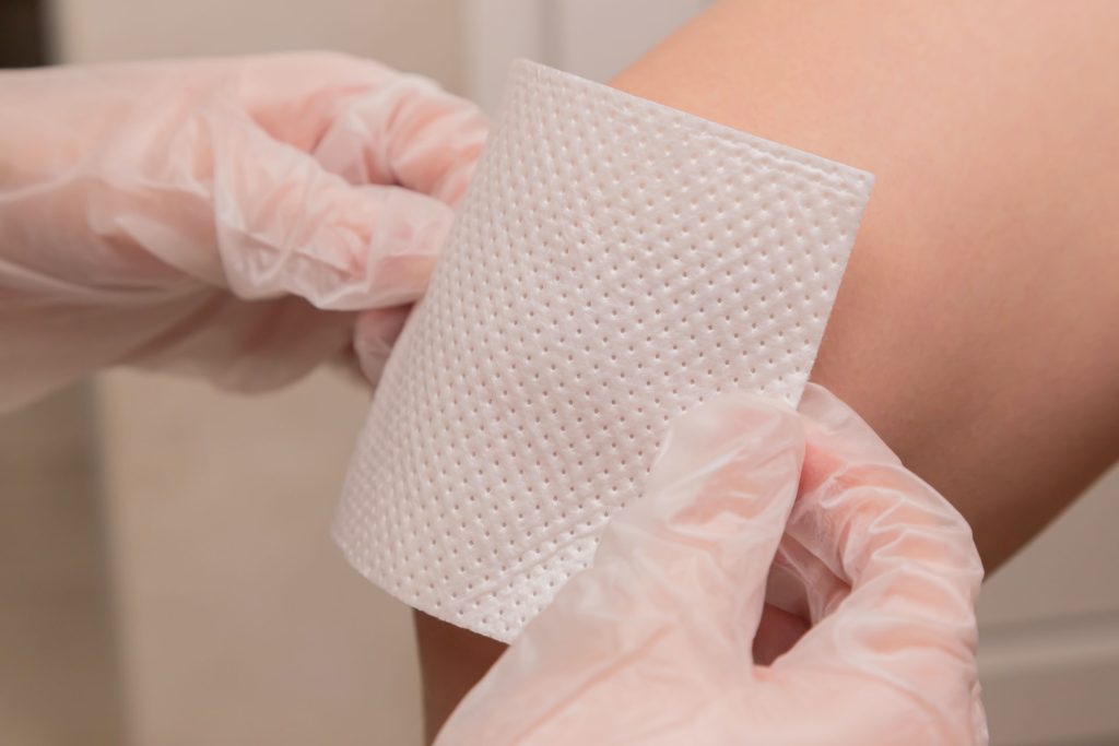
Photo by Diana Polekhina on Unsplash

A new wound dressing technology that can stop bleeding while preventing infection and scarring using a single material, has been developed. This technology also has potential applications in drug delivery, among other areas.

“Scarring is one of the worst consequences of severe wounds,” said Xiaoyang Wu, an associate professor in the Ben May Department of Cancer Research at the University of Chicago, noting that scar tissue formation is particularly common in human skin.

The researchers used a material science approach to develop a new method to overcome scarring, by impeding collagen synthesis by blocking transforming growth factor beta (TGF-β) – a cytokine that plays an important role in cell signaling, both in skin wound repair and tissue fibrosis.

“Increasing evidence suggests TGF-β is important in early phase wound repair for wound closure. But, later on, the signal may promote and enhance scarring,” Prof Wu said. This makes timing crucial. “We cannot simply block the signal, because that would slow down wound healing and would be dangerous for the patient,” he explained.

To get around this, the researchers came up with a delayed-release system combining a sutureless wound closure hydrogel material with a biodegradable microcapsule system, enabling them to control the release of the TGF-β inhibitor. “In this way, we can enhance skin wound repair and after 7-14 days can release the inhibitor that blocks the skin scarring process at the same time by using one material,” Prof Wu added.

The study results were recently published in Nature Communications.

At present, treatment of scarring is not ideal with little besides cosmetic surgery, and little can be done to prevent scar formation if a patient experiences a deep or messy wound. “The system we developed is very convenient for application,” said Wu, adding that the system has many possible future applications, such as drug delivery.

“We believe the novel system will have potential clinical importance in the future,” he said. To this end, the next steps include filing an investigational new drug (IND) application with the US Food and Drug Administration (FDA). Consistent manufacturing of the material is necessary and the researchers are exploring collaborations with pharmaceutical companies to move the research forward.

Since the system is a biocompatible material with adhesive properties, Wu said it has internal applications as well, adhering to and closing bleeding arteries and cardiac walls after irradiation with UV light. This was demonstrated in animal models, suggesting significant advantages as a traumatic wound sealant.

“Normal wound binding material does work well,” said Wu, noting that fibres are the most reliable material currently available, which, like surgical glue, is less biocompatible. “Biocompatibility is a significant advantage of our system,” he explained, “It is superior compared to current existing materials.”

Source: University of Chicago

New Wound Healing Scoring System Proposed

Photo by Tara Winstead from Pexels

Researchers have proposed a new scoring system for wound healing in mice based on parameters in each phase of healing.

The researchers described the system in an article in the peer-reviewed journal Stem Cells and Development.

Wound healing processes consist of a sequence of molecular and cellular events which occur after the onset of a tissue lesion in order to restore the damaged tissue. In order to evaluate the efficacy of new treatments, there is a need to monitor wound progression accurately and reproducibly over time. 

The parameters include re-epithelisation, epithelial thickness index, keratinisation, granulation tissue thickness, remodeling, and the scar elevation index. These parameters can be assessed using either Hematoxylin & Eosin or Masson’s Trichrome staining. Mari van de Vyver, from Stellenbosch University, and colleagues developed this histology scoring system for cutaneous wounds in mice. They then validated the system in four different types of murine skin wound models.

“This histological scoring system defines and describes the minimum recommended criteria for assessing wound healing dynamics,” state the authors. “The experience and ability of investigators to accurately identify structures in histology slides at different stages of healing is crucial for consistency and repeatability of measures to deliver meaningful results.”

“The development and validation of this scoring system in a randomized blinded investigation by researchers from Stellenbosch University (South Africa), Polish Academy of Sciences in Olsztyn (Poland), University of Texas Southwestern Medical Center (Texas, USA) and Obatala Sciences Inc. (New Orleans, USA) represents a truly international effort to advance the robust and accurate assessment of wound healing,” stated Graham C Parker, PhD, Editor-in-Chief of Stem Cells and Development and The Carman and Ann Adams Department of Pediatrics, Wayne State University School of Medicine, Detroit, MI.

Source: Mary Ann Liebert, Inc.

Journal information: van de Vyver, M., et al. (2021) Histology Scoring System for Murine Cutaneous Wounds. Stem Cells and Development.doi.org/10.1089/scd.2021.0124.

Why Nerves Fail to Regenerate

Image source: Pixabay

Though there are many reasons why nerves fail to regenerate, researchers from Ruhr-Universität Bochum (RUB) have made a breakthrough in their discovery a new mechanism which could lead to effective treatments.

Damage to nerve fibers in the central nervous system – brain, spinal cord, or optic nerve– often results in lifelong and severe disabilities, such as paraplegia or blindness. Though there are various known reasons why nerves fail to regenerate, treating them has not thus far not resulted in success.

Now, the RUB researchers have discovered that nerves release a protein at the injury site that attracts growing nerve fibres — and keeps them entrapped there. This prevents them from growing in the right direction to bridge the injury. Their findings are published in the journal Proceedings of the National Academy of Science (PNAS).

There are three known main causes for the inability of injured nerves of the central nervous system (CNS) to regenerate: insufficient activation of a regeneration program in injured nerve cells that stimulates the growth of fibres, so-called axons; scar formation at the injury site that is difficult for nerve fibres to penetrate; and an inhibitory effect of molecules in the nerve on regrowing axons. “Although experimental approaches have been found in recent decades to address these individual aspects by therapeutic means, even combinatorial approaches have shown only little success,” said Fischer. “So there must be other yet unknown causes for why nerve fibres in the CNS don’t regenerate.”

Using the optic nerve as a model, the research time has now shown another — quite surprising — cause for the regenerative failure in the CNS. The underlying mechanism is not based on inhibition of axon growth, as in the previously identified causes, but instead on a positive effect of a protein at the injury site on the nerve. This molecule is a so-called chemokine known as CXCL12. “The protein actually promotes the growth of axons and attracts regenerating fibers. It is, therefore, chemoattractive,” explained lead investigator Professor Dietmar Fischer. However, this chemoattraction turned out to be more hindrance than help after nerve injury in living animals.

Nerve fibres are trapped

The scientists showed that this protein is released at the nerve’s lesion site and, as a result, keeps the axons at the injured area through the chemoattractive effect. As a result, even some fibres that had already regenerated across the injury site reversed direction, growing backwards to the injury site. The regrowing fibers thus remained trapped due to CXCL12’s attractive effect.

The researchers figured out this effect when they knocked out the receptor for CXCL12 in the retinal nerve cells, rendering them blind to this protein. “Surprisingly, this led to greatly increased fibre growth in the injured optic nerves, and axons showed significantly less regrowth back to the injury site,” Dietmar Fischer points out.

New drug possibilities

The researchers then investigated where at the injury site the CXCL12 originated. They found out that about eight percent of the nerve cells in the retina produce this protein themselves, transport it along their fibers to the injury site in the optic nerve, and release it there from the severed axons. “It is still unknown why some of these nerve cells make CXCL12 and others make the receptor,” said Prof Fischer. “We don’t yet understand the physiological role of the protein, but we can see that it is a major inhibitor of neural repair.”

In further experiments, the researchers showed that knocking out CXCL12 in retinal nerve cells to prevent its release at the injury site equally improved axonal regeneration into the optic nerve. “These new findings open the opportunity to develop pharmacological approaches aimed at disrupting the interaction of CXCL12 and its receptor on the nerve fibres, to free them from their captivity at the site of injury,” concluded Prof Fischer.

His team is now investigating whether similar approaches can also promote the regeneration of axons in other areas of the injured brain or spinal cord.

Source: Ruhr-Universität Bochum

Journal information: Alexander M. Hilla, et al. CXCR4/CXCL12-mediated entrapment of axons at the injury site compromises optic nerve regeneration, in: PNAS, 2021, DOI: 10.1073/pnas.2016409118

Safety Commission Warns on Peloton Treadmill Hazard to Children

The US Consumer Product Safety Commission (CPSC) has put out a warning for owners of the popular Peloton Tread+ exercise machine following “multiple incidents of small children and a pet being injured beneath the machines.”

The warning comes weeks after Peloton CEO John Foley said a child died in an accident related to the machine. “While we are aware of only a small handful of incidents involving the Tread+ where children have been hurt, each one is devastating to all of us at Peloton, and our hearts go out to the families involved,” he said in a statement.

As a result, the CPSC launched an investigation into the treadmill, one that the commission says remains ongoing. The commission reported that it is aware of 38 other non-fatal safety incidents involving the device.
In the commission’s view, the Peloton Tread+ “poses serious risks to children for abrasions, fractures, and death” resulting from “children becoming entrapped, pinned, and pulled under the rear roller of the product.”

The announcement included a video of a child seemingly pulled underneath the treadmill while playing behind the machine.

The CPSC is advising those with children at home to stop using the Peloton Tread+ treadmill immediately and says that the hazard the machine imposes “cannot be avoided simply by locking the device when not in use.”

“Peloton has not yet agreed to do a recall or a stop sale,” Consumer Product Safety Commission spokesperson Joe Martyak told NPR. He continued, “We hope that will change.”

Generally, product recalls are done on a voluntary basis by companies, in concert with government.

Peloton responded to the CPSC, saying the warning was “inaccurate and misleading.”

“Like all motorized exercise equipment, the Tread+ can pose hazards if the warnings and safety instructions are not followed,” the company said. In response to further questions from NPR about a possible recall, a spokesperson for the company said “a recall has never been warranted.”

The spokesman added that, “The Peloton Tread+ is safe when operated as directed and in accordance with the warnings and safety instructions.”.

Source: NPR

New Adhesive Hydrogel For Soft Tissue Repair

Scientists have developed an injectable gel that serves as a biodegradable adhesive for various kinds of soft tissue injury.

Soft tissue tears are a common injury, and it is difficult for surgeons to secure the tissue back together, since stitches often do more harm than good. According to Dominique Pioletti, the head of the Laboratory of Biomechanical Orthopedics at EPFL’s School of Engineering, such surgeries often don’t produce the best results because the tissue doesn’t properly heal. 

Tears in tissue such as cartilage and the cornea, often fail to heal properly, and tissue repair strategies may be suboptimal. For example, loose pieces of cartilage are often excised for symptomatic relief, but the remaining cartilage in articulating joints is placed under greater burden and generates faster.

A long-standing goal for researchers around the world has been the development of an adhesive for soft tissue that can withstand the natural stresses and strains within the human body. Now, Pioletti’s group has come up with a novel family of injectable biomaterials that can adhere to various forms of soft tissue. Their gel-based bioadhesives, can be used in a variety of injury-treatment applications.
Like other hydrogels, this one has a high water content, 85%, and also has two key advantages: It is injectable anywhere in the human body, and it has high intrinsic adhesion without additional surface treatment. “What makes our hydrogel different is that it changes consistency while providing high adhesion to soft tissues,” said Peyman Karami, a postdoc at Pioletti’s lab who has developed the gel during his PhD. “It’s injected in a liquid form, but then sets when a light source is applied, enabling it to adhere to surrounding tissue.”

The hydrogel has an innovative design that allows its mechanical and adhesive properties to be tailored, making it an extremely versatile soft tissue glue that can be used throughout the human body.

To obtain these versatile properties in their hydrogel, the scientists took the base polymer and modified it with the compounds that play an important role in tissue adhesion. The first is known as Dopa and is derived from mussels. “Dopa is what lets mussels attach firmly to any kind of surface—organic or otherwise,” said Pioletti. The second is an amino acid that our bodies make naturally.

“The advantage of our hydrogel compounds is that, unlike some medical adhesives, they don’t interfere with the body’s chemical reactions, meaning our hydrogel is fully biocompatible,” said Karami.

The new hydrogel also possesses unique energy-dissipation characteristics that improve its adhesive capability. Karami added: “We had to achieve an adhesion mechanism for injectable hydrogels, through the resulting synergy between interfacial chemistry and hydrogel mechanical properties. The hydrogel is capable of dissipating the mechanical energy produced when the hydrogel deforms, so that it protects the interactions at the interface between the hydrogel and surrounding tissue.”

A further advantage of this hydrogel is that it can release drugs or cells to encourage tissue repair, which is especially beneficial for cartilage and other tissues that don’t regenerate on their own.

“Our in vitro tests showed that the hydrogel binds to many different kinds of tissue, including cartilage, meniscus, heart, liver, lung, kidney and cornea,” said Pioletti. “We’ve made a sort of universal hydrogel.”

The scientists have received a grant to research possible orthopedic applications of the gel, and hope to be able to release their innovation onto the market within the next five years.

Source: Medical Xpress

Journal information: An intrinsically‐adhesive family of injectable and photo‐curable hydrogels with functional physicochemical performance for regenerative medicine, Macromolecular Rapid Communications, DOI :10.100 2/marc.202000660

Positive Safety Evaluation for Tranexamic Acid

A large meta-analysis upheld the safety of tranexamic acid (TXA), even at higher doses.

TXA is an antifibrinolytic agent with a short half-life that is used for bleeding prevention and treatment, as in causes of trauma with open wounds. Current TXA is applied with caution due to perceived increased risk of seizures, MI, and other thrombotic complications.

The meta-analysis looked at 216 randomised trials involving 125 550 participants. The investigators found that the incidence of thromboembolic events, which included venous thrombosis, pulmonary embolism, venous thromboembolism, myocardial infarction (MI) or ischaemia, and cerebral infarction or ischaemia, was 2.1% of people receiving IV TXA and a similar 2.0% of peers getting placebo or another control, which was a non-significant difference.

TXA’s safety was inconclusive in those with neurological conditions, who showed increased heterogeneity and asymmetry in funnel plots, according to Patrick Meybohm, MD, of University Hospital Wuerzburg in Germany, and colleagues.

The review confirmed that TXA was linked to a significant reduction in overall mortality and bleeding mortality, but not nonbleeding mortality.

“The results of this study suggest that use of intravenous TXA may have utility in all medical fields, with some uncertainty for patients with neurological conditions,” the investigators concluded.

“Notably, we did not detect any dose-dependent association of TEs [thromboembolic events],” they stated. The included studies had participants with IV TXA administration at doses ranging 0.5-5g or 10-100mg/kg.

“There is little doubt that when used appropriately in the various patient populations evaluated with randomized clinical trials, TXA is effective. However, reasonable questions about thrombotic complications remain,” wrote John Holcomb, MD, of University of Alabama at Birmingham, and colleagues, in an invited commentary.

“Further research must focus on how to identify, as early as possible, the patients most likely to benefit from administration of TXA,” they urged.

One limitation mentioned by the investigators was the inclusion trials that evaluated thromboembolic events without ultrasound, so asymptomatic cases may have been excluded. Furthermore, many studies did not provide much information on thrombosis prophylaxis.

For Holcomb’s group, study’s main caveat was that it included “a notably heterogeneous population”, including a range of demographics and clinical conditions. Since the pooled studies were not “clinically homogeneous”, they violated “one of the cardinal tenets of systematic reviews and meta-analyses.”

Source: MedPage Today

Journal information (primary article): Taeuber I, et al “Association of intravenous tranexamic acid with thromboembolic events and mortality: a systematic review, meta-analysis, and meta-regression” JAMA Surg 2021; DOI: 10.1001/jamasurg.2021.0884.

Journal information (commentary): Holcomb JB, et al “Tranexamic acid and safety in the right patient” JAMA Surg 2021; DOI: 10.1001/jamasurg.2021.0929.

Social Plus Biological Factors Increase Knee Injuries Among Female Athletes

Writing in the British Medical Journal, scientists have argued that the study of knee injuries in female athletes is too focused on biological factors and not enough on social factors.

It is said that female athletes experience anterior cruciate ligament (ACL) injury, at a rate three to six times higher than their male counterparts.The ACL is one of the key ligaments that helps to stabilise the knee joint. This can be a career-ending injury, and occurs most often in sports involving sudden changes of direction such as basketball.

The authors of the paper argued that much of the focus still falls on biological and hormonal factors, with little attention paid to how sex-based factors are affected by the social concept of gender and how they could influence each other.

They suggested that gendered experiences are relevant in shaping female sport participation as well as disparities in injury outcomes. They demonstrated that, over a lifetime, gendered expectations of physical abilities (eg ‘throw like a girl’), to inequitable access to funding, training, and facilities for women’s sport (eg disparities in access to weight training).

Lastly, the authors suggested there may also be differences between post-injury rehabilitation for men and women recovering from an ACL injury. These social and environmental factors play a much bigger role in how sports injuries occur than once thought, they said, urging that there should be much more weight given to these issues.

Dr Sheree Bekker from the Department for Health at the University of Bath (UK) explained: “We wanted to unpack the biases and assumptions that we were seeing in research into and practice around sports injuries in girls and women. Specifically, we wanted to challenge the increasingly pervasive idea that this is simply a problem for girls/women because they are inherently prone to injury just because of their female biology. Approaching ACL injury prevention and management from a strictly biological view can propagate sexism in sport with detrimental consequences for girls and women.”

Dr Joanne Parsons from the University of Manitoba (Canada) added: “Over 20 years of research focussed on biological traits has failed to decrease the ACL injury rate in girls and women. To make a difference, we need to approach the problem in a different way. The recent challenges that the NCAA women’s teams faced with access to adequate training equipment is a perfect example of why we have to include society’s influence when talking about injury risk for girls/women.”

Paper co-author Dr Stephanie Coen of the University of Nottingham said: “By extending the focus from individual bodies and biology to the gendered environments contextualizing ACL injury, our approach identifies new opportunities to intervene and achieve better outcomes for girls and women, with implications beyond athletes. As childhood and youth physical activity levels influence those in adulthood, the sequelae of ACL injury can be lifelong and particularly concerning for girls and women who already participate in physical activity at lower rates than boys and men. There is a wider health equity issue at stake.”

Source: Medical Xpress

Journal information: Joanne L Parsons et al, Anterior cruciate ligament injury: towards a gendered environmental approach, British Journal of Sports Medicine (2021). DOI: 10.1136/bjsports-2020-103173