Tag: infection

Research Supports Letting A Moderate Fever Run its Course

Photo by Mockup Graphics on Unsplash

It may be better to let a mild fever run its course instead of automatically reaching for medication, new University of Alberta research suggests. Researchers found that, in fish models, untreated moderate fever helped them to quickly their infections, keep inflammation in check and repair damaged tissue. “We let nature do what nature does, and in this case it was very much a positive thing,” says Professor Daniel Barreda, immunologist and lead author on the study which is published in eLife.

Moderate fever is self-resolving, meaning that the body can both induce it and shut it down naturally without medication, Barreda explains. The health advantages of natural fever to humans still have to be confirmed through research, but the researchers say because the mechanisms driving and sustaining fever are shared among animals, it is reasonable to expect similar benefits are going to happen in humans.

That suggests the need to resist taking non-steroidal anti-inflammatory drugs at the first signs of a mild temperature, he says. “They take away the discomfort felt with fever, but you’re also likely giving away some of the benefits of this natural response.”

The study also sheds light on some benefits of moderate fever, which Barreda notes has been evolutionarily conserved across the animal kingdom for 550 million years. “Every animal examined has this biological response to infection.”

For the study, fish were given a bacterial infection and their behaviour was then tracked and evaluated using machine learning. Outward symptoms were similar to those seen in humans with fever, including immobility, fatigue and malaise. These were then matched to important immune mechanisms inside the animals.

The research showed that natural fever offers an integrative response that not only activates defences against infection, but also helps control it. The researchers found that fever helped to clear the fish of infection in about seven days – half the time it took for those animals not allowed to exert fever. Fever also helped to shut down inflammation and repair injured tissue.

“Our goal is to determine how to best take advantage of our medical advances while continuing to harness the benefits from natural mechanisms of immunity,” says Barreda.

Source: University of Alberta

Cytotoxic T Cells Become ‘Marathon Runners’ to Wage Long Immune Battles

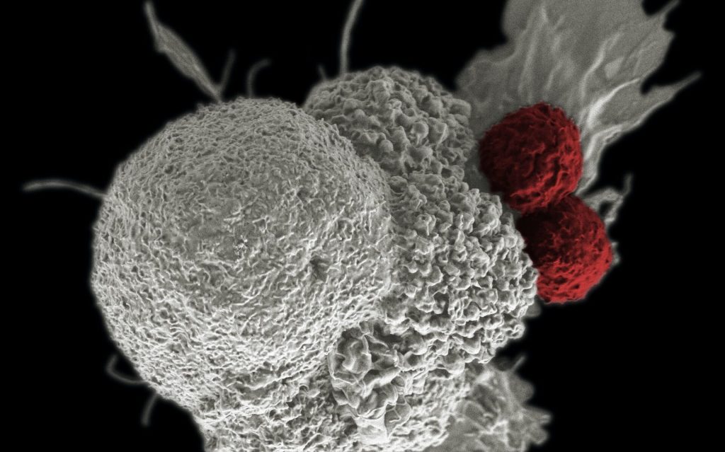
Shown here is a pseudo-colored scanning electron micrograph of an oral squamous cancer cell (white) being attacked by two cytotoxic T cells (red), part of a natural immune response. Photo by National Cancer Institute on Unsplash

When it comes to chronic infections and cancer, cytotoxic T cells play a central role in our defences. Research published in the journal Immunity has revealed that these cells can specialise into “sprinters” to fight a strong, short-term infection or into “marathon runners” for the long battle against chronic infections and cancer.

Professor Daniel Pinschewer at the Department of Biomedicine of the University of Basel led a study into understanding how cytotoxic T cells adapt to infection and cancer.

“These T cells can become specialised in two different ways: either as a kind of sprinter or as marathon runners,” explains Pinschewer. “However, the latter can also convert into sprinters at any time, in order to stamp out an infection.”

Chronic infections are a special case: the T cells are activated and a strong inflammatory response occurs at the same time. “This tends to ‘shock’ the T cells into developing into sprinters, which can only intervene effectively in the short term to remove infected cells,” says the virologist. “If all T cells behaved like that, our immune defences would break down pretty soon.”

Biological messenger counteracts the “shock”

The researchers examined how, in spite of this, the immune system is still able to provide enough T cells for the endurance race against chronic infections. According to their results, a biological messenger called interleukin-33 (IL-33) plays a key role. It allows the T cells to remain in their “marathon runner” state. “IL-33 takes away the shock of the inflammation, so to speak,” explains Dr Anna-Friederike Marx, lead author of the study.

In addition, the biological messenger causes the marathon T cells to proliferate, so that more endurance runners are available to combat the infection. “Thanks to IL-33, there are enough cytotoxic T cells around for the long haul that can still pull off a final sprint after their marathon,” says Marx.

The findings could help improve the treatment of chronic infections such as hepatitis C. It is conceivable that IL-33 could be administered to support an effective immune response. Thinking along the same lines, IL-33 could be one key to improving cancer immunotherapy, to enable T cells to wage an efficient and long-lasting offensive against tumour cells.

Source: University of Basel

Smartphone Pics of Post-surgical Wounds for Spotting Infections

Photo by Daniel Romero on Unsplash

Researchers have shown that smartphone pictures of post-surgical wounds taken by patients and then assessed by clinicians help spot infections early on.

These ‘surgery selfies’ were associated with a reduced number of GP visits and improved access to advice among patients who took them. This practice could help manage surgical patients’ care while they recover.

Death within 30 days of surgery is the third largest cause of mortality globally. More than a third of postoperative deaths are associated with surgical wound infections.

In the study, published in NPJ Digital Medicine, University of Edinburgh researchers conducted a randomised clinical trial involving 492 emergency abdominal surgery patients to determine if photos from smartphones and questions on symptoms of infection could be used to diagnose wound infections early.

One group of 223 patients were contacted on days three, seven and 15 after surgery and directed to an online survey, where they were asked about their wound and any symptoms they were experiencing. Then they were asked to take a picture of their wound and upload it.

A surgical team member assessed the photographs and patients’ responses were assessed for signs of wound infection. They followed up with patients 30 days after surgery to find out if they had been subsequently diagnosed with an infection.

A second group of 269 received standard care and were contacted 30 days after surgery to find out if they had been diagnosed with an infection.

No significant difference between groups was seen in the overall time it took to diagnose wound infections in the 30-days after surgery.

However, the smartphone group was nearly four times more likely to have their wound infection diagnosed within seven days of their surgery compared to the routine care group. They also had fewer GP visits and reported a better experience of trying to access post-operative care.

The research team is now conducting a follow-up study to determine how this can be best put into practice for surgical patients around the country. Artificial intelligence will also be used to help the clinical team in assessing the possibility of wound infection.

Professor Ewen Harrison, Professor of Surgery and Data Science, who led the research said: “Our study shows the benefits of using mobile technology for follow-up after surgery. Recovery can be an anxious time for everybody. These approaches provide reassurance – after all, most of us don’t know what a normally healing wound looks like a few weeks after surgery. We hope that picking up wound problems early can result in treatments that limit complications.”

Dr. Kenneth McLean, who co-led the research said: “Since the COVID-19 pandemic started, there have been big changes in how care after surgery is delivered. Patients and staff have become used to having remote consultations, and we’ve shown we can effectively and safely monitor wounds after surgery while patients recover at home – this is likely to become the new normal.”

Source: University of Edinburgh

Centenarians’ Unique Microbiomes Protect Against Bacterial Infections

Source: Miika Luotio on Unsplash

A new study has discovered that people who live to be 100 or older have a unique microbiome that may protect against certain bacterial infections  including those caused by multidrug-resistant bacteria. The findings, published in Nature, could point to new ways to treat chronic inflammation and bacterial disease.

A team of researchers studied microbes from  faecal samples of 160 Japanese centenarians who had an average age of 107. They found that centenarians, compared to people aged 85 to 89 and those between 21 and 55, had higher levels of several bacterial species that produce molecules called secondary bile acids. Secondary bile acids are generated by microbes in the colon and are thought to help protect the intestines from pathogens and regulate the body’s immune responses.

Next, the researchers treated common infection-causing bacteria in the lab with the secondary bile acids that were elevated in the centenarians. One molecule, called isoalloLCA, was found to strongly inhibit the growth of the  antibiotic-resistant bacterium Clostridioides difficile. Feeding mice infected with C. difficile diets supplemented with isoalloLCA similarly suppressed levels of the bacteria. The team also found that isoalloLCA potently inhibited or killed many other gram positive pathogens, suggesting that isoalloLCA may play a role in keeping the delicate equilibrium of microbial communities in a healthy gut.

“The ecological interaction between the host and different processes in bacteria really suggests the potential of these gut bugs for health maintenance,” said Plichta, a computational scientist at the Broad.

Additional studies from different regions around the world with more participants and longer duration could help find a causal link between longevity and bile acids. The bacteria identified in this study could help researchers in the meantime discover how to treat infections caused by antibiotic-resistant bacteria by manipulating bile acid.

“A unique cohort, international collaboration, computational analysis, and experimental microbiology all enabled this discovery that the gut microbiome holds the keys to healthy aging,” said co-first author Xavier, core institute member at the Broad. “Our collaborative work shows that future studies focusing on microbial enzymes and metabolites can potentially help us identify starting points for therapeutics.”

Source: Broad Institute of MIT and Harvard

Journal information: Sato Y, Atarashi K, et al. Unique bile acid-metabolizing bacteria in centenarians’ microbiome. Nature. Online July 29, 2021. DOI:10.1038/s41586-021-03832-5

New Biomarker for Soft Tissue Infections

This illustration depicted a three-dimensional (3D), computer-generated image, of a group of Gram-positive, Streptococcus pneumoniae bacteria. The artistic recreation was based upon scanning electron microscopic (SEM) imagery. Photo by CDC on Unsplash
This illustration depicted a three-dimensional (3D), computer-generated image, of a group of Gram-positive, Streptococcus pneumoniae bacteria. The artistic recreation was based upon scanning electron microscopic (SEM) imagery. Image by CDC on Unsplash

Researchers have identified a new and very promising biomarker for bacterial soft tissue infections, which previously lacked one. 

In bacterial soft tissue infections, rapid diagnosis is crucial in reducing the risk of severe injury or amputation. Vague symptoms and a varied patient presentations increase the risk of misdiagnosis.The study, by  Researchers at Karolinska Institutet in Sweden  and other research institutions, and published in the Journal of Clinical Investigation, may have implications for both diagnosis and treatment.

Last author Anna Norrby-Teglund, Professor, Department of Medicine, Karolinska Institutet, said: “There are currently no tools for safe, rapid diagnosis in life-threatening soft tissue infections. Our findings are consequently very interesting as the biomarkers identified are possible candidates for improved diagnostics. The results are also relevant for individualised treatment in the future.”

Necrotising soft tissue infections (NSTI) are bacterial infections which are characterised by rapid tissue degradation. Such infections, often caused by streptococci, while relatively uncommon, are extremely serious. In most cases they necessitate intensive care and can quickly become life-threatening.

Extensive surgery, intravenous antibiotics are often required to prevent the infection from spreading, and amputation may be required in extreme situations. Many patients also develop sepsis, which further complicates the course of the condition.

Early, correct diagnosis is crucial to save lives and avoid amputation, but this is complicated by factors such as vague symptoms including vomiting, fever and severe pain, as well as the heterogeneous group of patients. Despite recommendations for surgical evaluation in suspected NSTI, there is a considerable risk of misdiagnosis.

Currently, various laboratory tests, including white blood cell counts, are used as diagnostic tools, but suffer from low sensitivity. NSTI-specific biomarkers are therefore needed. The condition is classified into four types depending on the infecting organism.

Researchers at Karolinska Institutet, Haukeland University Hospital, Norway, and Copenhagen University Hospital, Denmark, have now been able to identify biomarkers specific to different patient groups with soft tissue infections.

Using machine learning, the researchers analysed 36 soluble factors in blood plasma from the 311 NSTI patients included in the international INFECT study. Control groups included patients with suspected NSTI and sepsis, respectively.

The analyses showed a new biomarker that accurately identifies patients with tissue necrosis.

“The new biomarker, thrombomodulin, proved to be superior to the laboratory parameters used clinically today. The analyses also identified biomarkers for patients with soft tissue infection caused by different types of bacteria, as well as patients who developed septic shock,” said first author Laura Palma Medina, researcher at the Department of Medicine, Karolinska Institutet (Huddinge).

Source: Karolinska Institutet

Journal information: Palma Medina, L.M., et al. (2021) Discriminatory plasma biomarkers predict specific clinical phenotypes of necrotizing soft-tissue infections. Journal of Clinical Investigation. doi.org/10.1172/JCI149523.